Received October 20, 2010 Accepted November 18, 2010
∙Woo Jin Choi, M.D.
Department of Orthopaedic Surgery, CHA Bundang Medical Center, CHA University, 351 Yatap-dong, Bundang-gu, Seongnam 463-712, Korea
Tel: +82-31-780-5289, Fax: +82-31-708-3578 E-mail: [email protected]
* 본 논문의 요지는 2010년도 대한정형외과학회 추계학술대회에서 발표되었음.
관절면을 침범한 설상형 종골골절의 수술적 치료: 관혈적 및 Essex-Lopresti 술식에 따른 비교
차의과학대학교 분당차병원 정형외과학교실, 차의과학대학교 구미차병원 정형외과학교실*
신동은⋅윤형구⋅한수홍⋅최우진⋅안창수*⋅옥현수
Operative Treatment of Tongue Type Intra-articular Calcaneal Fractures:
Comparison of the Open Reduction and Essex-Lopresti Technique
Dong-Eun Shin, M.D., Hyung-Ku Yoon, M.D., Soo-Hong Han, M.D., Woo Jin Choi, M.D., Chang-Soo Ahn, M.D.*, Hyun-Soo Ok, M.D.
Department of Orthopaedic Surgery, CHA Bundang Medical Center, CHA University, Seongnam, Korea Department of Orthopaedic Surgery, CHA Gumi Medical Center, CHA University, Gumi, Korea*
=Abstract=
Purpose: To analyze the clinical and radiological results of operative treatment in patients with tongue type intra-articular calcaneal fracture, and to compare the open reduction and Essex-Lopresti technique.
Materials and Methods: We examined a consecutive series of 42 patients who received surgical treatment for tongue type calcaneal fracture (24 cases of the open reduction and 18 cases of the Essex-Lopresti technique) and the postoperative data was compared with a minimum 1 year follow-up. The clinical outcome was analyzed using the American Orthopaedic Foot and Ankle Society (AOFAS) hindfoot scale and Salama's criteria. The preoperative, postoperative, and last follow-up changes in the Böhler angle was radiologically analyzed.
Results: There were no significant differences between the two groups in terms of the clinical and radiological results at the last follow-up. However, for the Sander's type 3 and 4 fractures, the open reduction group showed more improvement of AOFAS score and less reduction loss in the Böhler angle.
Conclusion: Although the clinical results were good irrespective of surgical technique, the open reduction and internal fixation can improve clinical outcome and reduce the reduction loss as compared with the Essex-Lopresti technique in the comminuted tongue type calcaneal fracture.
Key Words: Calcaneus, Tongue type fracture, Open reduction, Essex-Lopresti technique
서 론
종골은 체중에 대하여 견고하나 탄력성 있는 지주로서의 역할과 도약판(spring board)의 역할을 해주는 중요한 구조 물이다.1) 종골 골절은 일반적으로 관절을 침범한 골절과 침 범하지 않은 골절로 구분되는데, 거골하 관절을 침범한 경 우 골절편의 정확한 정복이 불가능하기 때문에 장기간의
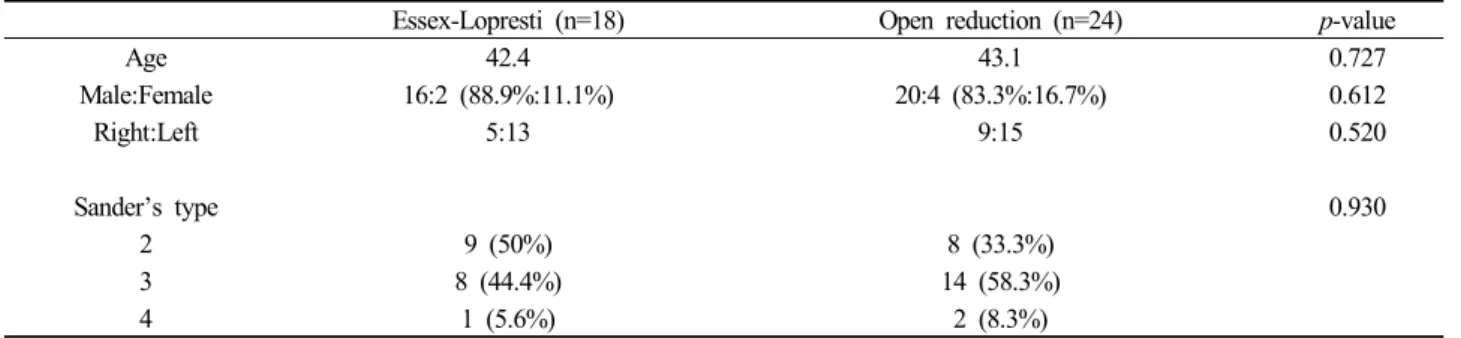
Table 1. Demographic Data
Essex-Lopresti (n=18) Open reduction (n=24) p-value
Age 42.4 43.1 0.727
Male:Female 16:2 (88.9%:11.1%) 20:4 (83.3%:16.7%) 0.612
Right:Left 5:13 9:15 0.520
Sander’s type 0.930
2 9 (50%) 8 (33.3%)
3 8 (44.4%) 14 (58.3%)
4 1 (5.6%) 2 (8.3%)
Figure 1. Essex-Lopresti technique.
치료에도 불구하고 동통과 기능장애를 초래하는 경우가 많 다. 치료는 보존적요법과 수술적 요법으로 대별할 수 있으 며, 수술적 요법에는 도수정복에 의한 Essex-Lopresti 술식2) 과 관혈적 정복에 의한 내고정술, 일차적 관절고정술 등이 있으나 여러 가지 수술방법에 대하여 많은 논란이 있어 왔 다. 그러나 좋은 결과를 얻기 위해서는 일반적인 골절치료 원칙과 마찬가지로 정상적인 해부학적 정복과 조기 관절운 동이 중요하며, 최근에 관혈적 정복술을 시행하여 좋은 결 과를 얻었다는 보고가 많다.3)
저자들은 관절을 침범한 설상형 종골 골절에서 수술적 치료를 시행하여 임상적 및 방사선학적 결과를 분석하고, Essex-Lopresti 술식과 관혈적 정복술에 따른 유용성을 비 교하고자 하였다.
대상 및 방법
1. 연구 대상
2004년 5월부터 2009년 5월까지 본원에 내원한 관절면 을 침범한 설상형 종골 골절 환자 중 수술적 치료를 시행 받고 최소 1년 이상 추시 관찰이 가능하였던 42예(Essex- Lopresti 술식 18예, 관혈적 정복술 24예)를 대상으로 하였 다(Table 1).
환자의 평균 나이 및 추시 기간은 전체 42.4세(16∼69 세)와 36.5개월(12~75개월), Essex-Lopresti 군의 경우 43.1 세(35~55세)와 50.2개월(12~60개월), 관혈적 정복술 군의 경우는 41.7세(16~69세)와 27.4개월(12~36개월)이었다. 성 별은 남자가 36명, 여자가 6명이었으며, Essex-Lopresti 군 의 경우 남자가 16명, 여자가 2명이었고, 관혈적 정복술군 의 경우 남자가 20명, 여자가 4명이었다.
Essex-Lopresti 방법과 관혈적 정복술의 선택은 시기적 으로 2004년 5월부터 2007년 4월까지는 Essex-Lopresti 방 법을, 2007년 5월부터 2009년 5월까지는 관혈적 정복술을
시행하였다.
2. 수술 방법 및 수술 후 처치
Essex-Lopresti 술식의 경우 보조자가 금속 강선을 물고 있는 요크를 이용하여 종골 장축을 따라 견인하면서 후족 부를 내반시킨 다음, 술자는 한 손은 Steinmann (S)-핀을 다른 손은 전족부를 잡고 엄지손가락 끝을 종골 골절부위 족저부에 대고 전족부는 족저부로 굽히고 S-핀을 족저부를 향해 굽힘으로서 후관절 골편이 정복되도록 하였다. 이때 보조자는 압박기를 이용하여 종골의 외측 돌출을 정복하면 서 후족부를 외반시켜 주었다. 영상 증폭기를 이용해서 정 복을 확인한 다음 다른 S-핀으로 골절을 고정하였다. 수술 후 단하지 석고고정을 6주간 하였고, S-핀은 수술 후 8주에 제거하였다.
관혈적 정복술의 경우 외측 도달법으로 비골 후방에서 시작하여 종입방 관절에 이르는 L-형의 피부절개를 가하고 피판을 골막과 함께 분리하여 종골의 외측면과 거골하관절 을 완전히 노출시켜 함몰된 후방관절과 골절을 정복하여 K-강선으로 임시적인 고정을 하고 측면 및 축면 방사선 촬 영으로 정복 상태를 확인하였다. 내고정은 대부분 F-형 금 속판과 나사를 이용하였으며 골절 정복 후 골 결손에 대하 여 골 이식을 시행하였다. 술 후 6주간 단하지 석고 고정을
Table 2. Salama’s Criteria
Excellent Patient satisfied, normal mobility of joint Asymptomatic no broadening of heel No pain
Good Patient satisfied with occasional pain Walking ability unaffected
Slight limitation of inversion-eversion Mild flat foot
Fair Patient not entirely satisfied (reserved) Pain after exertion
Walking ability reduced Limitation of tarsal movements Poor Patient not satisfied
Pain even on slight effort Walking ability markedly reduced Severe limitation of joint movements
Change of occupation
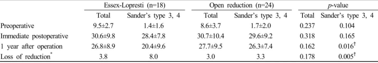
Table 3. Radiographic Data (Böhler angle)
Essex-Lopresti (n=18) Open reduction (n=24) p-value
Total Sander’s type 3, 4 Total Sander’s type 3, 4 Total Sander’s type 3, 4
Preoperative 9.5±2.7 1.4±1.6 8.6±3.7 1.7±2.0 0.237 0.104
Immediate postoperative 30.6±9.8 28.4±7.8 30.7±10.4 29.6±9.2 0.318 0.165
1 year after operation 26.8±8.9 20.4±9.6 27.7±9.5 26.3±7.4 0.162 0.016†
Loss of reduction* 3.8 8.0 3.0 3.3 0.178 0.005†
*Immediate postop - 1 year after operation; †p<0.05.
Figure 2. Open reduction and internal fixation with plate and screws.
하였고 방사선적 골 유합 상태에 따라 대게 8주 후부터 부 분 체중부하 보행을 허용하였다.
3. 방사선학적 및 임상적 평가
방사선학적 분석은 종골의 전후면, 측면 및 축면 방사선 촬영과 Broden’s view를 촬영하여 Essex-Lopresti 분류법2) 에 따라 설상형과 관절함몰형으로 분류하였고, 전산화 단층 촬영(Computed tomography)을 실시하여 Sanders 분류4)를 재적용하였다. 영상의학과 전문의 1인과 정형외과 전문의 1 인이 각각 3회씩 측정한 값의 평균값을 그 측정값으로 하였 으며 술 전, 후 및 최종 추시 Böhler 각을 측정하여 각각 수술 방법에 따라 나누어 분석하였다. Böhler 각은 종골의 측면 사진에서 전방돌기의 최고점과 후방관절면의 최고점 을 연결한 선과 후방 관절면의 최고점과 종골 조면의 최고 점을 연결한 선이 이루는 각으로 측정하였다.
임상적 결과의 판정은 American Orthopaedic Foot and Ankle Society (AOFAS) hindfoot scale5)과 환자의 만족도,
관절운동범위의 제한여부, 보행능력, 동통여부 등을 종합하 여 평가한 Salama 평가방법6)을 이용하였다(Table 2). 통계 학적 유의성의 검증은 SPSS (Statistical Package for the Social Sciences) version 12.0을 이용하였으며, 두 군 각각 의 수술 전 및 수술 후의 비교는 Mann-Whitney 검정을 적 용하였고, Essex-Lopresti군과 관혈적 정복술군간의 비교 분석은 Wilcoxon signed rank 검정을 이용하였다. 통계적 유의수준은 p값이 0.05 미만인 경우로 하였다.
결 과
Sanders에 따른 분류는 2형이 17예(2A 9예, 2B 8예), 3 형이 22예(3AB 6예, 3AC 9예, 3BC 6예), 4형이 3예로 2A 형이 가장 많았다. Essex-Lopresti 군의 Böhler 각은 술 전 평균 9.5±2.7도에서 술 후 30.6±9.8도로 향상되었고 술 후 1년에 26.8±8.9도로 약 3.8도의 정복 소실을 보였다. 관혈 적 정복술군에서는 술 전 평균 8.6±3.7도에서 술 후 30.7±
10.4도로 호전되었고 술 후 1년에 27.7±9.5도로 약 3.0도의 정복 소실을 보였다(Table 3). Böhler 각에서 수술 전, 수술 후, 최종 추시에 두 군 간에 통계적으로 유의한 차이는 없 었다(p>0.05). 또한 정복 소실률도 두 군 간에 통계적으로 유의한 차이가 없었다(p>0.05). 그러나 Sanders 분류의 3형 과 4형에 대한 정복 소실률을 비교한 경우에 Essex-Lopresti 군에서 통계적으로 유의하게 높은 정복 소실률을 보였다 (p<0.05) (Table 3).
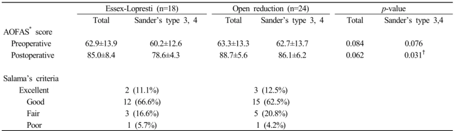
Table 4. Clinical Outcomes
Essex-Lopresti (n=18) Open reduction (n=24) p-value
Total Sander’s type 3, 4 Total Sander’s type 3, 4 Total Sander’s type 3,4 AOFAS* score
Preoperative 62.9±13.9 60.2±12.6 63.3±13.3 62.7±13.7 0.084 0.076
Postoperative 85.0±8.4 78.6±4.3 88.7±5.6 86.1±6.2 0.062 0.031†
Salama’s criteria
Excellent 2 (11.1%) 3 (12.5%)
Good 12 (66.6%) 15 (62.5%)
Fair 3 (16.6%) 5 (20.8%)
Poor 1 (5.7%) 1 (4.2%)
*AOFAS, American Orthopaedic Foot and Ankle Society; †p<0.05.
Essex-Lopresti 군의 AOFAS score는 술 전 평균 62.9±
13.9에서 최종 추시에 85.0±8.4로 호전되었으며 Salama 평 가는 우수 2예, 양호 12예, 보통 3예, 불량 1예였다. 관혈적 정복술군에서 AOFAS score는 술 전 평균 63.3±13.3에서 최종 추시에 88.7±5.6으로 호전되었고 Salama 평가에서 는 우수 3예, 양호 15예, 보통 5예, 불량 1예로 나타났다.
술 전, 술 후, 최종 추시에 각각 측정하여 비교한 결과 두 군 간에 통계적으로 유의한 차이를 보이지 않았다(p>0.05) (Table 4). 하지만, Sanders 분류의 3, 4형만을 따로 비교한 경우에는 Essex-Lopresti 군과 관혈적 정복술군의 술 후 AOFAS score가 각각 78.6±4.3과 86.1±6.2로 나와서 두 군 간에 통계적으로 유의한 차이를 보였다(p=0.031) (Table 4).
합병증으로는 Essex-Lopresti 군에서 핀 주위 감염 1예, 골 수염 1예, 거골하 관절의 관절염이 1예 발생하였고 관혈적 정복술군에서는 피부 절개 부위의 감염 2예, 피부괴사 1예, 골수염 1예, 거골하 관절의 관절염이 1예 발생하여 두 군 간에 유의한 차이는 없었다.
고 찰
전위된 관절 내 종골 골절의 치료방법에는 골절의 정복 없이 조기 운동시키는 방법, 도수정복 및 금속핀 내고정술, 관혈적 정복술 및 내고정술, 그리고 관절고정술 등에 이르 기까지 다양하며 최선의 치료 방법에 대해서는 많은 논란 이 있어왔다.2,7-12) 비관혈적 정복술 중에 주로 사용되고 있 는 도수정복 및 핀 고정술은 관혈적 정복술보다 간편하고 안 전한 방법으로써 Böhler,13) Hermann,14) Arnensen,15) Essex- Lopresti,2) Omoto 등7)에 의해 다양하게 주장되어 왔으며 그 중 Essex-Lopresti의 축성 핀 삽입에 의한 도수 정복술 (axial pin fixation)이 많이 사용되고 있다. Giachino와 Uhthoff,16)
Maxfield와 McDermott 등17)은 비관혈적 정복술로는 거골 하 관절을 침범하는 전위된 관절 내 골절의 정확한 정복을 거의 기대할 수 없고 관혈적 정복술에 의해서만 정확한 정 복을 얻을 수 있다고 하였으며, 핀 주위 감염을 대표적인 합병증으로 제시하였다. Ross와 Sowerby18)는 관혈적 정복 술의 목적은 해부학적 정복과 견고한 내고정 그리고 조기 관절운동이며 이러한 치료로써 비수술적 치료로 인하여 발 생하는 후기 합병증의 많은 부분을 예방할 수 있다고 하였 다. Palmer에 의해 보편화된 외측 도달법은 광범위한 절개 로 거골하 관절을 직접 노출시켜 정복할 수 있고, 신경혈관 손상의 위험이 없으며, 돌출된 외측 골편의 정복으로 비복 신경과 비골 건을 감압할 수 있고, 내고정하기가 좋으며, 종 입방 관절에 도달할 수 있는 등의 장점들이 있어 최근에 일 차적인 선택방법이 되었다.16,19)
본 연구에서 저자들은 Essex-Lopresti 술식을 시행할 때 충분한 전위정복을 얻고 유지할 수 있는지를 관혈적 정복 술과 비교 분석하였다. 임상적 평가로 이루어진 AOFAS score와 Salama 평가법은 두 군에서 유의한 차이를 보이지 않 았지만 Sanders 분류의 3, 4형만을 비교한 경우에는 AOFAS score가 관혈적 정복술을 시행한 군에서 통계적으로 유의 하게 호전되었다. 방사선학적 평가로 이루어진 수술 전, 후 및 최종 추시 관찰 시 측정한 Böhler 각은 두 군 사이에 통 계적으로 유의한 차이가 없었으나, Sanders 분류의 3, 4형만 을 따로 비교한 결과에서는 관혈적 정복술을 시행한 군의 정 복 소실률이 유의하게 낮았다. 이러한 이유로 Essex-Lopresti 술식을 이용할 경우 Sanders 분류의 3, 4형에서 골절편이 많아서 만족스러운 정복이 이루어지지 않으며 정복 후 골 유합 과정에서 관절 운동을 시작했을 때 정복의 유지력이 약하기 때문에 추시 관찰 시 정복의 소실이 일어났을 것이 라고 판단된다. 전위정복에 있어 대부분의 경우 수술 방법
에 상관없이 만족할 만한 전위정복을 얻을 수 있었다. Böhler 각은 종골 골절의 정복 시에 방사선 상 중요한 지표 가 되며 거골하 관절면을 회복시키고 종골이 옆으로 퍼지 지 않도록 정상적으로 회복시키는 것이 종골 골절의 치료 목적이라고 알려져 있다. 본 연구에서도 수술 방법에 무관 하게 42예 중 40예(95.2%)에서 만족할 만한 전위정복이 이 루어졌다. 그러나 그렇지 못한 2예(Essex-Lopresti 1예, 관 혈적 정복술 1예)는 Sanders 분류의 4형에 해당하는 골절 로서 분쇄가 심한 골절이었다. Chung 등20)은 설상형 골절 에서 관혈적 정복술을 시행하여 좋은 결과를 얻었으며 Sanders 등21)은 관혈적 정복술을 통한 결과를 보고하면서 예후 인자로 해부학적 정복과 골절편의 수를 제시하였다. 실제로 설상형 골절에서 술 전 전산화 단층촬영을 시행하 였을 때 Sanders 분류 2형과 더불어 3, 4형을 관찰할 수 있 는데 본 연구에서도 전체 42예 중 24예(57.1%)가 3형 이상 으로 분류되었다. Pennal과 Tadav10)는 분쇄가 심한 종골 골 절에 대해서는 일차적 거골하 관절 고정술을 시행하여 이 차적인 관절 고정술보다 더 좋은 결과를 얻었다고 하였다. Essex-Lopresti 술식에서 도수정복으로 만족할 만한 전위정 복이 이루어지지 않을 경우 효과적으로 Böhler 각을 회복 하고 유지하기 위해 추가적인 S-핀 등을 사용하여 정복을 시도할지, 아니면 관혈적 방법으로 전환하여 정복을 시도할 지가 관건이라고 생각한다. 따라서 관절면을 침범한 설상형 종골 골절에서 간편하고 안전한 Essex-Lopresti 술식으로 정복을 시도하고, 만일 만족스러운 정복을 얻지 못할 경우 관혈적 방법으로 전환하여 정복을 시도하는데 이때 Sanders 분류 3형 이상이면서 관절면의 분쇄가 심한 경우 처음부터 관혈적 정복술을 시도하는 것이 좋을 것으로 생 각된다. 관혈적 정복술의 경우 일반적으로 Essex-Lopresti 술식에 비하여 술 후 합병증이 많은 것으로 알려져 있으며 가장 흔한 합병증으로 창상감염에 의한 열개(wound dehiscence)가 저자에 따라서 약 25%까지 보고되고 있으나 본 연구에서는 발생하지 않았고 골수염이나 거골하관절의 관절염 등도 두 군간에 차이가 없었다. 본 연구는 대상규모 가 작고 두 군간의 추시 기간이 다른 한계점, 방사선학적 지표로 Böhler 각 한 가지만을 비교한 점이 있어 향후 더 큰 환자군을 대상으로 다양한 비교분석이 필요하다고 생각 한다.
결 론
관혈적 정복술 및 금속내고정술은 관절면을 침범한 설상 형 종골 골절의 치료에서 전통적인 Essex-Lopresti 술식과
더불어 효과적인 방법이며, 임상적, 방사선학적 결과에 있 어서 차이가 없으나 후방 관절면의 분쇄가 심한 Sanders 분 류 3, 4형에서는 관혈적 정복술 및 내고정술을 이용하여 정 확한 해부학적 정복 및 조기관절운동을 함으로써 Essex- Lopresti 술식에 비하여 만족스러운 결과를 얻을 것으로 사 료된다.
REFERENCES
1. Harty M. Anatomic considerations in injuries of the calcaneus.
Orthop Clin N Am. 1973;4:179-83.
2. Essex-Lopresti P. The mechanism, reduction technique, and results in fractures of the os calcis. Br J Surg. 1952;39:395- 419.
3. Burdeaux BD. Reduction of calcaneal fractures by the McReynolds medial approach technique and its experimental basis. Clin Orthop Relat Res. 1983;177:87-103.
4. Sanders R. Intra-articular fractures of the calcaneus: present state of the art. J Orthop Trauma. 1992;6:252-65.
5. Kitaoka HB, Alexander I, Adelaar RS, Nunley JA, Myerson MS, Sanders M. Clinical rating systems for the ankle-hindfoot, midfoot, hallux, and lesser toes. Foot Ankle Int. 1994;15:349-53.
6. Salama R, Benamara A, Weissman SL. Functional treatment of intra-articular fractures of the calcaneus. Clin Orthop.
1976;115:236-40.
7. Omoto H, Sakurada K, Sugi M, Nakamura K. A new method of manual reduction for intra-articular fracture of the calcaneus.
Clin Orthop. 1983;177:104-11.
8. Miller ME. Surgical management of calcaneal fractures:
indications and techniques. Instr Course Lect.1990;39:161- 65.
9. Hall MC, Pennal GF. Primary subtalar arthrodesis in the treatment of severe fractures of the calcaneum. J Bone Joint Surg. 1960;42:336-43.
10. Pennal GF, Tadav MP. Operative treatment of comminuted fractures of the os calcis. Orthop Clin N Am. 1973;4:197- 211.
11. McReynolds IS. Trauma to the os calcis and heel cord. In:
Jahss MH, ed. Disorders of the foot. Philadelphia:WB Saunders; 1982. 1497-1542.
12. Stephenson JR. Treatment of displaced intra-articular frac- tures of the calcaneus using medial and lateral approaches, in- ternal fixation, and early motion. J Bone Joint Surg. 1987;
69:115-30.
13. Böhler L. Diagnosis, pathology, and treatment of fractures of the os calcis. J Bone Joint Surg. 1931;13:75-89.
14. Hermann OJ. Conservative therapy for fracture of the os calcis. J Bone Joint Surg. 1937;19:709-18.
15. Arnensen A. Treatment of fractures of the os calcis with traction and manipulation. Acta Chir Scand. 1966;132:566- 73.
16. Giachino AA, Uhthoff HK. Intra-articular fracture of the
calcaneus. J Bone Joint Surg. 1989;71:784-7.
17. Maxfield JE, McDermott FJ. Experiences with the palmer open reduction of fractures of the calcaneus. J Bone Joint Surg. 1955;37:99-106.
18. Ross SDK, Sowerby MRR. The operative treatment of fractures of the os calcis. Clin Orthop. 1985;199:132-43.
19. Paley D, Hall H. Calcaneal fracture controversies: can we put humpty dumpty together again? Orthop Clin N Am. 1989;20:
665-77.
20. Chung HJ, Ahn JK, Bae SY, Jung H. Operative treatment of intraarticular calcaneal fractures using extensile lateral approach. J Korean Foot Ankle Soc. 2009;13:60-7.
21. Sanders R, Fortin P, DiPasquale T, Walling A. Operative treatment in 120 displaced intraarticular calcaneal fractures.
Results using a prognostic computed tomography scan classification. Clin Orthop Relat Res. 1993;290:87-95.