http://dx.doi.org/10.12790/jkssh.2013.18.3.111
THE HAND
서론
주상골 골절은 수근 골 골절 중 가장 흔히 발생하는 골절로 서 적절한 치료로써 90%-95%의 골유합을 얻을 수 있는 것으 로 알려져 있으나 비수술적 치료 시 약 5%-10%에서 불유합 이 일어난다1-5. 주상골 골절에 대하여 수술적 치료가 널리 행 해지고 있으며 무두 나사가 주상골 골절 시 고정에 사용되는
다른 기구들과의 비교에서 좋은 결과를 나타낸 것으로 알려져 있다6,7. 이에 대한 접근은 수장측과 수배측 도달법이 있으며 수장측 접근이 널리 쓰인다. 수배측 도달방법은 1989년에 소 개된 방법으로 근위부 1/3 부위로의 접근이 용이하며, 수근골 간 인대 손상의 확인과 치료, 수장측 접근보다 더 장축으로 나 사를 고정할 수 있으며 신전건 및 연부조직이 많지 않아서 견 인 시 수술 부위의 시야를 확보하기 더 쉬운 장점이 있다8. 주
Headless Autocompression Screw Fixation of
Scaphoid Fractures Using Open Dorsal Approach
Ho-Jung Kang, Yougun Won, Ji-Won Kwon, Il-Hyun Koh, Yun-Rak Choi
Department of Orthopaedic Surgery, Yonsei Univeristy College of Medicine, Seoul, Korea
Received:June 24, 2013 Revised:September 9, 2013 Accepted:September 13, 2013 Correspondence to:Ho-Jung Kang Department of Orthopaedic Surgery, Yonsei Univeristy College of Medicine, 50 Yonsei-ro, Seodaemoon-gu, Seoul 120-785, Korea TEL:+82-2-2019-3414
FAX:+82-2-2573-5393 E-mail:[email protected]
Purpose:We present the clinical and radiological results of open reduction and internal fixation for scaphoid fracture with retrograde headless screw fixation via dorsal approach.
Methods:This study carried out a survey targeting 15 patients who have a retro- grade headless screw fixation on nonunion of scaphoid fracture without previ- ous operation, 2 patients who have a retrograde headless screw fixation on nonunion of scaphoid fracture with previous operation and 8 patients who have a trans-scaphoid perilunate dislocation. We figured out a mechanism of injury, and clinical symptom, radiologic findings. The surgery was done with open dor- sal approach which is retrograde headless screw fixation internally, with or without bone graft. We analyzed the result by Maudsley method, in terms of bone union, duration for union, radiologic finding, clinical outcomes.
Results:After surgery, 22 of 25 patients had union result on fracture and other 3 patients had nonunion result. It took 12 weeks to achieve bone union on aver- age. Based on radiograhs, we had one case of partial avascular necrosis of proxi- mal fragment without clinical symptoms. We had one case of each scaphoid nonunion without previous operation, with operation and trans-scaphoid per- ilunate dislocation had arthritic change and non-symptomatic nonunion result.
In terms of clinical outcome, 22 patients showed satisfactory results and 3 patients had slight limitation of range of motion.
Conclusion:Retrograde headless screw fixation with or without bone graft for the treatment of scaphoid fracture is recommendable.
Keywords:Scaphoid fracture, Headless screw, Nonunion, Dorsal approach, Trans-scaphoid perilunate dislocation
This is an Open Access article distributed under the terms of the Creative Commons Attribution Non-Commercial License (http://creativecommons.org/ licenses/by- nc/3.0/) which permits unrestricted noncommercial use, distribution, and reproduction in any medium, provided the original work is properly cited.
상골 근위부 골절이나 불유합 및 전체 수근골 골절의 약 10%
발생빈도를 보이는 주상골 경유 월상골 주위 탈구의 경우 수 술적 접근 및 치료방법에 이견이 있으며, 불유합, 무혈성 괴사 등의 합병증이 빈발하는 것으로 알려져 있다. 특히 주상 근위 부의 작은 골편에 대해서 내고정술이나 골이식술 등을 시행하 기가 기술적으로 매우 어려워 다양한 치료 방법이 보고되고 있으며 수배측 접근을 통한 수술적 치료 후 양호한 결과가 보 고 되었다9-11.
저자들은 주상골 골절 치료에 있어 무두 나사를 수배측 접 근술로 주상골의 근위에서 원위로 역방향 내고정 및 골이식술 을 실시하였으며 그 치료 결과를 알아보고자 한다.
대상 및 방법
2000년 3월부터 2011년 7월까지 주상골 신선 골절에 대하
여 보존적 처치 후 발생한 불유합, 주상골 골절에 대하여 내고 정 시행 후 발생한 불유합 및 주상골 경유 월상골 주위 탈구로 수술받은 환자를 대상으로 연구하였고 그 중 경피적 핀 고정 술, 최소 절개 방법 혹은 최소침습적 방법은 제외하였다. 본원 에서 지속적으로 추시한 환자를 대상으로 손상 원인, 임상적 증상, 방사선학적 양상 등을 조사하였다. 대상 환자의 성별은 모두 남자였으며 수술 전 평균 연령은 30세(범위, 18-54세)였 다. 주상골 신선 골절 후 발생한 불유합 환자가 15예였으며 손 상 후 불유합 수술까지의 기간은 평균 30개월로 3-12개월이 8예, 12개월 이상인 경우가 7예였다. 주상골 경유 월상골 주위 탈구 환자가 8예, 내고정 시행 후 발생한 불유합 환자가 2예였 으며 첫 수술에서 전방 접근으로 내고정 시행되었고 재수술 시 전방으로 접근하여 내고정을 제거한 뒤 후방 접근하여 재 고정을 시행하였다. 1예에서는 첫 수술의 내고정기기가 해리되 어 전방 접근으로 제거한 뒤 후방 접근으로 재고정 및 골이식
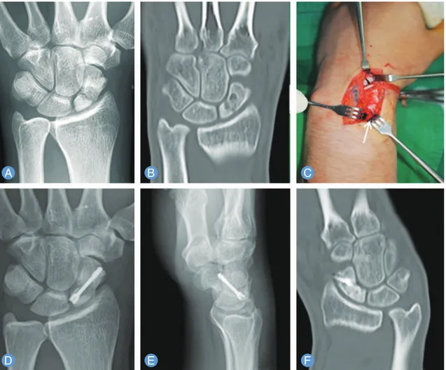
Fig. 1. (A)
Preoperative radiograph shows nonunion of scaphoid.
(B)Preoperative computed tomography (CT) shows a
radiolucent lesion with sclerotic and cystic changes of proximal pole of scaphoid.
(C)Harvesting autogenous radial bone
for graft under primary skin incision.
(D, E)Retrograde fixation of nonunion fragment with Herbert screw and autogenous
bone graft.
(F)One year after retrograde fixation follow-up CT shows complete radiologic union.
을 시행하였다. 첫 수술 후 재수술까지의 기간은 각각 2년, 13 년이었다. 주상골 골절의 분류는 Herbert 분류를 따랐으며12 주상골 골절의 신선 골절 및 내고정 후 발생한 17예 중에서 근 위부는 11예, 요부는 6예였으며, 원위부의 예는 없었다. 주상 골 경유 월상골 주위 탈구는 수배측 탈구와 수장측 탈구로 발 생할 수 있는데 각각 4예씩 확인되었으며 수배측 탈구에서 주 상골의 원위부, 요부, 근위부 골절은 각각 1예, 2예, 1예였으며 수장측 탈구에서는 1예, 3예였으며 근위부의 예는 없었다. 한 개의 무두 나사로 고정한 경우가 17예였으며(Fig. 1), 2개의 무
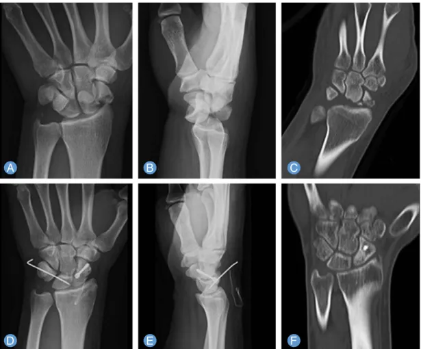
두 나사를 이용하여 고정한 경우가 2예였다. 이외에 무두 나사 와 K강선, mini 나사를 함께 사용한 경우가 6예였다. 수상 원 인으로는 손을 짚고 넘어진 경우가 10예, 추락에 의한 경우가 3예, 외력에 의한 직접 손상이 6예였다(Fig. 2). 추시 관찰은 수술 후 2개월마다 시행하였으며 평균 추시기간은 18개월(범 위, 12-16개월)이었습니다. 치료 결과에 대한 임상적인 평가 는 Maudsley와 Chen13의 평가 기준을 이용 하여 동통, 압통, 경직도를 평가하였고(Table 1), 작업 능력, 수근 관절의 동통 과 운동범위 및 방사선학적 유합 정도를 우수, 양호, 보통, 불 Fig. 2. (A, B)
Preoperative radiograph show trans-scaphoid perilunar dorsal dislocation.
(C)Preoperative computed
tomography (CT) shows comminuted fracture lines on waist of scaphoid.
(D, E)Open reduction and retrograde Using dorsal Herbert screw and a Kirschner’s wire.
(F)1.5 year after retrograde fixation follow-up CT shows complete radiog- ic union.
Table 1.
Method of assessment in clinical result (by Maudsley and Chen)
Excellent (-) (-) (-)
Good Mild (+) Mild
Fair Discomfort (+) Restriction in full motion
Poor (+) Interfering normal work Limitation
Assessment result Clinical
Pain Tenderness Stiffness
량의 4등급으로 분류하였다. 방사선 결과를 평가하기 위하여 수술 전과 수술 후 및 추시 관찰 때 마다 수근관절 전후면 및 측면 방사선과 수근 관절 척측, 요측 굴곡 및 30�외회전 방사 선 사진을 촬영하였으며, 방사선학적 결과를 평가하기 위하여 골절선의 유무, 전이 정도, 골절선의 경화 현상 또는 부분적인 낭종 형성 또는 골 흡수 소견 및 요골-주상골 간 퇴행성 변화 등을 관찰하였다. 최종 추시 측면 주상-월상골간 각도를 측정 하여 수근골간 인대의 손상으로 인한 완관절의 수근골간 불안 정성이 있는지를 관찰하였다. 모든 환자에서 수술 전에 컴퓨 터단층촬영을 시행하였고 수술 후 6개월에서 1년 사이에 다시 컴퓨터단층촬영을 시행하였다. 골절 간격이 소실되면서, 관절 선을 가로지르는 골소주가 관찰되면 골유합된 것으로 판단하 였다3,14.
결과
3예를 제외하고 임상적 및 방사선학적으로 골유합을 얻었으 며 수술 후 평균 유합 기간은 12주(범위, 6-16주)였다. 최종 추시의 결과 측정을 위해 사용한 Maudsley와 Chen13의 평가 법에 따르면 10예에서 통증 및 압통을 호소하지 않아 우수 소 견을 보였다. 12예에서는 수술 전보다는 증상이 호전되었으나 운동이나 휴식 시에 통증 및 압통을 호소하는 양호 소견을 보 였다. 2예에서는 운동이나 휴식 시 통증 압통 및 운동 범위의 제한이 나타나는 보통 소견을 보였다. 1예에서는 압통이 심하 고 일상 생활에 장애가 있을 정도의 통증을 보이는 불량소견 을 나타내었다. 54세 남환의 비 우세수에서 발생한 골절에 대 하여 보존적 치료하였으나 발생한 불유합에 대하여 수술 후 추시 18개월 째 주상골의 지연성 무혈성 괴사가 1예 합병증으 로 확인되었으나 환자는 증상을 나타내지는 않았다. 최종 추 시 시 무증상 불유합 3예, 방사선사진 소견에서 주상골 근위 골절 편의 경화성 소견 및 요-주상 관절에 초기 퇴행성 변화 (scaphoid nonunion advanced collapse, SNAC stage I)를 보이는 경우가 3예에서 발견되었다. 측면 주상-월상골 간 각 도는 평균 51�(범위, 43�-56�)로 전례에서 정상범위 내로 측 정되어 수근골간 불안정성은 관찰되지 않았다.
고찰
주상골 골절 수상 후 6개월이 지나도 골유합의 소견이 방사 선적으로 관찰되지 않는 경우 불유합으로 진단된다. 이로 인 해 수근부의 불안정성, 운동장애와 통증이 소실되지 않고, 방 치하게 되면 수근관절 주변의 퇴행성 변화(SNAC)로 관절염이
속발하게 되어 기능을 현저하게 저하시키므로, 적극적인 치료 를 요한다4,15,16. 주상골의 경우 혈액 공급은 외측 수장부, 수배 측, 원위부 세 개의 동맥군을 통해서 이루어지는데 원위부에 서 근위부로 혈액이 공급되는 구조를 가지고 있어 근위부는 원위부에 비하여 제한된 혈액 공급을 받고 있다. 이러한 해부 학적인 구조로 인해 근위부로 갈수록 불유합의 가능성이 높으 며 본 연구에서도 주상골 신선 골절 후 발생한 불유합과 내고 정술 후 발생한 불유합의 17예 중 11예가 근위부였고 나머지가 요부로 원위부의 예는 없었다. 주상골 골절의 관혈적 정복술 및 내고정을 위해서는 수장측으로 접근하는 방법과 수배측으 로 접근하는 방법이 있다. 수장측 접근을 통한 주상골 골절의 치료는 주상골의 70%-80%의 혈액 공급이 요동맥을 통한 수 배측으로 주행하는 것에 대한 손상을 주지 않으며 요부와 원 위부 골절에 대한 접근이 용이하고 곱사등 변형에 대한 교정 이 더 쉽다는 장점이 있다16-18. 단점으로는 근위부 골절에 대한 내고정이 어렵고 장측 요골수근인대의 손상을 줄 수 있다. 배 측으로의 접근은 원위 요골의 작은 골편에 대한 접근이 용이 하여 내고정하기 쉬우며 장측 요골수근인대의 손상이 적다.
또한 배측 접근으로는 혈관 부착 골이식을 할 수 있다. 단점은 원위부 1/3의 노출이 어렵고 곱사등 변형에 대한 교정이 어렵 다 또한 주상골의 주요 혈관에 대한 손상을 주기 쉽다1,19-21. 본 연구에서도 보존적 처치 후 발생한 불유합에 대하여 수술한 1 예에서 무혈성 괴사 소견이 확인되었다.
주상골 골절 후 무혈성 괴사가 동반된 경우나 골절부위가 근위부인 경우에는 혈관화 골이식을 내고정에 추가한 경우 좋 은 결과를 얻은 것으로 알려졌으나22,23, Robbins 등24은 장골에 서 채취하여 고식적인 방법으로 골이식을 시행하여 좋은 결과 를 얻은바 있으며 DeMaagd와 Engber8는 9예의 근위부 골절 후 발생한 불유합과 3예의 신선 전위 근위부 골절에 대하여 수 배측으로 접근하여 무두 나사를 이용하여 고정 혹은 추가적인 고식적 골이식을 시행하여 12예 중 11예에서 유합을 얻고 9예 에서 임상적으로 좋은 결과를 얻었다. 본 연구에서는 혈관화 골이식과 나사 고정 시 공여 혈관의 손상, 골편의 분쇄 가능성 이 있어 실시하지 않았으며 근위부의 골절 11예 중 11예 모두 에서 유합 소견을 보였고 임상적으로도 1예를 제외한 10예에 서 우수와 양호 소견을 보였다.
주상골 경유 월상골 주위 탈구의 수술적 치료에 대하여 관 혈적 정복술과 내고정술의 수술적 접근법에 대해서는 논란의 여지가 있는 데 수장측 도달법, 수배측 도달법, 수장측과 수배 측 도달법을 동시에 사용하는 방법이 여러 저자에 의해서 보 고되고 있다25,26. 수배측 접근법은 근위 수근열에 대한 직접적 인 노출이 쉬워 정복이 용이하다는 장점27외에도 후방 월삼 삼
각 골간 인대의 봉합이 가능하며 후방 관절막의 봉합이 용이 하고 주상골 골절의 고정 시 근위 골편을 더욱 단단하게 고정 할 수 있는 장점이 있다28,29. 이에 비해 수장측 도달법은 원위 부 주상골의 골절의 수술 시에 용이하고 수근관 유리술을 함 께 시행할 수 있는 장점이 있다. 또한 전방 접근법을 통해서는 월상골 주위 탈구의 경우 동반되는 월상 삼각 골간 인대의 파 열과 관절막 파열의 봉합이 보다 용이하며, 전방 접근을 통해 봉합이 가능한 월상 삼각 인대의 수장부는 배측 접근을 통해 봉합하게 되는 후방부에 비해 상대적으로 강하다는 장점이 있 다30. 그러나 Budoff31는 월상 삼각 골간 인대 부위의 전방 관 절낭의 파열이 있다 하더라도 해부학적 정복과 고정이 이루어 졌다면 수배측 접근법만으로도 충분한 회복을 얻을 수 있다고 보고하였다. 주상골 경유 월상골 주위 탈구에서 수배측 접근 을 통한 정복술과 내고정술은 높은 유합률을 보이는 것으로 알려져 있으며32주상골 경유 월상골 주위 탈구는 수상 후 수술 을 시행하는 시기 또한 중요하며 수상 후 가급적 빠른 시기에 고정하는 것이 유합률을 높일 수 있다고 알려져 있다33. 본 연 구에서 시행된 8예 중에서 7예(87.5%)에서 최종 추시의 결과 측정을 위해 사용한 Maudsley와 Chen13의 평가로 우수와 양 호로 확인되어 우수한 임상적 결과를 보였으며 8예 모두 골유 합 소견을 관찰 할 수 있어 영상학적으로도 우수한 결과를 확 인하였다. 이는 수상 초기에 관혈적 방법을 통한 해부학적 정 복과 내고정술을 시행함으로써 골유합 가능성을 높인 때문으 로 보인다.
결론
주상골 근위 골절 및 불유합과 주상 골절 및 월상골 주위 탈구 치료에 있어 무두 나사를 수배측 도달술로 접근하여 주 상골의 근위에서 원위로의 역방향 고정술 혹은 추가 자가골 이식술을 시행하는 것은 추천할 만한 치료법으로 생각된다.
REFERENCES
1. Cooney WP 3rd, Dobyns JH, Linscheid RL. Nonunion of the scaphoid: analysis of the results from bone grafting.
J Hand Surg Am. 1980;5:343-54.
2. Gelberman RH, Wolock BS, Siegel DB. Fractures and non-unions of the carpal scaphoid. J Bone Joint Surg Am. 1989;71:1560-5.
3. Bunker TD, McNamee PB, Scott TD. The Herbert screw for scaphoid fractures. A multicentre study. J Bone Joint Surg Br. 1987;69:631-4.
4. Jiranek WA, Ruby LK, Millender LB, Bankoff MS, Newberg AH. Long-term results after Russe bone-graft- ing: the effect of malunion of the scaphoid. J Bone Joint Surg Am. 1992;74:1217-28.
5. Leslie IJ, Dickson RA. The fractured carpal scaphoid.
Natural history and factors influencing outcome. J Bone Joint Surg Br. 1981;63:225-30.
6. Filan SL, Herbert TJ. Herbert screw fixation of scaphoid fractures. J Bone Joint Surg Br. 1996;78:519-29.
7. Barton NJ. Experience with scaphoid grafting. J Hand Surg Br. 1997;22:153-60.
8. DeMaagd RL, Engber WD. Retrograde Herbert screw fixation for treatment of proximal pole scaphoid nonunions. J Hand Surg Am. 1989;14:996-1003.
9. Green DP, O'Brien ET. Open reduction of carpal dislo- cations: indications and operative techniques. J Hand Surg Am. 1978;3:250-65.
10. Inoue G, Tanaka Y, Nakamura R. Treatment of trans- scaphoid perilunate dislocations by internal fixation with the Herbert screw. J Hand Surg Br. 1990;15:449-54.
11. Moneim MS, Hofammann KE 3rd, Omer GE.
Transscaphoid perilunate fracture-dislocation. Result of open reduction and pin fixation. Clin Orthop Relat Res. 1984;(190):227-35.
12. Herbert TJ, Fisher WE. Management of the fractured scaphoid using a new bone screw. J Bone Joint Surg Br.
1984;66:114-23.
13. Maudsley RH, Chen SC. Screw fixation in the manage- ment of the fractured carpal scaphoid. J Bone Joint Surg Br. 1972;54:432-41.
14. Matti H. Technik and resilte, meiner pseudoarthosen- operation. Z Chir. 1975;63:1442-53.
15. Berger RA. The anatomy of the scaphoid. Hand Clin.
2001;17:525-32.
16. Ruby LK, Stinson J, Belsky MR. The natural history of scaphoid non-union: a review of fifty-five cases. J Bone Joint Surg Am. 1985;67:428-32.
17. Gelberman RH, Menon J. The vascularity of the scaphoid bone. J Hand Surg Am. 1980;5:508-13.
18. Taleisnik J, Kelly PJ. The extraosseous and intraosseous blood supply of the scaphoid bone. J Bone Joint Surg Am. 1966;48:1125-37.
19. dos Reis FB, Koeberle G, Leite NM, Katchburian MV.
Internal fixation of scaphoid injuries using the Herbert screw through a dorsal approach. J Hand Surg Am.
1993;18:792-7.
20. Sukul DM, Johannes EJ, Marti RK. Corticocancellous grafting and an AO/ASIF lag screw for nonunion of the scaphoid: a retrospective analysis. J Bone Joint Surg Br.
1990;72:835-8.
21. Botte MJ, Mortensen WW, Gelberman RH, Rhoades CE, Gellman H. Internal vascularity of the scaphoid in cadavers after insertion of the Herbert screw. J Hand Surg Am. 1988;13:216-20.
22. Green DP. The effect of avascular necrosis on Russe bone grafting for scaphoid nonunion. J Hand Surg Am.
1985;10:597-605.
23. Fernandez DL, Eggli S. Non-union of the scaphoid.
Revascularization of the proximal pole with implanta- tion of a vascular bundle and bone-grafting. J Bone Joint Surg Am. 1995;77:883-93.
24. Robbins RR, Ridge O, Carter PR. Iliac crest bone graft- ing and Herbert screw fixation of nonunions of the scaphoid with avascular proximal poles. J Hand Surg Am. 1995;20:818-31.
25. Herzberg G, Forissier D. Acute dorsal trans-scaphoid perilunate fracture-dislocations: medium-term results.
J Hand Surg Br. 2002;27:498-502.
26. Apergis E, Maris J, Theodoratos G, Pavlakis D, Antoniou
N. Perilunate dislocations and fracture-dislocations.
Closed and early open reduction compared in 28 cases.
Acta Orthop Scand Suppl. 1997;275:55-9.
27. Kardashian G, Christoforou DC, Lee SK. Perilunate dis- locations. Bull NYU Hosp Jt Dis. 2011;69:87-96.
28. Trumble T. Carpal fracture-dislocations. Rosemont:
American Academy of Orthopaedic Surgeons; 2002.
29. Moneim MS. Management of greater arc carpal frac- tures. Hand Clin. 1988;4:457-67.
30. Ritt MJ, Bishop AT, Berger RA, Linscheid RL, Berglund LJ, An KN. Lunotriquetral ligament properties: a com- parison of three anatomic subregions. J Hand Surg Am.
1998;23:425-31.
31. Budoff JE. Treatment of acute lunate and perilunate dislocations. J Hand Surg Am. 2008;33:1424-32.
32. Bedi A, Jebson PJ, Hayden RJ, Jacobson JA, Martus JE.
Internal fixation of acute, nondisplaced scaphoid waist fractures via a limited dorsal approach: an assessment of radiographic and functional outcomes. J Hand Surg Am. 2007;32:326-33.
33. Trumble T, Nyland W. Scaphoid nonunions. Pitfalls and pearls. Hand Clin. 2001;17:611-24.
주상골 골절에 대한 관혈적 배측 접근법을 이용한 무두 자가압박 나사 고정술
강호정∙원유건∙권지원∙고일현∙최윤락
연세대학교 의과대학 정형외과학교실
목적:저자들은 주상골 골절 치료에 있어 무두 나사를 수배측 접근술로 주상골의 근위에서 원위 방향으로 역방향 내고정 을 실시하였으며 결과를 알아보고자 한다.
방법:주상골 골절 후 불유합 15예, 주상골 골절 내고정 후 발생한 불유합으로 재수술을 시행한 2예 및 주상골 경유 월상 골 주위 탈구로 수술한 8예의 환자를 대상으로 손상 원인, 임상적 증상, 방사선학적 양상 등을 조사하였다. 수술은 수배 측 도달법을 이용하여 근위부에서 원위부 방향으로 무두 나사로 내고정하였다. 수술 후 방사선학적 결과 및 임상적 결과 를 Maudsley와 Chen기준으로 평가하였다.
결과:급성 및 불유합 주상골 골절 25예 중 총 22예에서는 유합을 얻었다. 임상적 평가에서는 88%에서 우수 및 양호, 12%에서 보통 및 불만족의 결과를 얻었다.
결론:주상골 불유합과 주상 골절 및 월상골 주위 탈구 치료에 있어 수배측 도달술로 접근하여 주상골의 무두 나사로 내 고정 시행하는 것은 추천할 만한 치료법으로 생각된다.
색인단어:주상골 골절, 불유합, 무두 나사, 수배측 접근, 주상골 경유 월상골 주위 탈구
접수일2013년 7월 15일수정일2013년 8월 20일 게재확정일2013년 9월 3일
교신저자강호정
서울시 서대문구 연세로 50 연세대학교 의과대학 정형외과교실
TEL02-2019-3414 FAX02-2573-5393 [email protected]