Introduction
The posterior cruciate ligament (PCL) is an important structure that helps to maintain the stability of the knee during flexion and rotation. Since the PCL is strong, avulsion fractures at the at
tachment site of PCL occur commonly. It is generally agreed that
avulsion fractures of the PCL should be anatomically reduced and fixed for complete restoration of PCL function
1).
In most cases, conservative treatments lead to unsatisfactory re
sults mainly due to functional disability and fracture nonunion
2). Many surgeons believe the displaced or unstable tibial avul
sion fracture of PCL should be reduced and fixed anatomically through surgeries with various techniques
3). Surgical treatments for PCL avulsion fractures of the tibia include arthroscopic repair as well as open reduction and internal fixation. Open reduction and fixation through the traditional posterior approach is techni
cally easier than arthroscopic surgery, does not have requirement for specialized equipment, has a relatively short learning curve
4); whereas it has a potential risk of significant soft tissue damage and neurovascular damage, as the tibial attachment of PCL is lo
cated in an area difficult to access
5). Recently, due to its deep loca
tion and the complexity of the adjacent anatomy, minimally inva
sive arthroscopic techniques are gaining interest
6). The additional
Open Posterior Approach versus Arthroscopic Suture Fixation for Displaced Posterior Cruciate Ligament Avulsion Fractures: Systematic Review
JaeGwang Song, MD
1, KyungWook Nha, MD
2, and SeWon Lee, MD
31Department of Orthopedic Surgery, Suncheon Joongang Hospital, Suncheon; 2Department of Orthopedic Surgery, Ilsan Paik Hospital, College of Medicine, Inje University College of Medicine, Goyang; 3Department of Orthopedic Surgery, Yeouido St. Mary’s Hospital, College of Medicine, The Catholic University of Korea, Seoul, Korea
Purpose: To compare the clinical outcomes between the open posterior approach and arthroscopic suture fixation for displaced posterior cruciate ligament (PCL) avulsion fractures.
Methods: A literature search was performed on MEDLINE, EMBASE, and the Cochrane Library databases. The inclusion criteria were as follows:
papers written in English on displaced PCL avulsion fractures, clinical trial(s) with clear description of surgical technique, adult subjects, a followup longer than 12 months and modified Coleman methodology score (CMS) more than 60 points.
Results: Twelve studies were included with a mean CMS value of 72.4 (standard deviation, 7.6). Overall, 134 patients underwent the open posterior approach with a minimum 12month followup, and 174 patients underwent arthroscopic suture fixation. At final followup, the range of Lysholm score was 85–100 for the open approach and 80–100 for the arthroscopic approach. Patients who were rated as normal or nearly normal in the International Knee Documentation Committee subjective knee assessment were 92%–100% for the open approach and 90%–100% for the arthroscopic approach. The range of sidetoside difference was 0–5 mm for both approaches.
Conclusions: Both arthroscopic and open methods for the treatment of PCL tibialside avulsion injuries resulted in comparably good clinical outcomes, radiological healing, and stable knees.
Keywords: Knee, Posterior cruciate ligament, Avulsion, Arthroscopy, Open pISSN 2234-0726 · eISSN 2234-2451
Knee Surgery & Related Research
Received October 3, 2017; Revised February 28, 2018;
Accepted May 27, 2018
Correspondence to: SeWon Lee, MD
Department of Orthopedic Surgery, Yeouido St. Mary’s Hospital, College of Medicine, The Catholic University of Korea, 63ro 10, Yeongdeungpo
gu, Seoul 07345, Korea
Tel: +82237791068, Fax: +8227830252 Email: [email protected]
275
This is an Open Access article distributed under the terms of the Creative Commons Attribution NonCommercial License (http://creativecommons.org/licenses/bync/4.0/) which permits unrestricted noncommercial use, distribution, and reproduction in any medium, provided the original work is properly cited.
Copyright © 2018 KOREAN KNEE SOCIETY
www.jksrr.org
advantages of the arthroscopic approach are direct visualization of fragment reduction and concomitant intraarticular injuries in the form of meniscal tears; further, osteochondral loose frag
ments or ligament injuries may be addressed at the time of the operation
510). Despite comparable biomechanical properties of open and arthroscopic techniques
11), there is a paucity of com
parative clinical studies (open vs. arthroscopic) in the literature.
The present systematic review was conducted to compare the clinical outcomes between open reduction and screw fixation and arthroscopic suture fixation for displaced tibial PCL avulsion fractures. Our initial hypothesis was that arthroscopic suture fixa
tion would provide superior outcomes with less complications.
Materials and Methods
1. Search Strategy
Two of the authors (JGS and SWL) independently performed comprehensive online literature searches of the MEDLINE, EMBASE, and Cochrane Library databases between May 10, 2016 and May 20, 2016. For each database, search formula was modified individually. Database search terms included “pos
terior cruciate ligament” OR “PCL” AND “fracture” OR “tibia”
OR “avulsion(s)” OR “bone” OR “arthroscopic” OR “open” OR
“approach” OR “surgical” OR “fixation”. The same 2 authors independently screened the title and abstract of each returned article and then reviewed the full text of each article that had been selected on the basis of the inclusion and exclusion criteria (Table 1). In the case of two or more studies by the same author, we determined whether the patients were duplicated or not. If duplicated, we included only the study with a longer followup period. Reference lists and bibliographies of the selected articles were also reviewed additionally.
2. Quality Assessment
The methodological quality of each of the studies included in the analysis was evaluated by 2 of the authors individually ac
cording to the Coleman methodology score
12). Each study was assessed for each of the methodology’s 10 criteria, resulting in a final score ranging anywhere from 0 to 100. A perfect score of 100 indicated a study design that largely avoids the influence of chance, various biases, and confounding factors. Each author scored the methodological quality of the studies twice with a 10
day interval between assessments. In the case of disagreement, the 2 authors debated the controversial score until reaching a consensus. To ensure the reliability of reported findings, data were extracted only from studies with ≥60 points Coleman score.
3. Data Abstraction
The studies were evaluated by 2 authors (JGS and SWL) for methodological quality. To extract data from the papers, we used a standardized form including the following items: first author, publication year, publishing journal, study type, demographic factors, sample sizes, and results of research. Data were then extracted and crosschecked for accuracy. Subjects in the studies were divided into 2 treatment groups: those undergoing the open posterior approach and those undergoing arthroscopic fixation.
Study data including (1) demographic data of patients (includ
ing age and sex distribution), (2) time to operation, (3) associ
ated injuries, (4) surgical approach, (5) fixation method (suture, screw, or any device), and (6) followup are summarized in Table 2. The clinical outcome data extracted from studies included (1) overall clinical results, (2) remained instability, and (3) complica
tions, as summarized in Table 3. The clinical outcome measures specifically recorded in all included studies were (1) Lysholm score at final followup, (2) International Knee Documentation
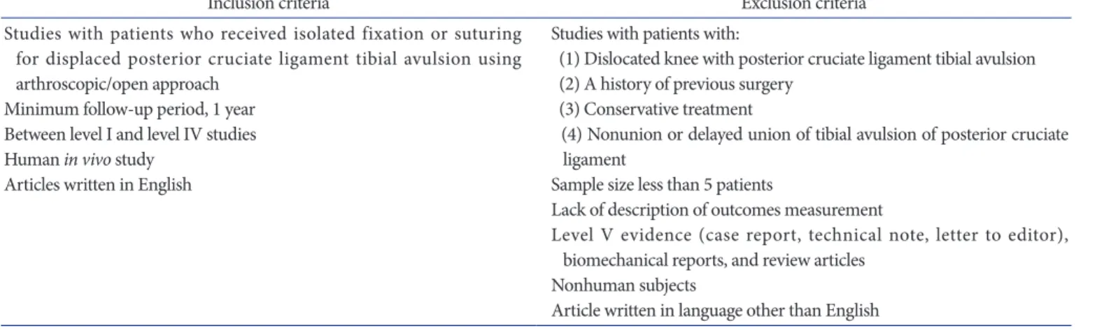
Table 1. Inclusion and Exclusion Criteria
Inclusion criteria Exclusion criteria
Studies with patients who received isolated fixation or suturing for displaced posterior cruciate ligament tibial avulsion using arthroscopic/open approach
Minimum followup period, 1 year Between level I and level IV studies Human in vivo study
Articles written in English
Studies with patients with:
(1) Dislocated knee with posterior cruciate ligament tibial avulsion (2) A history of previous surgery
(3) Conservative treatment
(4) Nonunion or delayed union of tibial avulsion of posterior cruciate ligament
Sample size less than 5 patients
Lack of description of outcomes measurement
Level V evidence (case report, technical note, letter to editor), biomechanical reports, and review articles
Nonhuman subjects
Article written in language other than English
Table 2. Patients’ Demographic Information and Surgical Procedure
Study No. of
patients Sex (M/F)
Mean age (yr)
Time to operation
(day)
Associated
injuries Operative
time (min) Surgical
approach Fixation
device fragment
size Followup
(mo) Posterior open approach
Singer and Halawa15) 16 16/0 34.5 8 Excluded 32 (25–40) Modified posterome dial approach (medial head of gast rocnemius was split)
Pull out suture using
Ethibond 18 (12–28)
Chen et al.13) 24 15/9 35.8 2 to 7 Injuries in
other ligaments and menisci (6)
52.5 Minimally invasive posterior approach (macroendoscopic technique)
Cannulated screw
fixation 8–12 mm: 7
12–26 mm: 11 Comminuted frac ture
with two frag ments over 8 mm
33.6 (2460)
Inoue et al.14) 16 (exclud ing PCL injuries without bony involvement)
6/25 44 7.2 days in group O;
8.3 days in group N
Excluded N/A Traditional posterior approach and post
eromedial approach (the proportion was not presented)
Cannulated cancellous
screws with a washer The size of the fragments was 1.5×1.5 cm or greater.
36 (24–96)
Yang et al.4) 16 (includ ing 2 chronic cases)
10/6 28 <2 weeks in 14; >4 weeks in 2
LCL injury (1), ACL injury (1), medial meniscus injury (1)
N/A Traditional posterior approach
14: malleolar screw 2: pull out screw due to small fragment size
N/A, but it was recorded that 2/16 was fixed with pullout suture due to small fragment size
38 (24–58)
Chiarapattanakom et al.3) 10 6/4 30 10 Excluded N/A Posteromedial approach Unicortical cancellous
screw fixation.
If fragment size was small, spike washer was added.
N/A 40 (22–58)
Arthroscopic approach
Zhao et al.7) 29 21/8 32 12 Excluded 55 (45–75) Pullout suture using
Yshaped bone tunnel and titanium button
2 No. 6 polyester
sutures N/A 32 (24–41)
Huang et al.5) 18 13/5 28 4.8 N/A 35 (21–55) Anterior arthroscopy
assisted fixation guided with a tibial PCL guide
One or two antegrade
screws Inclusion: the fracture fragment size was greater than 20 mm
34 (24–49)
Gui et al.8) 28 19/9 35.3 3.4 LCL (1), MM
(4), LM (4), MCL (1)
67 (45–90) Pullout suture using
single tunnel PDS 20 single fragment
(mean, 16 mm) 8 comminution
(largest fragment,
<10 mm)
40 (26–61)
Chen et al.9) 36 24/12 35.6 5 LM (2), MM
(3), MCL (2), LCL (2)
N/A Pullout suture using
double tunnel No. 5 Ethibond Various
fragmentation sizes (range, 10×6×5 to 30×32×15 mm;
mean, 15×17×9 mm)
36 (24–45)
Chen et al.10) 22 20/2 37 13 N/A 70.5 Pullout suture using
double tunnel No. 5 Ethibond N/A 24.5 (19–28)
Comparative study Sabat et al.1)
Open 27 25/2 28.4 6.2 MCL (2), LCL
(2)
N/A Modified posteromedial approach (medial head of gastrocnemius was split)
Partial threaded cannlated screw and washer
N/A 12
Arthroscopic 20 18/2 26.6 8.4 MCL (1), LCL
(1), LM (1), MM (2), ACL (6)
N/A Single tunnel pullout
suture No. 2 Orthocord, tied
over suture disk N/A 12
Pardiwala et al.16)
Open 25 N/A N/A N/A N/A N/A Posteromedial approach 4 mm cannulated
cancellous screw and washer or No. 5 Ethibond (fragment size: small or comminuted)
N/A 39 (24–58)
Arthroscopic 25 N/A N/A N/A N/A N/A Pullout suture using
double tunnel No. 5 Ethibond or
No. 2 Fiberwire N/A 39 (24–58)
Values are presented as mean (range or standard deviation).
PCL: posterior cruciate ligament, N/A: not available, LCL: lateral collateral ligament, MM: medial meniscus, LM: lateral meniscus, MCL: medial collateral ligament, ACL: anterior cruciate ligament.
Committee (IKDC) at final followup, (3) posterior draw test on physical examination, and (4) sidetoside difference on KT2000 at final followup.
Results
1. Literature Search
The electronic search initially identified 1,092 articles. Critical
application of the inclusion and exclusion criteria subsequently reduced that number to 12; 5 studies on open posterior approach, 5 studies on arthroscopic fixation, and 2 directly comparative studies of the open posterior approach and arthroscopic fixation.
The search strategy is outlined in Fig. 1, and an overview of the study characteristics is presented in Table 4.
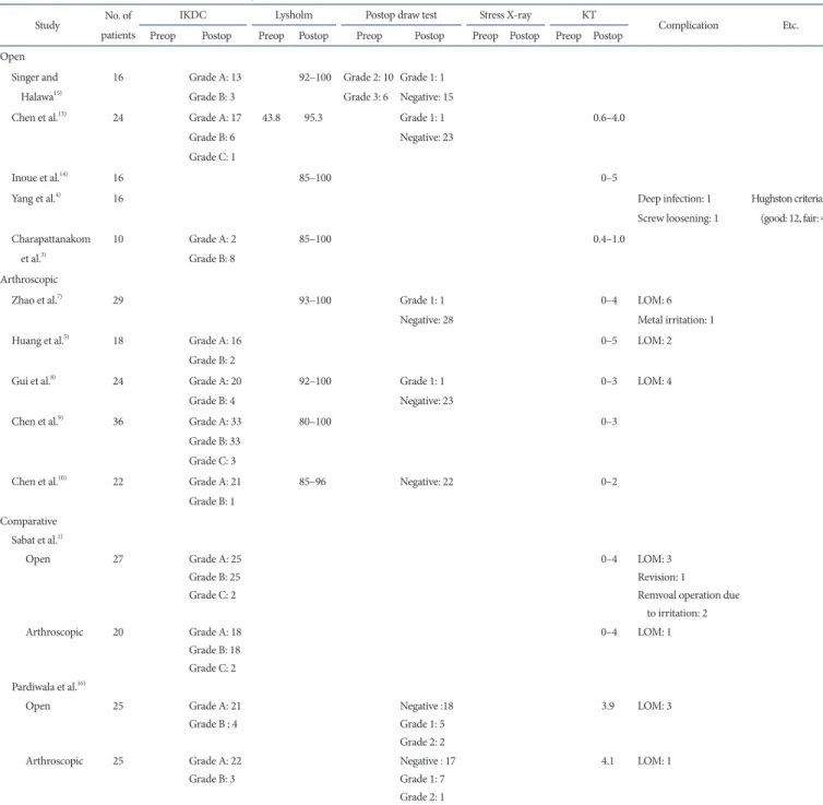
Table 3. Overall Clinical Outcomes and Complications in Studies
Study No. of
patients
IKDC Lysholm Postop draw test Stress Xray KT
Complication Etc.
Preop Postop Preop Postop Preop Postop Preop Postop Preop Postop
Open Singer and
Halawa15)
16 Grade A: 13
Grade B: 3
92–100 Grade 2: 10 Grade 3: 6
Grade 1: 1 Negative: 15
Chen et al.13) 24 Grade A: 17
Grade B: 6 Grade C: 1
43.8 95.3 Grade 1: 1
Negative: 23
0.6–4.0
Inoue et al.14) 16 85–100 0–5
Yang et al.4) 16 Deep infection: 1
Screw loosening: 1
Hughston criteria (good: 12, fair: 4) Charapattanakom
et al.3)
10 Grade A: 2
Grade B: 8
85–100 0.4–1.0
Arthroscopic
Zhao et al.7) 29 93–100 Grade 1: 1
Negative: 28
0–4 LOM: 6 Metal irritation: 1
Huang et al.5) 18 Grade A: 16
Grade B: 2
0–5 LOM: 2
Gui et al.8) 24 Grade A: 20
Grade B: 4
92–100 Grade 1: 1
Negative: 23
0–3 LOM: 4
Chen et al.9) 36 Grade A: 33
Grade B: 33 Grade C: 3
80–100 0–3
Chen et al.10) 22 Grade A: 21
Grade B: 1
85–96 Negative: 22 0–2
Comparative Sabat et al.1)
Open 27 Grade A: 25
Grade B: 25 Grade C: 2
0–4 LOM: 3 Revision: 1
Remvoal operation due to irritation: 2
Arthroscopic 20 Grade A: 18
Grade B: 18 Grade C: 2
0–4 LOM: 1
Pardiwala et al.16)
Open 25 Grade A: 21
Grade B : 4
Negative :18 Grade 1: 5 Grade 2: 2
3.9 LOM: 3
Arthroscopic 25 Grade A: 22
Grade B: 3
Negative : 17 Grade 1: 7 Grade 2: 1
4.1 LOM: 1
IKDC: International Knee Documentation Committee, Postop: Postoperative, Preop: preoperative, LOM: limitation of motion.
2. Quality Assessment
The mean modified Coleman methodology score of the includ
ed studies was 73.4±8.1 (range, 61 to 89). The mean Coleman
methodology score
12)for each criterion is shown in Table 5.
Initial search:
Total 1,092 studies identified (PubMed: 296, EMBASE: 902, Cochrane: 10)
Potentially relevant:
35 studies selected for full review
Meeting entry criteria:
12 articles
1,047 Articles discarded after review of titles and abstracts
Full-text articles excluded: 23 Duplication
Review articles
Technical notes without reporting outcomes Articles with sample size less than 5
Articles with mean follow-up less than 12 months Non-human studies
Ariticles written in language other than English
Fig. 1. Flowchart of selection process.
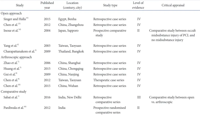
Table 4. Characteristics of Included Studies
Study Published
year Location
(contury, city) Study type Level of
evidence Critical appraisal Open approach
Singer and Halla15) 2015 Egypt, Benha Retrospective case series IV Chen et al.13) 2012 China, Zhangzhou Retrospective case series IV Inoue et al.14) 2004 Japan, Sapporo Prospective comparative
study II Comparative study between occult
midsubstance injury of PCL and no midsubstance injury Yang et al.4) 2003 Taiwan, Taoyuan Retrospective case series IV
Charapattanakom et al.3) 2009 Thailand, Bangkok Retrospective case series IV Arthroscopic approach
Zhao et al.7) 2006 China, Shanghai Retrospective case series IV Huang et al.5) 2015 China, Chongqing Retrospective case series IV
Gui et al.8) 2009 China, Nanjing Retrospective case series IV
Chen et al.9) 2012 Taiwan, Taoyuan Therapeutic case series IV
Chen et al.10) 2015 China, Wuhan Retrospective case series IV
Comparative study
Sabat et al.1) 2016 India, New Delhi Retrospective
comparative series III Comparative study between open vs. arthroscopic
Pardiwala et al.16) 2012 India Prospective randomised
comparative series II PCL: posterior cruciate ligament.
3. Data Abstraction
1) Surgical approach and technique (1) Open posterior approach
Five studies and 2 comparative studies reported the outcomes of patients undergoing the open posterior approach
1,3,4,1316). Overall, 134 patients underwent the open posterior approach with a mini
mum 12month followup. Open posterior approach included the traditional open posterior approach and its modifications.
(2) Direct posterior approach
The traditional open posterior approach was originally de
scribed by Abbott and Carpenter
4,17,18). It is a direct posterior approach using the interval between the heads of the medial and lateral gastrocnemius muscles, and it requires the identification and protection of the tibial nerve, artery, and vein. However, division of the medial head of the gastrocnemius is commonly recommended to enhance exposure of the PCL avulsion, which could lead to postoperative weakness of this muscle and may unnecessarily increase the morbidity of the operation. The tra
ditional open posterior approach was performed in 2 of the in
cluded studies
4,14).
Yang et al.
4)used this approach in 18 patients including 2 chronic cases. Inoue et al.
14)used the direct posterior approach although a modified posteromedial approach, described by Burks and Schaffer
19), was also used in the study without specific description of the proportion. Among its modifications, Chen et al.
13)suggested that the direct posterior approach under mac
roendoscopic assistance through a single minimal incision by a posterior midline approach is feasible for reduction of fragments and screw fixation.
(3) Posteromedial approach
The posteromedial approach was introduced by Burks and Schaffer
19)because of the complexity of the direct posterior ap
proach and the need for dissection of the neurovascular bundle in the popliteal fossa. An interval between the medial border of the gastrocnemius and the semimembranosus tendon is used to expose the posterior joint capsule. This minimally invasive approach provides satisfactory exposure of the fracture site in a safe, simple, and less timeconsuming manner for treatment of PCL injuries. Among the studies included in our analysis, this approach was used by Chiarapattanakom et al.
3)and Pardiwala et al.
16)(in their open group). It avoids dissection of the neurovascu
lar structures in the popliteal fossa as well, but it does not provide
Table 5. Overall Coleman Methodology Score for Each Criterion Criteria (maximal score)OpenArthroscopicComparative Singer and Halawa15)Chen et al.13)Inoue et al.14)Yang et al.4)Charapattanakom et al.3)Zhao et al.7)Huang et al.5)Gui et al.8)Chen et al.9)Chen et al.10)Sabat et al.1)Pardiwala et al.16) Part A Study size (10)044004044444 Mean followup (5)255555555525 No. of procedures (10)101010101010101010101010 Type of study (15)10010000100100010 Diagnostic certainty (5)555555555555 Surgery description (5)555555555555 Rehabilitation description (10)101010101010101010101010 Part B Outcome criteria (10)101010101010101010101010 Procedure for outcomes (15)36116116111115111111 Selection process (15)151010101213101215151515 Total score706580616868767289757285
adequate exposure to the lateral base of the PCL and the capsule.
The mass of the retracted tissue makes it difficult to place a screw perpendicular to the fracture plane, which could potentially lead to less stable fixation.
(4) Modified posteromedial approach
Other authors used a modified posteromedial approach, which splits the fibers of the medial gastrocnemius muscle to expose the PCL avulsion fracture. The lateral half of the fibers could protect the neurovascular elements in the popliteal space. This approach is anatomic and saves the medial head. Among the included stud
ies, Singer and Halawa
15)and Sabat et al.
1)(in their open group) used this approach.
(5) Arthroscopic fixation
Five studies and two comparative studies reported the outcomes of the arthroscopic approach
1,5,710,16). Overall, 174 patients un
derwent the arthroscopic approach with a minimum 12month followup.
Even though fracture reduction was done arthroscopically in all studies, each fixation method was different. Zhao et al.
7)made a Yshaped bone tunnel and fixed the pullout suture with a tita
nium button. Three articles (including one comparative study) described fixation was achieved using a pullout suture through double tunnels
9,10,16). Gui et al.
8)performed pullout suture fixa
tion through a single tunnel. Huang et al.
5)introduced antegrade screw fixation using a PCL guide after arthroscopic fracture re
duction. It was selectively performed for patients with a fragment size larger than 20 mm.
4. Clinical Outcomes