INTRODUCTION
The treatment options for mandibular condyle frac- tures include closed and open techniques for reduction and fixation. Open techniques can be accomplished with
extraoral and transoral approaches, whereas closed methods include intermaxillary fixation with bone or dental ligations or both. And, Open reduction does not necessarily mean rigid fixation. Open reduction merely means that a fracture has been anatomically reduced with verification via direct visualization through an open approach. Subsequent to reduction, some form of fixation may be used to stabilize the fracture1). Although it has now been recognized that ORIF(Open reduction intermaxillary fixation) provides better functional recon- struction of mandibular condyle fractures than
* Corresponding author Soon-Min Hong
Dept. of OMFS, Kangdong Sacred Heart Hospital, Hallym University Gil-dong 445, Kangdong-ku, Seoul, 134-010, Korea
Tel: 82-2-2224-2333 Fax: 82-2-483-9647 E-mail: [email protected]
Open versus closed reduction of mandibular condyle fractures : A systematic review of comparative studies
Jong Sik Kim*, Hyun-Soo Seo*, Ki-Young Kim*, Yun-Jung Song**, Seonah Kim***, Soon-Min Hong*, Jun-Woo Park*
*Department of Oral and Maxillofacial Surgery, Kangdong Sacred Heart Hospital, Hallym University, Korea
**Department of Operative dentistry, Hallym University Dental Hosptal, Korea
***Department of Operative dentistry, Kangdong Sacred Heart Hospital, Hallym University, Korea
Objective : The objective of this review was to provide reliable comparative results regarding the effectiveness of any interventions either open or closed that can be used in the management of fractured mandibular condyle
Patients and Methods : Research of studies from MEDLINE and Cochrane since 1990 was done. Controlled vocabulary terms were used. MeSH Terms were “ Mandibular condyle”AND “Fractures, bone”. Only comparative study were considered in this review using the “limit”function. According to the criteria, two review authors independently assessed the abstracts of studies resulting from the searches. The studies were divided according to some criteria, and following were measured: Ramus height, condyle sagittal displace- ment, condyle Towns’s image displacement, Maximum open length, Protrusion & Lateral excursion, TMJ pain, Malocclusion, and TMJ disorder.
Results : Many studies were analyzed to review the post-operative result of the two methods of treatment. Ramus height decreased more in when treated by closed reduction as opposed to open reduction. Sagittal condyle displacement was shown to be greater in closed reduction. Condyle Town’s image condyle displacement had greater values in closed reduction. Maximum open length showed lower values in closed reduction. In protrusive and lateral movement, closed reduction was less than ORIF. Closed reduction showed greater occurrence of malocclusion than ORIF. However, post-operative pain and discomfort was greater in ORIF.
Conclusion :In almost all categories, ORIF showed better results than CRIF. However, the use of the open reduction method should be considered due to the potential surgical morbidity and increased hospitalization time and cost. To these days, Endoscopic surgical techniques for ORIF (EORIF) are now in their infancy with the specific aims of eliminating concern for damage to the facial nerve and of reducing or eliminating facial scars.
Before performing any types of treatment, patients must be understood of both of the treatment methods, and the best treatment method should be taken on permission.
Key words
Mandibular condyle fracture, Open reduction, Close reduction, Systematic review Abstract
CRMMF(Closed reduction and maxillomandibular fixa- tion), attempts have been made to limit the potential adverse postoperative sequelae associated with ORIF.
There is a lot of methods or fixation schemes that have been used to stabilize mandibular condyle fractures in open reduction. There are use of a urethral sound, condylectomy, intraosseous or transosseous wire fixa- tion, intramedullary pins, traction screw osteosynthesis with combination nut at angle, long screw placement, onlay-inlay splint, miniaturized dynamic compression plates designed for zygoma fractures, free graft with wire fixation after extracorporeal avulsion, disk repair with silicone rubber implantation, axial anchor screws, rigid plates and screws, bioabsorbable plates and screws, etc.1).
Today, for dislocated fractures, open approaches are considered as the treatment of choice in many units.
However, for moderately displaced condylar fractures, open treatment is still controversial. Specific indications for open reduction based on the degree of dislocation and concomitant subjective symptoms2). Previously reported retrospective studies demonstrated a better anatomical position after operative treatment3). Haug RH et al. reported for the absolute indications of ORIF4). It was considerated patient preference, manipulation which can not re-establish pretraumatic occlusion and/or excursions, addressing other fractures affecting the occlusion, and stability of the occlusion limited.
After treatment for condyle fractures, there were vari- ous complications. In the use of the open reduction method, potential surgical morbidity and increased hos- pitalization time and cost should be consideral5). Whereas, After closed functional treatment, considerable malalignment (notably in the anterior posterior direc- tion), distinctive changes in condylar form (flattening of the articular condylar surface) and resorption of the frac- tured condyle were frequently seen. De Riu et al. sug- gested that, in the long term, incomplete anatomical restoration in non-surgical methods can cause facial asymmetry and inclination of the occlusal plane, as well as functional occlusal problems, such as premature con- tact in protrusion and lateral protrusion6). Complications such impaired masticatory function and pain located to the affected joint or masticatory muscles were seen sig- nificantly more frequent in patients treated surgically5). Whereas, Patients treated by closed techniques had a sig- nificantly greater percentage of malocclusion compared with patients treated by open reduction. It is possible
that the main reason for development of many of the complications was the inability of the patient to over- come the different neuromuscular and other functional problems induced by an unrepositioned subcondylar fracture8).
There were a lot of controversies over a medical treat- ment in a condyle fracture than the other parts of trauma on a face. There have been many studies about the choic- es of the treatment compared with after effects, compli- cations, etc. The objective of this review was to provide reliable comparative results regarding the effectiveness of any interventions either open or closed that can be used in the management of fractured mandibular condyle.
MATERIALS AND METHODS
1. CRITERIA FOR CONSIDERING STUDIES FOR THIS REVIEW
(1) Types of studies
Only comparative studies were considered in this review. Therefore, there were a number of types, as ran- domized clinical trials, controlled clinical trials, research report, and case report.
(2) Types of participants
There were no limits on age or gender. It did not classi- fy or exclude according to reasons for the fracture. It also did not have any restriction by laterality or both sides of the fractures. It included the cases of having other frac- tures in other parts.
(3) Types of intervention
Any form of open and closed method of reduction and fixation was included.
2. SEARCH METHOD
(1) Database searched
We searched in MEDLINE and Cochrane (studies pub- lished after 1990).
(2) Search term
The controlled vocabulary terms (MeSH) were used a. Pubmed : 1,040 studies was found by a computer
search with the keywords, “Mandibular condyle”
AND “Fractures, bone“ [MeSH]. I have searched
again for a comparative study in the previous result using a search function ‘limit’and I have found 65 of them.
b. Cochrane : I have searched with the keywords,
“mandibular condyle”AND “mandibular fractures.”
As a result, I have gotten the same results as Pubmed.
(3) Language
There was a “English”language restrictions on the included studies.
3. METHODS OF THE REVIEW
(1) Evaluation for searching result a. Description of studies
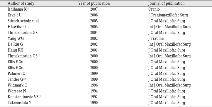
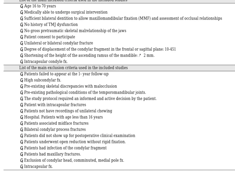
Two review authors independently assessed the abstracts of studies resulting from the searches. we have excluded the papers other than the ones compared with the results of open and closed operations like the ones that compared with fractures displaced and not dis- placed, condyle fix for each part, and operation materials such as a miniplate, etc. 17 papers have been selected by these standards(Table 1). In Table 2, it shows a list of the main inclusion criteria and exclusion criteria used in the included studies.
b. Characteristics of the interventions
The surgical methods for ORIF used in included stud- ies show in Table 3.
(2) Assessment of methodological quality and quantity The master data set included variables categorizing the quality and quantity
a. Study method
- Study quality (Table 4)
b. Participants
- Sample size categories (Table 5)
- Graph showing the quantity and quality of the studies used in the analyses (Fig. 1)
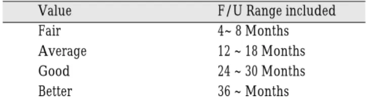
i) Follow-up losses (Table 6, Fig. 2)
ii) Evaluation by whether it has a clear statement of a reason for not having f/u in it or not - There are 3 studies which have the statement of a rea-
son for not having f/u among 17 in total (Table 7).
(3) Intervention a. Treatment option
We have checked how ORIF and Closed reduction are described about participants. There are some papers which choose an operation method with clear standard.
They have chosen whether they operate ORIF or not
Table 1.List of the included studies
Author of study Year of publication Journel of publication
Ishihama K18) 2007 Cranio
Eckelt U 2006 J Craniomaxillofac Surg
Stiesch-scholz et al 2005 J Oral Maxillofac Surg
Hlawitschka 2005 Int J Oral Maxillofac Surg
Throckmorton GS 2004 J Oral Maxillofac Surg
Yang WG 2002 J Trauma
De Riu G 2002 Int J Oral Maxillofac Surg
Haug RH 2001 J Oral Maxillofac Surg
Throckmorton GS19) 2000 Int J Oral Maxillofac Surg
Ellis E 3rd 2000 J Oral Maxillofac Surg
Ellis E 3rd 2000 J Oral Maxillofac Surg
Palmieri C 1999 J Oral Maxillofac Surg
Santler G20) 1999 J Oral Maxillofac Surg
Widmark G 1996 Int J Oral Maxillofac Surg
Worsaae N 1994 J Oral Maxillofac Surg
Konstantinovic VS12) 1992 J Oral Maxillofac Surg
Takenoshita Y 1990 J Oral Maxillofac Surg
based on a subjective symptom and excursion in a study2). Also, there are the cases that let a patient choose it. They have let their patients choose their operation method in a study10).
b. Doctor
We evaluated them if it is operated in one hospital or multi-centre. In a study, Eckelt U et al. reported that they operated in 7 hospitals3). Also, We checked how many operating surgeons are there. All the operation were done by a senior author in a study5).
Table 3.The surgical methods for ORIF used in included studies The surgical methods for ORIF used in included studies
●Internal bone plate and screw
●Transosseous wire osteosynthesis (0.3mm stainless steel wires)
●Titanium screw & compress screw, micromesh, absorbable polylactide screw
●Mini-dynamic compression plate & screw
●Endoscopie
●Miniplate, lag screw, osteosynthesis
●Short risdon / chamry miniplate, screws, stainless wires, Kirschner pin
Table 2.list of the main inclusion criteria and exclusion criteria used in the included studies List of the main inclusion criteria used in the included studies
●Age 16 to 70 years
●Medically able to undergo surgical intervention
●Sufficient bilateral dentition to allow maxillomandibular fixation (MMF) and assessment of occlusal relationships
●No history of TMJ dysfunction
●No gross pretraumatic skeletal malrelationship of the jaws
●Patient consent to participate
●Unilateral or bilateral condylar fracture
●Degree of displacement of the condylar fragment in the frontal or sagittal plane: 10-451
●Shortening of the height of the ascending ramus of the mandible: × 2 mm.
●Intracapsular condyle fx.
List of the main exclusion criteria used in the included studies
●Patients failed to appear at the 1- year follow-up
●High subcondylar fx.
●Pre-existing skeletal discrepancies with malocclusion
●Pre-existing pathological conditions of the temporomandibular joints.
●The study protocol required an informed and active decision by the patient.
●Patient with intracapsular fractures
●Patients not have recordings of unilateral chewing
●Hospital. Patients with age less than 16 years
●Patients associated midface fractures
●Bilateral condylar process fractures
●Patients did not show up for postoperative clinical examination
●Patients underwent open reduction without rigid fixation.
●Patients had infection of the condylar fragment
●Patients had maxillary fractures.
●Exclusion of condylar head, comminuted, medial pole fx.
●Intracapsular fx.
(4) Outcomes
Check how many surveyors are there and how many times they operate to reduce errors. Almost all studies did not mention surveyors. A physician and two sur- geons did the evaluation in a study7). They blindly oper- ated to reduce a bias.
4. STATISTICS ANALYSIS METHOD
(1) Analysis of a logistic recurrence
It is a popular method when analyzing a relationship between dependent and independent variables.
(2) Process
We compared with the average value of the results from each operation in various studies. We let ORIF (a Table 7.The statement of a reason for not having follow-up
Study No. Reason for loss
2 Patient refusing
6 An error of measuring person number was seen
54 Died, not possible to locate, moved to other parts of the country, not respond to the f/u call Table 5.Sample size categories
Category Defining
Small 30 이하
Medium 31 to 100
Large 101 이상
Table 6.Follow-up losses
Value F/U Range included
Fair 4~ 8 Months
Average 12 ~ 18 Months
Good 24 ~ 30 Months
Better 36 ~ Months
Table 4.Study quality
Category Definig property
Fair Case report
Average Clinical Trials Retrospective study
Good Prospective study
Better RCT
Fig. 1. Graph showing the quantity and quality of the studies used in the analyses.
Fig. 2. Evaluation follow-up losses.
patient group) be 1 and Closed reduction (a comparative group) be 0, which is a basis for a dependent value. We presented OR value for a relationship between an opera- tion and a measurement parts. The estimates of effect of an intervention were expressed as odds ratios (ORs) together with 95 % confidence intervals. (CIs)
RESULT
If you analyze a probability of heed in B and OR values from a result table, there is a big difference in an average value and a unit for each study. We did not think of them as a good average value from the result table as long as the values are somewhat different by a weight value of data in logistic recurrence analysis and it is ana- lyzed with an average data to do recurrence analysis. It was not a good idea to understand that you make a recurrence analysis data logically fitting into an average value. It was needed to analyze how to see + and - and a size of B value generally. We thought of that as an increase if a calculation is + and a decrease if it is -. It shows OR value increases several times (Table 10).
1. Changes in radiographic images
(1) Ramus
As a result, closed reduction reduces after an operation showing B = .063 and OR = 3.138 to look at ORIF and closed reduction in Ramus height through a logistic recurrence analysis.
Hlawitschka M et al. performed to evaluate and com- pare the results of open and closed treatments of diaca- pitular fractures of the mandible. Following ORIF patients showed better radiological results with regard to the mandibular ramus height, resorption and pathologi- cal changes to the condyle, compared to the patient group after closed functional treatment9).
Yang WG et al. compared the functional results of uni- lateral mandibular condylar process fractures treated either by open reduction or by closed treatment5). Co- mparison of displacement parameters in subcondylar fractures between open reduction and closed treatment groups revealed a statistically significant difference in vertical shortening and coronal angulation5). Patients undergoing open reduction had more severe displace- ment than those undergoing closed treatment5). Ellis E 3rd et al. reported that patients whose were treated by closed methods had significantly shorter posterior facial Table 8.Result Analysis
B S.E. Wald P-value OR
95% confidence interval for EXP(B) The Upper The Lower
limit limit
1. Ramus independent variable .263 .264 .996 .318 .063 .045 1.289
height constant 1.143 1.556 .540 .462 .263
2. Sagittal independent variable -2.113 1.049 4.057 .044 .121 .015 .945
displacement constant .866 1.406 0.379 .538 2.377
3. Town’s independent variable -0.279 .114 5.926 .015 .775 .605 .947
Image constant 3.296 1.610 4.192 .041 .050
4. TMJ pain independent variable .009 .010 .236 .013 .017 .002 .945
constant .021 1.633 .122 .023 .001
5. Maximum independent variable 8.246 4.467 .000 .991 .012 .000 .122
opening constant 3.275 3.417 .000 .991 .
6. protrusive independent variable 12.590 1.818 .000 .991 .003 .000 .135
movement constant 7.556 77.112 .000 .992 6
7. lateral independent variable .008 .016 .233 .629 .092 .090 1.024
movement constant -1.075 .845 1.617 .204 .341
8. malocclusion independent variable -0.701 .438 2.559 .110 2.016 .854 4.760
constant -12.076 6.762 3.189 .074 .000
and ramus heights on the side of injury, and more tilting of the occlusal and bigonial planes toward the fractured side, than patients whose fractures were treated by open methods10).
(2) Displacement in Sagittal view
Closed reduction is higher after an operation showing B = -2.113, OR = .121 by looking at ORIF and closed reduction in Sagittal displacement.
Palmieri et al. compared mandibular and condylar mobility after open or closed treatment for fractures of the mandibular condylar process11). Measures of condylar process displacement at the initial (pretreatment) time showed that patients who subsequently were in the open treatment group had, on average, twice the amount of displacement in the coronal plane than those who subse- quently underwent closed treatment11). The condylar dis- placement was surgically eliminated. Therefore, the ini- tial (post-traumatic, pretreatment) amount of displace- ment does not seem to affect motion outcomes for patients with condylar neck or subcondylar fractures treated by open reduction if the normal condylar posi- tion can be restored with surgery.
(3) Displacement in Town’s image
Closed reduction is higher after an operation showing OR = .775 by looking at ORIF and closed reduction of a malposition angle in Town’s image.
In Palmieri et al. study, Comparison of displacement variables between the closed and open groups showed that there was still a statistically significant difference in the coronal position of the condylar processes and in the amount of vertical overlap11). Konstantinovic et al. report- ed that The radiographic examinations showed a statisti- cally better position of the surgically reduced condylar process fractures12). However, There was no significant clinical difference between patients with surgically and those with conservatively treated unilateral condylar process fractures12).
2. Temporomandibular joint disorder
Closed reduction is frequently after an operation show- ing B = 0.009, OR = .017 by looking at ORIF and Closed reduction in TMJ pain. Yang WG at el. Reported that the closer the fracture site is to the TMJ, the greater the prob- ability of TMJ injuries. Temporomandibular joint
injuries, such as capsular rupture, disc disruption, and condylar head dislocation, often accompany condylar fractures5). Therefore, TMJ symptoms occurred more fre- quently in the condylar subgroups than in the subcondy- lar subgroups5). In this study, patients treated with open reduction or closed treatment did not reveal a signifi- cantly functional difference5). For subcondylar fractures, open reduction provides satisfactory functional results in patients with severely displaced fractures5). In the patients treated open reduction, the incidence of TMJ pain and significant chin deviation seemed less com- pared with closed treatment5).
3. Changes in motion
(1) Maximum opening
Closed reduction is lower after an operation showing B
= -8.246 and OR = .012 by looking at ORIF and closed reduction in a maximum opening.
Eckelt U et al. compared operative and conservative treatment of displaced condylar fractures of the mandible. The range of movement was assessed by max- imal mouth opening, protrusion and lateral excursion (Fig. 8). In the closed treatment group the average inter- incisal distance postoperatively was 40.9 mm(SD 6.7) and in the operatively treated group 46.5mm (SD 5.3).
Throckmorton GS et al. determined the rate of recovery of mandibular motion in patients treated for fractures of the mandibular condylar process13). Patients treated open will have reduced maximum opening initially, but may reach normal levels of opening sooner than patients treated without surgery. Patients treated open recover more of their maximum opening, and recover more quickly than patients treated closed13). Widmark G et al.
compared the results between two groups of patients in 1 year after trauma2)Surgical correction normalized faster in opening incisor pathways during mastication4).
(2) Protrusive movement
Closed reduction is lower after an operation showing B
= 12.59 and OR = .003 by looking at ORIF and closed reduction in a protrusive movement.
Eckelt U et al. observed that in the closed treatment group the average range of protrusion was significantly less (p0.0005) with 4.7mm (SD 2.5) when compared with 7.3mm (SD 2.0) in the operatively treated group3).
(3) Lateral movement
Closed reduction is lower after an operation showing B
= .008 and OR = .092 by looking at ORIF and closed reduction in a lateral movement.
Haug RH et al. reported no statistically significant dif- ferences were noted between groups for range of right and left lateral excursion4). However, Throckmorton GS et al. reported that lateral excursion toward the non-frac- ture side remained significantly smaller in all patients treated closed at 3 years after fracture13).
4. Malocclusion
Closed reduction is frequent after an operation show- ing B = -.701 and OR = 2.016 by looking at ORIF and closed reduction in a malocclusion.
Yang WG et al compared the functional results of uni- lateral mandibular condylar process fractures treated either by open reduction or by closed treatment5). Generally, acceptable facial symmetry and occlusion were obtained in all patients no matter which treatment was used5). However, De Riu G et al. Reported that open reduction gave better occlusal results, anatomic restora- tion and faster recovery rates than non-surgical tech- niques6). And In this study, there was no difference greater than 2 mm between maximum intercuspation and centric relationship (MI-RC) in the surgically treated group6).
DISCUSSION
The objective of this review was to provide reliable comparative results regarding the effectiveness of any interventions either open or closed that can be used in the management of fractured mandibular condyle9). Because the condylar displacement was surgically elimi- nated and fixed anatomically by open reduction, we could expect to have more good result than closed reduc- tion. In a study, neither the degree of dislocation of the proximal fragment, concomitant mandibular fractures, nor the absence of posterior occlusal support seemed to influence the results of comparison between open and closed reduction8).
In our study, Ramus height decreased more in when treated by closed reduction as opposed to open reduc- tion. Sagittal condyle displacement was shown to be greater in closed reduction. Condyle Towns’s image condyle displacement had greater values in closed
reduction. Maximum open length showed lower values in closed reduction. In protrusive and lateral movement, closed reduction was less than ORIF. Closed reduction showed greater occurrence of malocclusion than ORIF.
However, post-operative pain and discomfort was greater in ORIF.
In almost all categories, we could know that ORIF showed better results than closed reduction. However, the use of the open reduction method should be consid- ered due to the potential surgical morbidity and increased hospitalization time and cost. Therefore, Endoscopic surgical techniques for ORIF (EORIF) are now in their infancy with the specific aims of eliminating concern for damage to the facial nerve and of reducing or eliminating facial scars. Lee et al.16)found that 37 of 40 patients treated with EORIF went on to uneventful heal- ing. However, Lauer and Schmelzeisen17) noted that in 1 of 3 patients, loose hardware required early removal due to insufficient fixation.
At present, in temporomandibular joint problems, there were not sufficient long-term data to support open reduction to prevent future joint problems. Patients treat- ed for condylar process fractures by closed methods fre- quently develop a new articulation more inferiorly in the fossa, often at the bottom of the articulareminence15). In fact, arthritic changes, including remodeling, could occur with both open and closed reduction with about the same degree of frequency15).
To these days, there is a number of technical and surgi- cal controversies relating to the type of interventions that could be used. We should consider that before our per- forming any types of treatment, patients must be under- stood of both of the treatment methods, and the best treatment method should be taken on permission.
REFERENCES
1. M. Todd Brandt RH, Haug: Open Versus Closed Reduction of Adult Mandibular Condyle Fractures : A Review of the Literature Regarding the Evolution of Current Thoughts on Management. J Oral Maxillofac Surg 2003;61:1324-32.
2. Widmark G, Bagenholm T, Kahnberg KE, Lindahl L: Open reduction of subcondylar fractures. A study of functional rehabilitation. Int J Oral Maxillofac Surg 1996;25(2):107-11.
3. Eckelt U, Schneider M, Erasmus F, Gerlach KL, Kuhlisch E, Loukota R, et al.: Open versus closed treatment of fractures of the mandibular condylar process-a prospective random- ized multi-centre study. J Craniomaxillofac Surg 2006;34(5):306-14.
4. Haug RH, Assael LA: Outcomes of open versus closed treatment of mandibular subcondylar fractures. J Oral Maxillofac Surg 2001;59(4):370-5; discussion 75-6.
of unilateral mandibular condylar process fractures after open and closed treatment. J Trauma 2002;52(3):498-503.
6. De Riu G, Gamba U, Anghinoni M, Sesenna E: A compari- son of open and closed treatment of condylar fractures: a change in philosophy. Int J Oral Maxillofac Surg 2001;
30(5):384-9.
7. Ellis E, 3rd, Simon P, Throckmorton GS: Occlusal results af- ter open or closed treatment of fractures of the mandibular condylar process. J Oral Maxillofac Surg 2000;58(3):260-8.
8. Worsaae N, Thorn JJ: Surgical versus nonsurgical treatment of unilateral dislocated low subcondylar fractures: a clini- cal study of 52 cases. J Oral Maxillofac Surg 1994;52(4):353- 60; discussion 60-1.
9. Hlawitschka M LR, Eckelt U: Functional and radiological results of open and closed treatment if intracapsular (diaca- pitular) condylar fractures of the mandible. Int J Oral Maxillofac Surg. 2005;34(6):597-604.
10. Ellis E, 3rd, Throckmorton G. Facial symmetry after closed and open treatment of fractures of the mandibular condy- lar process. J Oral Maxillofac Surg 2000;58(7):719-28; dis- cussion 29-30.
11. Palmieri C, Ellis E, 3rd, Throckmorton G. Mandibular mo- tion after closed and open treatment of unilateral mandibu- lar condylar process fractures. J Oral Maxillofac Surg 1999;57(7):764-75; discussion 75-6.
12. Konstantinovic VS, Dimitrijevic B: Surgical versus conserv- ative treatment of unilateral condylar process fractures:
clinical and radiographic evaluation of 80 patients. J Oral Maxillofac Surg 1992;50(4):349-52; discussion 52-3.
13. Throckmorton GS, Ellis E, 3rd. Recovery of mandibular motion after closed and open treatment of unilateral mandibular condylar process fractures. Int J Oral Maxillofac Surg 2000;29(6):421-7.
14. Stiesch-Scholz M, Schmidt S, Eckardt A: Condylar motion after open and closed treatment of mandibular condylar fractures. J Oral Maxillofac Surg 2005;63(9):1304-9.
15. Takenoshita Y, Ishibashi H, Oka M: Comparison of func- tional recovery after nonsurgical and surgical treatment of condylar fractures. J Oral Maxillofac Surg 1990;48(11):1191- 5.
16. Lee C SM, Young DM: Cranial nerve VII region of the trau- matized facial skeleton : Optimizing fracture repair with the endoscope. J Trauma 2000;48(423).
17. Lauer G SR: Endoscope-assisted fixation of mandibular condylar process fractures. J Oral Maxillofac Surg 1999;
57(36).
18. Ishihama K, Iida S, Kimura T, Koizumi H, Yamazawa M, Kogo M: Comparison of surgical and nonsurgical treat- ment of bilateral condylar fractures based on maximal mouth opening. Cranio 2007;25(1):16-22.
19. Throckmorton GS, Ellis E, 3rd, Hayasaki H. Masticatory motion after surgical or nonsurgical treatment for unilater- al fractures of the mandibular condylar process. J Oral Maxillofac Surg 2004;62(2):127-38.
20. Santler G, Karcher H, Ruda C, Kole E: Fractures of the condylar process: surgical versus nonsurgical treatment. J Oral Maxillofac Surg 1999;57(4):392-7; discussion 97-8.