1. Introduction
1)The mixotrophic dinoflagellate Alexandrium minu- tum Halim produces the potent neurotoxin and is re- sponsible for paralytic shellfish poisoning (PSP)1,2). About 30 species are included within the genus Alexandrium Halim and the majority of PSP events have been caused by A. tamarense-complex and the A.
minutum species group2,3). The PSP outbreaks occur globally and the range and the frequency of the blooms seem to be increasing world-wide2). The first known bloom of Alexandrium species and PSP event in Korean waters occurred in Chinhae Bay in 19784), since then the PSP events have recurred along the southern coasts of Korea.
The classification of species within Alexandrium is primarily based on details of the thecal plate pattern.
Most Alexandrium species have relatively thin and
Corresponding Author : Tae-Gyu Park, Ecology and Oceanog- raphy Division, National Fisheries Research and Development Institute (NFRDI), Busan 619-705, Korea
Phone: +82-51-720-2235 E-mail: [email protected]
smooth thecal plates, and their generic characteristics include the thecal plate formula Po, 4, 6, 5, 2, 6c and 9-10s5). For morphological identification of Alexandrium species, light microscopy and/or scanning electron mi- croscopy (SEM) analyses on laboratory-cultured or field-derived cells are required. These conventional identification methods are labor intensive and not suit- able for rapid sample processing. To overcome these difficulties, molecular identification methods have been applied for quantitative detection of target organisms6,7). A number of approaches have been used for identi- fication of dinoflagellates. Real-time quantitative PCR offers highly sensitive and rapid analysis as well as high-sample throughput analysis. This method has been used for detection and quantification of a number of dinoflagellates including Cryptoperidiniopsis brodyi Steidinger et Litaker, Cochlodinium polykrikoides Margalef, Karlodinium veneficum (Ballantine) J. Larsen, and Pfiesteria piscicida Steidinger et Burkholder6~10). For understanding the ecological roles of A. minu- tum, information on the abundances of this dino- flagellate in environments is needed. However its tem-
Temporal Changes in Abundances of the Toxic Dinoflagellate Alexandrium minutum (Dinophyceae) in Chinhae Bay, Korea
Tae-Gyu Park and Yang-Soon Kang
Ecology and Oceanography Division, National Fisheries Research and Development Institute (NFRDI), Busan 619-705, Korea
(Manuscript received 16 June, 2009; revised 7 August, 2009; accepted 7 September, 2009)
Abstract
Marine dinoflagellate Alexandrium minutum producing paralytic shellfish toxins is responsible for paralytic shellfish poisoning (PSP). To investigate its temporal distributions in Chinhae Bay where PSP occurs annually, SYBR GreenⅠ based A. minutum-specific real-time PCR probe was developed on the LSU rDNA region.
Assay specificity and sensitivity were tested against related species, and its specificity was further confirmed by sequencing of field-derived samples. Ten months field survey in 2008 (a total 100 surface water samples) by using the real-time PCR probe showed that A. minutum was detected at very low densities of 1-4 cells L-1 in May and June being spring in Chinhae Bay, Korea.
Key Words : Alexandrium minutum, Dinoflagellate, PSP toxins, Real-time PCR
poral and geographic distributions are poorly understood.
In the present study, A. minutum-specific real-time PCR assay using a TaqMan probe was developed based on the large subunit (LSU) rDNA. Temporal and spatial distributions of A. minutum in surface waters of Chinhae Bay, Korea were investigated using the newly designed real-time PCR probe.
2. Materials and Methods 2.1. Cultures
Cultures were obtained from NFRDI's collection of microalgae, University of Tasmania, and CCMP (Provasoli-Guillard National Center for Culture of Marine Phytoplankton) (Table 1). Mixotrophic/photo- trophic cultures were maintained in f/2 medium of 28
of salinity
‰ 11)without sodium silicate at 24oC, with cool white fluorescent lamps of 100 µmol photons m-2s-1on a 12 : 12-h light : dark cycle. Heterotrophic dinoflagellates were grown in f/2 medium of 15‰of salinity at 24oC in the dark and Rhodomonas salina (Wislouch) Hill et Wetherbee was supplied to them as
food.
2.2. Collection of surface water samples from Chinhae Bay
Surface water samples of 250 mL were collected monthly at 10 stations from February to November 2008 in Chinhae Bay, Korea (Fig. 1). The water sam- ples were filtered onto a 1.2 µm pore-size, 25 mm di- ameter glass microfibre GF/C filter (Whatman, Ltd.
Maidstone, England). The filtered sample was placed in a 2 mL microcentrifuge tube, and stored at 70oC un- til DNA extraction. To prevent degradation of the tar- get DNA, filtering samples were processed on a re- search vessel.
2.3. DNA extraction
A phenol-chloroform extraction protocol was used for extraction of genomic DNAs from the surface wa- ter samples and laboratory cultures12). Filter samples were suspended in 900 µL of TE buffer (10 mM Tris-HCl, pH 7.5; 1 mM EDTA, pH 8.0) and ground with wooden applicator sticks. The sample was boiled
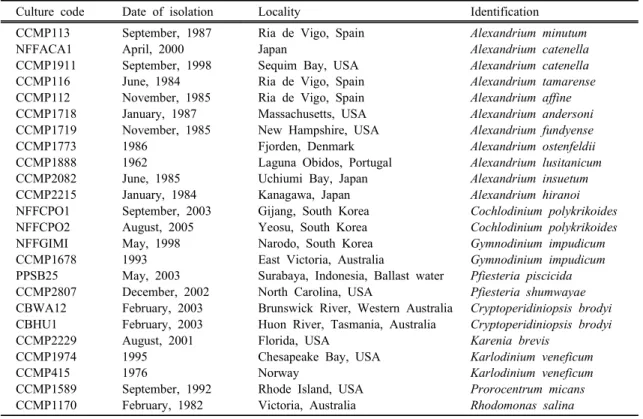
Table 1. Cultures used in this study
Culture code Date of isolation Locality Identification
CCMP113 NFFACA1 CCMP1911 CCMP116 CCMP112 CCMP1718 CCMP1719 CCMP1773 CCMP1888 CCMP2082 CCMP2215 NFFCPO1 NFFCPO2 NFFGIMI CCMP1678 PPSB25 CCMP2807 CBWA12 CBHU1 CCMP2229 CCMP1974 CCMP415 CCMP1589 CCMP1170
September, 1987 April, 2000 September, 1998 June, 1984 November, 1985 January, 1987 November, 1985 1986
1962 June, 1985 January, 1984 September, 2003 August, 2005 May, 1998 1993 May, 2003 December, 2002 February, 2003 February, 2003 August, 2001 1995 1976
September, 1992 February, 1982
Ria de Vigo, Spain Japan
Sequim Bay, USA Ria de Vigo, Spain Ria de Vigo, Spain Massachusetts, USA New Hampshire, USA Fjorden, Denmark Laguna Obidos, Portugal Uchiumi Bay, Japan Kanagawa, Japan Gijang, South Korea Yeosu, South Korea Narodo, South Korea East Victoria, Australia
Surabaya, Indonesia, Ballast water North Carolina, USA
Brunswick River, Western Australia Huon River, Tasmania, Australia Florida, USA
Chesapeake Bay, USA Norway
Rhode Island, USA Victoria, Australia
Alexandrium minutum Alexandrium catenella Alexandrium catenella Alexandrium tamarense Alexandrium affine Alexandrium andersoni Alexandrium fundyense Alexandrium ostenfeldii Alexandrium lusitanicum Alexandrium insuetum Alexandrium hiranoi Cochlodinium polykrikoides Cochlodinium polykrikoides Gymnodinium impudicum Gymnodinium impudicum Pfiesteria piscicida Pfiesteria shumwayae Cryptoperidiniopsis brodyi Cryptoperidiniopsis brodyi Karenia brevis
Karlodinium veneficum Karlodinium veneficum Prorocentrum micans Rhodomonas salina
Fig. 1. Locations where surface water samples were col- lected for this study. Water samples were obtained from 10 stations in Chinhae Bay from February to November 2008.
at 100oC for 5 min, followed by adding 900 µL of phe- nol : chloroform : isoamyl alcohol (25 : 24 : 1). The sample was then mixed thoroughly and centrifuged at 14,000 rpm for 10 min. The supernatant transferred to a new tube, and 30 µL of 3 M sodium acetate (pH 5.2) and 700 µL of 99.5% of ethanol (20oC) were add- ed, followed by incubation of the sample at 20oC for 30 min. The DNA samples were centrifuged at 14,000 rpm for 20 min at 4oC, and was rinsed twice with 70%
of ethanol. They were then dried and dissolved in 100 µL of TE buffer.
2.4. Design of a SYBR Green -based real-time Ⅰ PCR probe specific for A. minutum
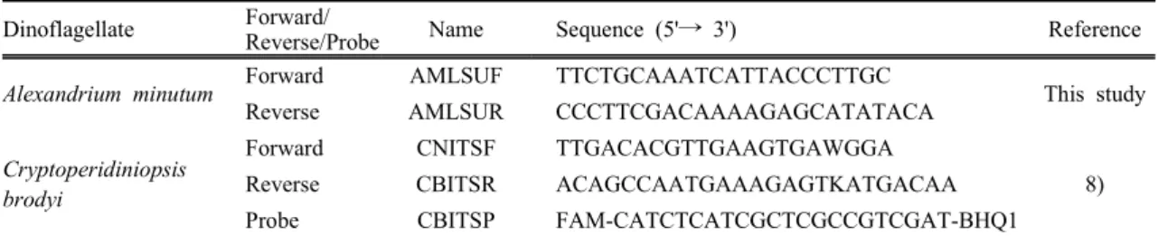
The LSU rDNA sequences of A. minutum available from GenBank were aligned with sequences of otherorganisms (about 100 species were compared) using the program ClustalX13). Unique sequences were man- ually searched from the alignments for designing a SYBR GreenⅠ-based species-specific real-time PCR probe. The A. minutum specific probe forms 129-bp amplicon in size, and it was designed on the D2 of LSU rDNA region because the region was highly vari- able between species but conserved within the species (Table 2). The sequences of the primers had at least 20-bp mismatches with sequences of closely related organisms. The sequences of the primers were also checked against published sequences in GenBank by BLAST homology search. Optimal melting temperature and secondary structure of primer and probe sequences were predicted by Primer 3 (Whitehead Institute and Howard Hughes Medical Institute, Maryland) and Oligo analyzer 3 (Intergrated DNA Technologies, Inc., Iowa) software. The primer set was synthesized by Sigma-Proligo (Paris, France).
2.5. Assay specificity of the A. minutum -selective real-time PCR probe
A total of 24 species was used for testing assay specificity (Table 1). Specificity of the forward and reverse primers was tested using standard format PCR which does not require a fluorogenic probe. PCR am- plicons of the target DNA (129-bp) in a 2% agarose gel stained with ethidium bromide was inspected to con- firm the absence of nonspecific reactions. Subsequently, specificity of the SYBR GreenⅠ-based real-time PCR probe was tested against other relatives. When ap- plied to field samples, positive samples by real-time PCR were further analyzed by gel electrophoresis us- ing 2% agarose gels for confirmation of assay specificity.
Table 2. Primers for species-specific real-time PCR Dinoflagellate Forward/
Reverse/Probe Name Sequence (5'→ 3') Reference
Alexandrium minutum Forward AMLSUF TTCTGCAAATCATTACCCTTGC
This study
Reverse AMLSUR CCCTTCGACAAAAGAGCATATACA
Cryptoperidiniopsis brodyi
Forward CNITSF TTGACACGTTGAAGTGAWGGA
8)
Reverse CBITSR ACAGCCAATGAAAGAGTKATGACAA
Probe CBITSP FAM-CATCTCATCGCTCGCCGTCGAT-BHQ1
2.6. Verification of assay specificity by sequencing of environmental samples
One of positives (Station 1, May 2008) was used for sequencing analysis. The rDNA (129-bp in size) of positive sample was amplified with primers AMLSUF- AMLSUR in a standard PCR platform using Takara EX Taq DNA polymerase with 39 cycles of 94oC for 1 min, 60oC for 1 min 30 s, 72oC for 2 min. The DNA band was visualized on 2% agarose gel stained with ethidium bromide, and the PCR product was cloned and insert-containing plasmid DNA was purified.
Three clones of the purified product were selected for sequencing, and their analyses were conducted with pUC/M13 primers (Promega, Madison, WI, USA) by Macrogen (Seoul, Korea). Expectation values (E-val- ues) and sequence similarity of the field sequences were estimated by a nucleotide BLAST program.
2.7. Real-time PCR conditions
Primers AMLSUF-AMLSUR had a similar melting temperature (Tm) of approximately 60oC when tested by Primer 3 computer program. Subsequently, 60oC was chosen for the annealing temperature. The follow- ing reagents were added for TaqMan based real-time PCR: 5 µL of platinum quantitative PCR supermix- UDG (Invitrogen, Eugene, Oregon, USA), primers at a final concentration of 0.2 µM, fluorogenic probe at a final concentration of 0.15 µM, 1 µL of template DNA, and PCR grade water to a final volume of 10 µL. For SYBR GreenⅠbased real-time PCR, the fol- lowing components were added: 5 µL of platinum SYBR Green quantitative PCR supermix-UDG (Invitrogen, Eugene, Oregon, USA), primers at a final concen- tration of 3.0 µM, 1 µL of template DNA, and PCR grade water to a final volume of 10 µL. The thermal cycling conditions consisted of 2 min at 50oC (uracil DNA glycosylase incubation for prevention of the reamplification of carryover PCR products) and 2 min at 95oC following by 40 cycles of 15 s at 95oC and 60 s at 60oC. DNA melting curves were monitored from 70oC to 85oC in 1oC increments using a 5 s hold at each step. The assay was analyzed in the FAM chan- nel (excitation/emission maxima of 470 nm-510 nm) of the Rotor Gene instrument. Fluorescence data were
collected at the end of each cycle by the real-time PCR instrument (Rotor Gene 6000, Corbett Research, Sydney, Australia), and they were analyzed using the Rotor Gene software (v1.7 build 61).
2.8. Standard PCR conditions
Standard PCR was performed using Takara EX Taq DNA polymerase (Takara Mirus Bio, Madison, WI, USA) with 32 cycles of 94oC for 1 min, 60oC for 1 min 30 s, 72oC for 1 min. Positive DNA bands were visualized on 2% agarose gel stained with ethidium bromide. The following reagents were added for stand- ard PCR: 0.1 µL of EX Taq DNA polymerase, primers at a final concentration of 300 µM, 2 µL of EX Taq buffer, 2.5 mM of dNTP mixture, 0.5 µL of template DNA, and PCR grade water to a final volume of 20 µL.
2.9. Standard curves for enumeration of cell num- bers
Laboratory-cultured A. minutum was used for con- struction of a standard curve. Cell numbers were esti- mated by light microscopy using a Sedgwick-Rafter counting chamber before harvesting the cells (total 25,530 cells). Genomic DNA was extracted, and 10-fold serial dilutions of the DNA extracts were used to construct the standard curve. The curve was con- structed by triplicate measurements using real-time PCR. The cell number of A. minutum in surface water samples was calculated as CT values, and was meas- ured by comparison with the standard curve. The accu- racy of the standard curves was evaluated using known concentrations of A. minutum (18,300, 3,660 and 732 cells; n = 3) spiked into sterile-filtered field samples (0.2 µm membrane filter; MFS, California). After DNA extraction, cell numbers estimated by real-time PCR were compared to the cell numbers estimated by light microscopy.
2.10. PCR inhibitor removal/template DNA dilution from surface waters
A real-time PCR probe specific for Cryptoperidiniopsis brodyi Steidinger et Litaker was used for checking the inhibitors because C. brodyi has not been reported in Korean waters. Serial DNA dilutions of field sam-
ples(non-dilution, 10-fold, and 50-fold dilutions) spiked with C. brodyi DNA (CBWA12; 1.5 ng µL-1) were amplified using real-time PCR, and their CTval- ues were compared. C. brodyi DNA without the addi- tion of the field DNA was used for a positive control.
Water samples collected in May from Chinhae Bay were used for template DNA. The absence of inhibitors was confirmed in 10-fold and 50-fold DNA dilutions.
Ten other field samples were randomly chosen and were tested to confirm the absence of the inhibitors.
Subsequently, 10-fold dilution of field-derived DNA was used for removal of PCR inhibitors.
3. Results
3.1. Specificity of A. minutum -selective real-time PCR assay
Specificity of forward and reverse primers was test- ed using SYBR GreenⅠ format real-time PCR. When the PCR product was visualized on agarose gel, only one amplicon of the expected size (129-bp) for the tar- get species was produced and there was no non-specif- ic reactions against related species (Fig. 2). Assay spe- cificity was then tested in SYBR GreenⅠ format re- al-time PCR against related species. Since SYBR GreenⅠ format real-time PCR is amplicon sequence non-specific method, this dye detects any double-strand
Fig. 2. Agarose gel analysis showing A. minutum-selective real-time PCR product (129-bp). Assay specificity was tested against related species (Table 1). Lanes:
1, 2 kbp-ladder molecular size marker; 2, A. minu- tum 3, A. tamarense 4, A. affine 5, A. hiranoi 6, A. andersoni 7, A. fundyense 8, A. ostenfeldii 9, A. lusitanicum 10, A. insuetum 11, A. catenella 12, no-template control.
DNA molecules. The formation of non-specific ampli- cons was checked by melting curve analysis. A. minu- tum gave a strong positive fluorescent signal after 13 cycles, while related species produced faint signals on- ly after 30 cycles (result not shown). The mean value of melting temperature (oC) for positives was 79.7 and standard deviation was 0.05, while that of negative controls/related species was 75 to 77oC (Fig. 3) in- dicating that the weak signals from the negative con- trol observed after 30 cycles were likely to be primer dimers. Assay specificity was further confirmed by se- quencing of a field-derived sample positive for A. min- utum-specific real-time PCR assay. The partial LSU rDNA sequences (129-bp) obtained from Station 1, May, 2008 were identical to documented A. minutum sequences (GenBank accession number, AY831408) and low expectation values (E-value) of less than 1e-5 were estimated by a nucleotide BLAST program in- dicating that assay was specific for A. minutum.
3.2. Standard curve and detection limit
A standard curve was constructed using 10-fold seri- al dilutions of DNA extracts from A. minutum. A strong linear correlation between log (known cell num- ber) and CT value was yielded for the assay (correlation coefficient R2 of 0.990). The values of slope (M) and intercept (B) were -3.161 and 24.409, respectively (Fig. 4). Detection limit of the assay was 2.5 cells per reaction within the dynamic range of cell numbers in the linear. The accuracy of standard curve was also tested. When DNA extracts from known cell numbers of A. minutum (18,300, 3,660 and 732 cells)Fig. 3. Melting curves of SYBR GreenⅠ-based assay with A. minutum and A. insuetum DNA extracts and no-template. Melting temperature (oC) of A. minu- tum was 79.7 ± 0.05 (n=3).
Fig. 4. Linear relationship between the CTvalues and the cell numbers for A. minutum (R2 = 0.990). The standard errors from three measurements are shown as error bars.
spiked with environmental samples were compared to the cell standard curve, cell numbers of the 10-fold di- luted spiked cells (2,450 ± 620, 511 ± 141, and 58 ± 49 cells, respectively; n = 3; P > 0.05 by Student's t test) did not significantly differ from those of standard curve (1,830, 366 and 73 cells).
3.3. Temporal changes in A. minutum abundances in Chinhae Bay
Of 100 water samples analyzed, only 3 were pos- itive for A. minutum (Table 3). The DNA of A. minu- tum was detected at 1 to 4 cells L-1in May and June.
The absence of A. minutum in the other samples was checked by melting curve analysis and gel electro- phoresis using 2% agarose gels. Melting curve analysis was conducted for all samples, and gel electrophoresis was performed with 30 samples randomly chosen from negatives. All of the samples except for the 3 positives showed melting temperature of less than 77oC and no positive bands on gel indicating that there was no false negative reaction.
4. Discussion
4.1. Quantitative detection of A. minutum in natu- ral environments
Information on geographic and temporal dis- tributions of dinoflagellates is necessary to understand their bloom dynamics. Although Alexandrium species are responsible for paralytic shellfish poising for deca- des, their accurate detection has been problematic due to difficulty in rapid identification of these species. To address this issue, a number of molecular methods have been developed for quantitative detection of the cells. Real-time PCR is a validate method that offers highly sensitive and accurate detection of dinoflagellates.
This method also reduces the analytical time to as little as 3 h from the time a sample arrived at the laboratory.
The SYBR GreenⅠformat real-time PCR has been used for a number of dinoflagellates including the ge- nus Alexandrium14). The A. minutum specific real-time PCR probe developed in this study provided a high sensitivity that allows the detection limit of less than 2.5 cells per reaction, which is similar to the results reported in other studies6-8). When applied to environ- mental samples, false-negative results caused by PCR inhibitors coextracted with target DNA may be problematic. Surface water contains PCR inhibitors such as phenolic compounds, heavy metals, and humic acids15). Dilution of template DNA obtained from 250 mL of surface water effectively removed the inhibitors indicating that there were no false negative reactions in the present study. For further confirmation of assay specificity, identification of field samples by culture dependent and independent methods may be desirable.
Since a mono culture of A. minutum was not estab- lished during this survey, field-derived samples were
Table 3. Occurrences of A. minutum in Chinhae Bay, Korea during February and November 2008 measured by species specific real-time PCR assay. Surface water samples were collected from 10 stations at 1 month intervals (a total of 100 water samples). The primer set for real-time PCR is shown in Table 2. Only positive detection is shown. Values are the mean ± standard deviation of triplicate wells
Sampling site Sampling date and cells liter-1
1 8 21
Feb.
N/D N/D N/D
Mar.
N/D N/D N/D
Apr.
N/D N/D N/D
May 4±3 N/D 1±0.4
Jun.
N/D 1±0.6
N/D
Jul.
N/D N/D N/D
Aug.
N/D N/D N/D
Sep.
N/D N/D N/D
Oct.
N/D N/D N/D
Nov.
N/D N/D N/D
sequenced for further confirmation of assay specificity.
The sequence result indicates that this assay provides A. minutum specific reaction.
4.2. Temporal distributions of A. minutum
For 10 months survey in Chinhae Bay, A. minutum was only found at very low densities (1-4 cells L-1) in May and June. Since the difficulty in rapid identi- fication of this species in natural samples, cell numbers estimated by real-time PCR did not compared to those counted by light microscopy. To verify the accuracy in the quantification of cells by the real-time PCR, known concentrations of A. minutum spiked into ster- ile-filtered field samples were compared to the stand- ard curve, and the result indicated that A. minutum in field samples was accurately quantified in the present study. One possibility to explain the low cell density is the real-time PCR probe developed in this study may not detect all of the genotypes in the species. There has been known several genotypes in A. minutum worldwide, and there are high genetic variations within the LSU rDNA locus of this species16). Since the high genetic variation, the real-time PCR probe from this study showed high assay specificity and may reduce false-positive reactions when applied to natural samples. This assay can detect most of genotypes in- cluding strains from the U.S., Hong Kong, China, North Atlantic Ocean, Spain, Italy and France, which have identical sequences with those of A. minutum-spe- cific primers. However, the primer set showed se- quence differences with strains from Cape Town har- bor in South Africa (GenBank accession numbers, DQ453522, DQ453524) and Fleet Lagoon in the U.K.(AY268598, AY705869) indicating that numbers of A.
minutum may be underestimated by the real-time PCR probe if there exists various genotypes in Chinhae Bay.
Other sequence regions such as mitochondrial cyto- chrome b successfully applied to detection of Pfiesteria species17) may be an alternative choice for designing a species-specific probe. PSP events have caused the closure of shellfish harvesting grounds in Chinhae Bay when shellfish exceed the quarantine toxin level of 80 µg STXeq 100 fresh weight g-1 18). Since the lack of information on the correlation between A. minutum
abundances and PSP events in Chinhae Bay, it is diffi- cult to understand its association with PSP or its bloom dynamics. However, the present study showed the oc- currences of A. minutum in spring when PSP occurs in Chinhae Bay.
5. Conclusion
In summary, the present study developed A. minu- tum-specific real-time PCR probe based on the LSU rDNA region. The assay was sensitive and specific to the target cells, and it was successfully applied to in- vestigate temporal changes in abundances of this species. The occurrences of A. minutum at very low density indicate that this dinoflagellate is a minor com- ponent of the plankton community in Chinhae Bay.
Acknowledgments
We thank Jin-Hee Choi of National Fisheries Research and Development Institute (NFRDI) for tech- nical support on molecular works, and Chul-Woo Park of NFRDI for providing water samples. We are also grateful to Heon-Meen Bae, Sang-Ok Chung, Young- Tae Park and Chang-Kyu Lee of NFRDI for valuable advice and suggestions. This work was funded by a grant from NFRDI (RP-2009-ME-030).
References
1) Hallegraeff G. M., 1993, A review of harmful algal blooms and their apparent global increase, Phycologia, 32, 79-99.
2) Hallegraeff G. M., 2003, Harmful algal blooms: a global overview. In Hallegraeff G. M., Anderson D.
M., Cembella A. D. (ed.), Manual on Harmful Marine Microalgae, United Nations Educational, Scientific and Cultural Organization, Paris, 25-49.
3) Cembella A. D., 1998, Ecophysiology and metabolism of paralytic shellfish toxins in marine microalgae, In Anderson D. M., Cembella A. D., Hallegraff G. M.
(ed.), Physological Ecology of Harmful Algal Blooms, Springer Verlag, Berlin, Heidelberg, 381- 404.
4) Cho C. H., 1978, On the Gonyaulax red tide in Jinhae Bay. Bull. Korean Fish. Soc., 11, 111-114.
5) Balech E., 1995, The genus Alexandrium Halim (Dinoflagellata), Sherkin Island Marine Station, Ireland.
6) Bowers H. A., T. Tengs, H. B. Glasgow, J. M.
Burkholder, P. A. Rublee and D. W. Oldach, 2000, Development of real-time PCR assays for rapid de- tection of Pfiesteria piscicida and related dino- flagellates, Appl. Environ. Microbiol., 66, 4641-4648.
7) Zhang H. and S. Lin, 2005, Development of a cob-18S rDNA gene real-time PCR assay for quanti- fying Pfiesteria shumwayae in the natural environ- ment, Appl. Environ. Microbiol., 71, 7053-7063.
8) Park T. G., M. F. de Salas, C. J. S. Bolch and G.
M. Hallegraeff, 2007, Development of a real-time PCR probe for quantification of the heterotrophic di- noflagellate Cryptoperidiniopsis brodyi (Dinophyceae) in environmental samples, Appl. Environ. Microbiol., 73, 2552-2560.
9) Park T. G., Y. T. Park and Y. Lee, 2009a, Development of a SYTO9 based real-time PCR probe for detection and quantification of toxic dinoflagellate Karlodinium veneficum (Dinophyceae) in environmental samples, Phycologia, 48, 32-43.
10) Park T. G., G. H. Park, Y. T. Park, Y. S. Kang, H.
M. Bae, C. H. Kim, H. J. Jeong and Y. Lee, 2009b, Identification of the dinoflagellate community during Cochlodinium polykrikoides (Dinophyceae) blooms using amplified rDNA melting curve analysis and re- al-time PCR probes. Harmful Algae, 8, 430-440.
11) Guillard R. R. L. and J. H. Ryther, 1962, Studies of marine planktonic diatoms.Ⅰ Cyclotella nana Hustedt. and Detonula confervacea cleve, Can. J. Microbiol., 8, 229-239.
12) Hosoi-Tanabe S. and Y. Sako, 2005, Species-specific detection and quantification of toxic marine dino- flagellates Alexandrium tamarense and A. catenella by real-time PCR assay, Mar. Biotechnol., 7, 506-514.
13) Thompson J. D., T. J. Gibson, F. Plewnaik, F.
Jeanmougin and D. G. Higgins, 1997, The CLUSTALX windows interface: flexible strategies for multiple se- quence alignment aided by quality tools, Nucleic Acids Res., 25, 4876-4882.
14) Galluzzi L., A. Penna, E. Bertozzini, M. Vila, E.
Garcés and M. Magnani, 2004, Development of a re- al-time PCR assay for rapid detection and quantifica- tion of Alexandrium minutum (a Dinoflagellate), Appl.
Environ. Microbiol., 70, 1199-1206.
15) Wilson I. G., 1997, Inhibition and facilitation of nu- cleic acid amplification, Appl. Environ. Microbiol. 63, 3741-3751.
16) Hansen G., N. Daugbjerg and J. M. Franco, 2003., Morphology, toxin composition and LSU rDNA phy- logeny of Alexandrium minutum (Dinophyceae) from Denmark, with some morphological observations on other European strains. Harmful Algae, 2, 317-335.
17) Lin S., H. Zhang and A. Dubois, 2006, Low abun- dance distribution of Pfiesteria piscicida in Pacific and Western Atlantic as detected by mtDNA-18S rDNA real-time polymerase chain reaction, J. Plank.
Res., 28, 667-681.
18) NFRDI, 2008, Data on shellfish poisoning. http://
www.nfrdi.re.kr (written in Korean).