INTRODUCTION
The annual volume of coronary revascularizations has contin- uously increased between 2006 and 2010 in Korea, although this trend differs for individual procedure types. Nevertheless, a high percentage of drug-eluting stent (DES) procedures and a high rate of in-hospital mortality at low-volume hospitals have been noted.1 One comparative study between second-genera- tion DES in Korea2 showed target lesion revascularization (TLR) rates of 1.1% and 0.7%.2
One of the most important problems after a percutaneous
Routine Angiographic Follow-Up versus Clinical Follow-Up after Percutaneous Coronary
Intervention in Acute Myocardial Infarction
Yong Hoon Kim
1*, Ae-Young Her
1*, Seung-Woon Rha
2, Byoung Geol Choi
3, Minsuk Shim
3, Se Yeon Choi
3, Jae Kyeong Byun
3, Hu Li
3, Woohyeun Kim
2, Jun Hyuk Kang
2, Jah Yeon Choi
2, Eun Jin Park
2, Sung Hun Park
4, Sunki Lee
2, Jin Oh Na
2, Cheol Ung Choi
2, Hong Euy Lim
2, Eung Ju Kim
2, Chang Gyu Park
2, Hong Seog Seo
2, and Dong Joo Oh
21Division of Cardiology, Department of Internal Medicine, Kangwon National University School of Medicine, Chuncheon;
2Cardiovascular Center, Korea University Guro Hospital, Seoul;
3Department of Medicine, Korea University Graduate School, Seoul;
4Division of Cardiology, Department of Internal Medicine, Cardiovascular Center, Nowon Eulji Medical Center, Eulji University, Seoul, Korea.
Purpose: Differences in the utility of routine angiographic follow-up (RAF) and clinical follow-up (CF) after percutaneous coro- nary intervention (PCI) in acute myocardial infarction (AMI) are not well understood. The present study aimed to compare the 3-year clinical outcomes of RAF and CF in AMI patients who underwent PCI with drug-eluting stents (DES).
Materials and Methods: A total of 774 consecutive AMI patients who underwent PCI with DES were enrolled. RAF was per- formed at 6 to 9 months after index PCI (n=425). The remaining patients were medically managed and clinically followed (n=349);
symptom-driven events were captured. To adjust for any potential confounders, a propensity score matched analysis was per- formed using a logistic regression model, and two propensity-matched groups (248 pairs, n=496, C-statistic=0.739) were generat- ed. Cumulative clinical outcomes up to 3 years were compared between RAF and CF groups.
Results: During the 3-year follow-up period, the cumulative incidences of revascularization [target lesion revascularization: haz- ard ratio (HR), 2.40; 95% confidence interval (CI), 1.18–4.85; p=0.015, target vessel revascularization (TVR): HR, 3.33; 95% CI, 1.69–6.58; p=0.001, non-TVR: HR, 5.64; 95% CI, 1.90–16.6; p=0.002] and major adverse cardiac events (MACE; HR, 3.32; 95% CI, 1.92–5.73; p<0.001) were significantly higher in the RAF group than the CF group. However, the 3-year incidences of death and myocardial infarction were not different between the two groups.
Conclusion: RAF following index PCI with DES in AMI patients was associated with increased incidences of revascularization and MACE. Therefore, CF seems warranted for asymptomatic patients after PCI for AMI.
Key Words: Acute myocardial infarction, coronary angiography, outcomes
pISSN: 0513-5796 · eISSN: 1976-2437
Received: December 23, 2016 Revised: March 2, 2017 Accepted: March 9, 2017
Corresponding author: Dr. Seung-Woon Rha, Cardiovascular Center, Korea Uni- versity Guro Hospital, 148 Gurodong-ro, Guro-gu, Seoul 08308, Korea.
Tel: 82-2-2626-3020, Fax: 82-2-864-3062, E-mail: [email protected]
*Yong Hoon Kim and Ae-Young Her contributed equally to this work.
•The authors have no financial conflicts of interest.
© Copyright: Yonsei University College of Medicine 2017
This is an Open Access article distributed under the terms of the Creative Com- mons Attribution Non-Commercial License (http://creativecommons.org/licenses/
by-nc/4.0) which permits unrestricted non-commercial use, distribution, and repro- duction in any medium, provided the original work is properly cited.
Yonsei Med J 2017 Jul;58(4):720-730 https://doi.org/10.3349/ymj.2017.58.4.720
coronary intervention (PCI) is restenosis. It occurs in 10–30%
of patients treated with bare metal stents (BMS) and 5–15% of patients treated with DES.3,4 Restenosis can cause recurrence of symptoms, rehospitalization, and/or TLR or target vessel re- vascularization (TVR).5 Clinical restesnosis occurs less than angiographic restenosis, and repeat revascularization is per- formed in only 50–70% of patients in whom angiographic re- stenosis has been confirmed.5 This discrepancy may be due to the fact that restenotic lesions do not always compromise enough of the lumen area to cause ischemic symptoms. Current U.S. and European guidelines recommend clinical follow-up (CF) after PCI, and angiography is reserved for evaluating patients who have recurrent symptoms or objective evidence of myocardial ischemia.6,7 PCI of functionally insignificant stenosis shows little added benefit above continued optical medical therapy.8 According to recent reports,9 routine follow-up coronary angi- ography (CAG) after primary PCI in ST-segment elevation myo- cardial infarction (STEMI) is associated with doubling the rate of revascularization without an improvement in death or myo- cardial infarction (MI), and therefore, cannot be recommend- ed. However, there is limited data on whether routine follow-up CAG, regardless of patient symptoms after a successful PCI with DES, in overall acute myocardial infarction (AMI) patients is ben- eficial or not, particularly in Koreans. We aimed to compare 3-year clinical outcomes of routine angiographic follow-up (RAF) and CF in AMI patients who underwent PCI with DESs.
MATERIALS AND METHODS
This study was designed as a single-center, prospective, all-com-
er registry study to reflect “real world” practice since 2004.
Data were collected by a trained study-coordinator with a stan- dardized case report form. This study was approved by the local Ethics Committee, and all subjects provided informed written consent. This study was performed in accordance with the ethi- cal standards stipulated in the 1964 Declaration of Helsinki.
Study population
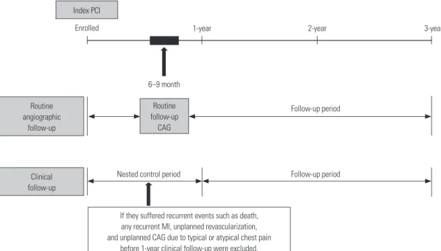
From January 2004 to May 2011, a total of 883 AMI patients were enrolled. Patients who suffered from recurrent events, such as death, any recurrent MI, unplanned revascularization, or unplanned CAG, due to typical or atypical chest pain be- fore CF, especially during 1-year after index PCI, were exclud- ed due to the nested control period (Fig. 1). In general, most of the procedure related complications occurred within 1 year af- ter index PCI. Because we wanted to investigate the beneficial effects of RAF and CF on major adverse cardiac events (MACE) during the follow-up period, these patients were excluded.
During the nested control period, our study strictly defined pa- tients who underwent CAG as scheduled (6–9 months after index PCI) without having any of the above conditions as the RAF group. Follow-up strategies were planned at the day of in- dex PCI, and RAF was scheduled 6–9 months after index PCI, at the operator’s discretion. Finally, a total of 774 eligible AMI patients who successfully underwent PCI with DES without clinical events within 1 year after the index PCI were enrolled in our study. We classified them into either the RAF group (n=
425) or CF group (n=349) according to the two different fol- low-up strategies (Fig. 2). The choice of follow-up modality af- ter index PCI was decided in accordance with physician’s pref- erence. A total eight interventional cardiologists at our single Index PCI
Routine angiographic
follow-up
Clinical follow-up
If they suffered recurrent events such as death, any recurrent MI, unplanned revascularization, and unplanned CAG due to typical or atypical chest pain
before 1-year clinical follow-up were excluded.
Routine follow-up
CAG
Nested control period Follow-up period
Follow-up period 6–9 month
1-year
Enrolled 2-year 3-year
Fig. 1. Schematic presentation of flow sheet of this study. PCI, percutaneous coronary intervention; CAG, coronary angiography; MI, myocardial in- farction.
center participated in our registry. Coincidentally, four physi- cians insisted on RAF, while the other four physicians insisted on CF. None of the physicians changed their choice of follow- up modality until the end of study, such that the enrolled pa- tients never crossed-over. Although this may introduce some inherent limitations, we feel this better reflects real and routine hospital clinical practice. After propensity score-matching (PSM) analysis, two PSM groups (248 pairs, n=496, C-statistic=0.739) were generated, and the baseline characteristics of the two groups were balanced.
PCI procedure and medical treatment
Diagnostic CAG and PCI were performed through either the femoral or radial artery after administration of unfractionated heparin (70–100 IU/kg). Each patient’s activated clotting time was maintained above 250 seconds during the procedure. Re- vascularization was considered clinically indicated when the patient had angina and/or signs of ischemia and restenosis
≥50% in diameter by angiography or restenosis ≥70% in diam- eter, even in the absence of signs and symptoms. The use of cilostazol (Pletaal®, Otsuka Pharmaceutical Co., Tokyo, Japan) or platelet glycoprotein IIb/IIIa receptor blockers was left to the discretion of the individual operators. A successful PCI was defined as the achievement of an angiographic residual steno- sis less than 30% and a final thrombolysis in myocardial infarc- tion blood flow grade of 3. During hospitalization, enrolled pa- tients were to take cardiovascular beneficial medications, including beta-blockers (BB), angiotensin converting enzyme inhibitors (ACEI), angiotensin receptor blockers (ARB), calci- um channel blockers (CCB), and lipid lowering agents. After discharge, the patients were encouraged to stay on the same medications they received during hospitalization. Dual anti- platelet therapy, which comprised a combination of aspirin (100
mg/day) and clopidogrel (75 mg/day), was recommended for at least 12 months to patients who underwent PCI.
Study definitions and clinical follow-up
The recording of cardiovascular risk factors and past medical histories were based on patient self reports. We defined the oc- currences of MACE as total death, recurrent non-fatal myocar- dial infarction, TLR, TVR, or non-TVR. The primary endpoint was composite patient-based outcomes. All deaths were clas- sified cardiac in origin unless a non-cardiac cause could be doc- umented. Re-AMI was defined as the presence of clinical symp- toms, electrocardiographic changes, or abnormal imaging findings of MI in combination with an increase in creatine ki- nase myocardial band fraction above the upper normal limits or an increase in troponin-T/troponin-I to greater than the 99th percentile of the upper normal limit. TLR was defined as a re- vascularization of the target lesion due to restenosis or reoc- clusion within the stent or 5 mm in and adjacent to the distal or proximal segment. TVR was defined as a revascularization of the target vessel or any segment of the coronary artery con- taining the target lesion. Non-TVR was defined as a revascular- ization of any segment of the non-target coronary artery. TLR- MACE was defined as the composite of cardiac death, recurrent Q-wave MI, and TLR. TVR-MACE was defined as the compos- ite of total death, recurrent any MI (Q-wave MI and non-Q wave MI), and TVR. Total MACE was defined as the composite of TVR-MACE and non-TVR. TLR-MACE was considered a le- sion-based clinical outcome, while total MACE and TVR-MACE were deemed patient-based clinical outcomes. We attempted to compare the cumulative incidences of TLR, TVR, and non- TVR by the Kaplan-Meier analysis in order to evaluate their con- tributions to MACE. All participants were required to visit the outpatient clinic of the cardiology department at the end of the first month and then every 3 to 6 months after the index PCI procedure, as well as whenever angina-like symptoms occurred.
Clinical restenosis was suspected when the patients showed new development of one of the following symptoms: recur- rent resting or exertional chest pain, electrocardiographic ST- segment changes during resting or provocation test, elevation of cardiac enzyme [troponin-I, troponin-T, or creatine kinase- MB (CK-MB) level], and abnormal result of imaging study.7 The cumulative incidences of various MACE during the 3-year fol- low-up period were compared between the two groups.
Statistical analysis
For continuous variables, differences between groups were evaluated with the unpaired t-test or Mann-Whitney rank test.
Data are expressed as mean±standard deviations. For discrete variables, differences are expressed as counts and percentag- es, and were analyzed with χ2 or Fisher’s exact test between the groups as appropriate. To adjust for potential confound- ers, propensity score marching (PSM) analysis was performed using a logistic regression model. We tested all available vari- A total 1235 AMI patients underwent coronary angiography
from Jan 2004 to May 2011
Among them, 883 AMI patients were enrolled
RAF (n=425) CF (n=349)
Exclusion
- Cardiac death (n=71) - Non-cardiac death (n=33) - Not participate (n=248)
Exclusion due to nested control period - Death (n=3)
- Recurrent MI (n=1)
- Unplanned revascularization (n=83) - Unplanned CAG (n=106) A total of 774 AMI patients were finally enrolled
Fig. 2. Flow chart of study number of patients. AMI, acute myocardial in- farction; MI, myocardial infarction; CAG, coronary angiography; RAF, rou- tine angiographic follow-up; CF, clinical follow-up.
ables that could be of potential relevance: sex (men), age, left ventricular ejection fraction, STEMI, cardiogenic shock, cardio- vascular diseases risk factors [hypertension, diabetes, dyslipid- emia, chronic kidney disease, cerebrovascular accident (CVA),
peripheral vascular disease (PVD), history of coronary artery disease, current smokers, and current alcoholics], laboratory findings [hemoglobin, CK-MB, troponin-T, high sensitivity C- reactive protein (hs-CRP), lipid profiles, fasting serum glucose,
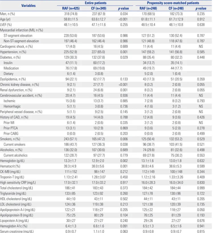
Table 1. Clinical Characteristics and Laboratory Findings
Variables Entire patients Propensity score-matched patients
RAF (n=425) CF (n=349) p value RAF (n=248) CF (n=248) p value
Men, n (%) 318 (74.8) 237 (67.9) 0.034 170 (68.5) 182 (73.3) 0.235
Age (yr) 59.8±11.5 63.6±12.7 <0.001 61.8±11.1 61.7±12.9 0.912
LVEF (%) 48.1±10.5 47.1±11.6 0.255 48.5±10.4 48.1±10.8 0.638
Myocardial infarction (MI), n (%)
ST-segment elevation 228 (53.6) 187 (53.6) 0.986 127 (51.2) 130 (52.4) 0.787
Non-ST-segment elevation 197 (46.4) 162 (46.4) 0.986 121 (48.8) 118 (47.6) 0.787
Cardiogenic shock, n (%) 17 (4.0)0 16 (4.5)0 0.689 11 (4.4)0 11 (4.4)0 NS
Hypertension, n (%) 225 (52.9) 227 (65.0) 0.001 147 (59.2) 141 (56.8) 0.585
Diabetes, n (%) 129 (30.3) 132 (37.8) 0.029 88 (35.4) 80 (32.2) 0.448
Insulin 47 (11.1) 60 (17.2) 34 (13.7) 35 (14.1)
Medication 76 (17.8) 69 (19.8) 49 (19.7) 44 (17.7)
Dietary 6 (1.4)0 3 (0.8)0 5 (2.0)0 1 (0.4)0
Dyslipidemia, n (%) 94 (22.1) 62 (17.7) 0.133 43 (17.3) 51 (20.5) 0.359
Chronic kidney disease, n (%) 9 (2.1)0 27 (7.7)0 <0.001 8 (3.2)0 2 (0.8)0 0.055
Renal dysfunction, n (%) 9 (2.1)0 24 (6.8)0 0.001 8 (3.2)0 2 (0.8)0 0.055
Cerebrovascular accident, n (%) 20 (4.7)0 16 (4.5)0 0.936 11 (4.4)0 11 (4.4)0 NS
Ischemic 15 (3.6)0 13 (3.7)0 0.885 7 (2.8)0 8 (3.2)0 0.793
Hemorrhagic 5 (1.1)0 3 (0.8)0 0.736 4 (1.6)0 3 (1.2)0 NS
Peripheral vessel disease, n (%) 5 (1.1)0 9 (2.5)0 0.145 3 (1.2)0 2 (0.8)0 NS
History of CAD, n (%) 19 (4.5)0 14 (4.0)0 0.788 12 (4.8)0 9 (3.6)0 0.426
Prior MI 6 (1.4)0 2 (0.6)0 0.335 3 (1.2)0 2 (0.8)0 NS
Prior PTCA 13 (3.1)0 10 (2.9)0 0.869 9 (3.6)0 5 (2.0)0 0.278
Prior CABG 0 (0.0)0 2 (0.5)0 0.203 0 (0.0)0 2 (0.8)0 0.499
Smokers, n (%) 243 (57.1) 165 (47.2) 0.006 125 (50.4) 132 (53.2) 0.529
Current smokers 186 (43.7) 127 (36.3) 0.038 96 (38.7) 103 (41.5) 0.521
Alcoholics, n (%) 136 (32.0) 107 (30.6) 0.689 74 (29.8) 81 (32.6) 0.498
Current alcoholics 122 (28.7) 97 (27.7) 0.779 69 (27.8) 75 (30.2) 0.553
Hemoglobin (g/dL) 13.3±1.70 12.9±2.00 0.002 13.1±1.60 13.0±1.80 0.632
Hematocrit (%) 39.3±4.90 38.0±5.60 0.001 38.8±4.60 38.6±5.30 0.599
CK-MB (mg/dL) 111±152 098±147 0.212 112±149 100±148 0.344
Troponin-T (ng/dL) 1.13±2.41 1.28±3.07 0.450 1.12±2.16 1.33±3.26 0.409
High sensitivity CRP (mg/L) 17.0±32.1 17.0±33.2 0.917 16.0±28.3 16.0±34.0 0.830
Total cholesterol (mg/dL) 186±410 183±430 0.373 184±420 184±440 0.999
Triglyceride (mg/dL) 133±850 123±920 0.260 127±780 130±960 0.722
HDL cholesterol (mg/dL) 44±10 43±11 0.502 44±11 43±11 0.205
LDL cholesterol (mg/dL) 124±360 119±380 0.213 121±380 120±390 0.735
Apolipoprotein A-I (mg/dL) 122±210 118±270 0.205 125±220 118±270 0.050
Apolipoprotein B (mg/dL) 75±25 80±29 0.104 76±25 81±29 0.193
Lipoprotein A (mg/dL) 30±27 27±27 0.240 29±26 27±27 0.676
Hemoglobin A1c (%) 6.4±1.3 6.6±1.6 0.091 6.5±1.3 6.5±1.6 0.941
Serum creatinine (mg/dL) 0.9±0.7 1.1±1.0 0.063 0.9±0.8 0.9±0.7 0.815
RAF, routine angiographic follow-up; CF, clinical follow-up; LVEF, left ventricular ejection fraction; CAD, coronary artery disease; PTCA, percutaneous transluminal coronary angioplasty; CABG, coronary artery bypass graft; CK, creatine kinase; CRP, C-reactive protein; HDL, high-density lipoprotein; LDL, low-density lipoprotein;
NS, not significant (>0.999).
Values are mean±SD or n (%). The p value for continuous data from analysis of variance. The p value for categorical data from chi-square test.
hemoglobin A1c and serum creatinine], number of target ves- sels, number of diseased vessels, total number of stent per pa- tient, American College of Cardiology (ACC)/American Heart
Association (AHA) B1/B2/C lesions, type of DES, and post-PCI medications (aspirin, clopidogrel, cilostazol, prasugrel, BB, CCB, ACEI, ARB, diuretics, lipid lowering agents, and proton pump Table 2. Angiographic Characteristics
Variables Entire patients Propensity score-matched patients
RAF (n=425) CF (n=349) p value RAF (n=248) CF (n=248) p value Treated vessels, n (%)
Left main 14 (3.2)0 8 (2.2)0 0.404 5 (2.0)0 5 (2.0)0 NS
Left artery descending 222 (52.2) 205 (58.7) 0.070 131 (52.8) 129 (52.0) 0.857
Left circumflex 131 (30.8) 97 (27.7) 0.358 74 (29.8) 75 (30.2) 0.922
Right coronary artery 172 (40.4) 123 (35.2) 0.136 95 (38.3) 99 (39.9) 0.713
ACC/AHA lesion type, n (%)
Type B1 6 (1.4)0 10 (2.8)0 0.157 5 (2.0)0 5 (2.0)0 NS
Type B2 71 (16.7) 58 (16.6) 0.974 41 (16.5) 43 (17.3) 0.811
Type C 348 (81.8) 281 (80.5) 0.628 202 (81.4) 200 (80.6) 0.819
Bifurcation type (Lefevre), n (%) 162 (38.1) 130 (37.2) 0.804 93 (37.5) 99 (39.9) 0.580
Type 1 77 (18.1) 64 (18.3) 43 (17.3) 53 (21.4)
Type 2 38 (8.9)0 22 (6.3)0 22 (8.9)0 15 (6.1)0
Type 3 11 (2.6)0 11 (3.2)0 7 (2.8)0 10 (4.0)0
Type 4 8 (1.9)0 3 (0.9)0 4 (1.6)0 1 (0.4)0
Type 5 20 (4.7)0 18 (5.1)0 11 (4.4)0 12 (4.8)0
Type 6 8 (1.9)0 12 (3.4)0 6 (2.5)0 8 (3.2)0
Left main disease, n (%) 21 (4.9)0 20 (5.7)0 0.626 10 (4.0)0 12 (4.8)0 0.663
Multi-vessel disease, n (%) 103 (24.2) 72 (20.6) 0.233 49 (19.7) 53 (21.3) 0.657
1 vessel disease 322 (75.7) 277 (79.3) 199 (80.2) 195 (78.6)
2 vessel disease 90 (21.1) 58 (16.6) 39 (15.7) 44 (17.7)
3 vessel disease 13 (3.0)0 14 (4.0)0 10 (4.0)0 9 (3.6)0
Number of vessels 1.2±0.5 1.2±0.5 0.475 1.2±0.5 1.2±0.5 0.793
Number of lesions 1.5±0.8 1.5±0.8 0.635 1.5±0.8 1.5±0.8 0.959
IABP, n (%) 58 (13.6) 41 (11.7) 0.106 28 (11.3) 33 (13.3) 0.564
Ostial (≤5 mm) lesion, n (%) 77 (18.1) 66 (18.9) 0.777 43 (17.3) 44 (17.7) 0.906
Diffuse long lesion (>3 cm), n (%) 191 (44.9) 160 (45.8) 0.802 117 (47.1) 114 (45.9) 0.787
Small vessel (≤2.25 mm), n (%) 23 (5.4)0 18 (5.1)0 0.875 10 (4.0)0 13 (5.2)0 0.522
Calcified lesion, n (%) 47 (11.0) 62 (17.7) 0.008 30 (12.0) 32 (12.9) 0.786
Type of DES, n (%)
Sirolimus-eluting 124 (29.1) 63 (18.0) <0.001 58 (23.3) 51 (20.5) 0.448
Paclitaxel-eluting 149 (35.0) 68 (19.4) <0.001 66 (26.6) 58 (23.3) 0.407
Zotarolimus-eluting 108 (25.4) 135 (38.6) <0.001 84 (33.8) 83 (33.4) 0.924
Endeavor Sprint 55 (12.9) 49 (14.0) 0.656 36 (14.5) 37 (14.9) 0.899
Endeavor Resolute 53 (12.4) 88 (25.2) <0.001 48 (19.3) 47 (18.9) 0.909
Everolimus-eluting 68 (16.0) 104 (29.7) <0.001 51 (20.5) 64 (25.8) 0.167
Xience V/Promus 47 (11.0) 80 (22.9) <0.001 37 (14.9) 48 (19.3) 0.190
Promus element 21 (4.9)0 23 (6.5)0 0.324 14 (5.6)0 17 (6.8)0 0.578
Xience prime 1 (0.2)0 3 (0.8)0 0.332 1 (0.4)0 1 (0.4)0 NS
Biodegradable-polymer-biolimus-eluting 10 (2.3)0 5 (1.4)0 0.355 6 (2.4)0 5 (2.0)0 0.760
Others 7 (1.6)0 1 (0.2)0 0.079 1 (0.4)0 1 (0.4)0 NS
Procedure time (min) 44±34 39±24 0.058 41±32 40±25 0.755
Total doses of unfractionated heparin (international units) 4114±1605 3952±1767 0.205 3860±1287 4110±1908 0.099
Final activated clotting time 230±730 237±850 0.268 234±710 235±850 0.889
RAF, routine angiographic follow-up; CF, clinical follow-up; ACC/AHA, American College of Cardiology/American Heart Association; IABP, intra-aortic balloon pump; DES, drug-eluting stent; NS, not significant (>0.999).
Values are mean±SD or n (%). The p value for continuous data from analysis of variance. The p value for categorical data from chi-square test.
inhibitors). The logistic model by which the propensity scores were estimated showed good predictive value (C-statistic=
0.739). Patients in the RAF group were then one-to-one matched to those in the CF group according to propensity scores with the nearest available pair matching method. Subjects were matched with a caliper width equal to 0.01. The procedure yielded 248 well-matched pairs. Cox-proportional hazard mod- els were used to assess the adjusted hazard ratio (HR) compar- ing the two groups in PSM population. For all analyses, a two- sided p<0.05 was considered statistically significant. All data were processed with Statistical Package for the Social Sciences
version 20.0 (IBM SPSS, Inc., Chicago, IL, USA).
RESULTS
The final study population included 774 eligible AMI patients who successfully underwent PCI with DESs. After PSM analy- sis, two PSM groups (248 pairs, n=496, C-statistic=0.739) were generated. The patient’s baseline clinical characteristics, labo- ratory findings, and angiographic characteristics are summa- rized in Tables 1 and 2. In the unmatched study population, the
Table 3. Post-Percutaneous Coronary Intervention Medications
Variables Entire patients Propensity score-matched patients
RAF (n=425) CF (n=349) p value RAF (n=248) CF (n=248) p value
Aspirin, n (%) 394 (92.7) 336 (96.2) 0.033 234 (94.3) 235 (94.7) 0.843
Clopidogrel, n (%) 395 (92.9) 328 (93.9) 0.561 231 (93.1) 232 (93.5) 0.857
Cilostazol, n (%) 110 (25.8) 80 (22.9) 0.341 57 (22.9) 61 (24.5) 0.673
Prasugrel, n (%) 2 (0.4)0 4 (1.1)0 0.417 2 (0.8)0 2 (0.8)0 NS
Beta blockers, n (%) 244 (57.4) 218 (62.4) 0.154 146 (58.8) 149 (60.0) 0.784
Calcium channel blockers, n (%) 155 (36.4) 105 (30.0) 0.061 85 (34.2) 78 (31.4) 0.503
ACEI, n (%) 129 (30.3) 108 (30.9) 0.859 76 (30.6) 72 (29.0) 0.695
ARBs, n (%) 166 (39.0) 144 (41.2) 0.534 94 (37.9) 103 (41.5) 0.409
Diuretics, n (%) 91 (21.4) 76 (21.7) 0.902 55 (22.1) 57 (22.9) 0.830
Lipid lowering agents, n (%) 378 (88.9) 315 (90.2) 0.552 224 (90.3) 226 (91.1) 0.757
Proton pump inhibitors, n (%) 31 (7.2)0 39 (11.1) 0.061 24 (9.6)0 22 (8.8)0 0.757
RAF, routine angiographic follow-up; CF, clinical follow-up; ACEI, angiotensin converting enzyme inhibitors; ARBs, angiotensin receptor blockers; NS, not significant (>0.999).
Values are numbers and percentages. The p value for categorical data from chi-square test.
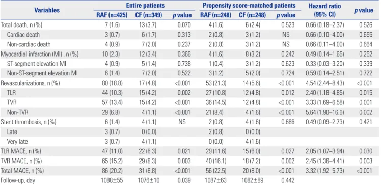
Table 4. Cumulative Events Up to Three Years after the Nested Control Period
Variables Entire patients Propensity score-matched patients Hazard ratio
(95% CI) p value RAF (n=425) CF (n=349) p value RAF (n=248) CF (n=248) p value
Total death, n (%) 07 (1.6) 13 (3.7) 0.070 04 (1.6) 06 (2.4) 0.523 0.66 (0.18–2.37) 0.526
Cardiac death 03 (0.7) 06 (1.7) 0.313 02 (0.8) 03 (1.2) NS 0.66 (0.10–4.00) 0.655
Non-cardiac death 04 (0.9) 07 (2.0) 0.237 02 (0.8) 03 (1.2) NS 0.66 (0.11–4.00) 0.664
Myocardial infarction (MI) , n (%) 10 (2.3) 12 (3.4) 0.366 04 (1.6) 08 (3.2) 0.242 0.49 (0.14–1.65) 0.252
ST-segment elevation MI 04 (0.9) 05 (1.4) 0.738 01 (0.4) 03 (1.2) 0.623 0.33 (0.03–3.20) 0.339
Non-ST-segment elevation MI 06 (1.4) 07 (2.0) 0.522 03 (1.2) 05 (2.0) 0.724 0.59 (0.14–2.51) 0.722
Revascularizations, n (%) 80 (18.8) 17 (4.8) <0.001 53 (21.3) 14 (5.6) <0.001 4.54 (2.44–8.43) <0.001
TLR 44 (10.3) 15 (4.2) 0.002 27 (10.8) 12 (4.8) 0.012 2.40 (1.18–4.85) 0.015
TVR 57 (13.4) 15 (4.2) <0.001 36 (14.5) 12 (4.8) <0.001 3.33 (1.69–6.58) 0.001
Non-TVR 29 (6.8) 04 (1.1) <0.001 21 (8.4) 04 (1.6) <0.001 5.64 (1.90–16.6) 0.002
Stent thrombosis, n (%) 06 (1.4) 04 (1.1) NS 02 (0.8) 04 (1.6) 0.686 0.49 (0.09–2.73) 0.421
Late 03 (0.7) 00 (0.0) 02 (0.8) 00 (0.0)
Very late 03 (0.7) 04 (1.1) 00 (0.0) 04 (1.6)
TLR MACE, n (%) 47 (11.0) 22 (6.3) 0.021 29 (11.6) 15 (6.0) 0.027 2.05 (1.07–3.94) 0.030
TVR MACE, n (%) 65 (15.2) 29 (8.3) 0.003 40 (16.1) 18 (7.2) 0.002 2.45 (1.36–4.41) 0.003
Total MACE, n (%) 86 (20.2) 31 (8.8) <0.001 56 (22.5) 20 (8.0) <0.001 3.32 (1.92–5.73) <0.001
Follow-up, day 1088±55 1076±10 0.039 1087±63 1082±89 0.442
RAF, routine angiographic follow-up; CF, clinical follow-up; MACE, major adverse cardiovascular events; TLR, target lesion revascularization; TVR, target vessel revascularization; CI, confidence interval; NS, not significant (>0.999).
Values are numbers and percentages. The p value for categorical data from chi-square test or Cox-proportional hazard models.
mean age (mean±SD) of the RAF group was 59.8±11.5 years, and that of the CF group was 63.6±12.7 years (p<0.001). The RAF group had a higher number of smokers and higher levels of se- rum hemoglobin and hematocrit, compared with the CF group, while the CF group was more likely to have hypertension, chronic kidney disease, and renal dysfunction. There was no difference in the proportion of patients with STEMI, non-STE- MI, cardiogenic shock, dyslipidemia, prior CVA, PVD, prior MI, prior PCI, prior coronary artery bypass graft, lipid profile, hs- CRP, fasting blood glucose, and hemoglobin A1c (Table 1). An- giographic characteristics in the unmatched population were similar between the two groups, including treated vessels, ACC/
AHA lesion type, bifurcation type (Lefevre), left main disease, multi-vessel disease, ostial lesion, diffuse long lesion, and small vessel disease. Types of DES deployed between the two groups were different. Sirolimus-eluting (CypherTM, Corporation, John- son and Johnson, Warren County, NJ, USA) stent and paclitax- el-eluting (TaxusTM, Boston Scientific, Natick, MA, USA) stents were more frequently deployed in the RAF group; Zotarolimus- eluting (ResoluteTM, Medtronic Inc., Santa Rosa, CA, USA) stents and everolimus-eluting stents (Xience VTM/PromusTM, Boston Scientific) were more common in the CF group (Table 2). The unmatched CF group were more likely than the RAF group to have received aspirin after PCI (96.2% vs. 92.7%, p=0.033). The use of other medications, including clopidogrel, cilostazol (Pletaal®, Otsuka Pharmaceutical Co., Tokyo, Japan), prasug- rel (Effient®, Daiichi Sankyo Company Ltd. UK/Eli Lilly and Company Ltd., Tokyo, Japan), BB, CCB, ACEI, ARB, diuretics, and lipid lowering agents, were similar between the two groups (Table 3). Table 4 shows the cumulative clinical outcomes be- tween one to three years in patients with RAF and CF. The inci-
dences of total death and MI were not significantly different be- tween the two groups. However, the incidence of repeat revascularization (TLR, TVR, non-TVR) in the RAF group was significantly higher than that in the CF group regardless of PSM [TLR: HR, 2.40; 95% confidence interval (CI), 1.18–4.85; p=0.015, TVR: HR, 3.33; 95% CI, 1.69–6.58; p=0.001, and non-TVR: HR, 5.64; 95% CI, 1.90–16.6; p=0.002].
The incidence of MACE was also higher in the RAF group than the CF group unmatched and after PSM (HR, 3.32; 95%
CI, 1.92–5.73; p<0.001). However, the incidence of death (HR, 0.66; 95% CI, 0.18–2.37; p=0.526) or MI (HR, 0.49; 95% CI, 0.14–
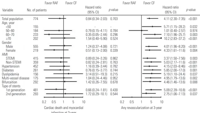
1.65; p=0.252) was not significantly different between the two groups. Results of propensity score adjusted Cox-regression analysis for MACE up to 3 years in various subgroups are shown in Fig. 3. Subgroup analysis for the two different follow-up meth- ods revealed that CF group had more favorable results in view of revascularization rates during the 3 years among all sub- groups.
DISCUSSION
Despite expected beneficial effects, RAF following index PCI with DES in AMI patients was associated with higher inci- dences of repeat revascularizations, including TLR, TVR, and non-TVR, without significant differences in the incidence of death or recurrent AMI during a 3-year CF period. These in- creased revascularization incidences, due to possible ‘oculo- stenotic reflex’ in the RAF group, resulted in a higher MACE in- cidence in our study. The RAF group’s total incidence of MACE was more than three times higher than that for the CF group.
Fig. 3. Propensity score adjusted Cox-regression analysis for cardiac death and myocardial infarction, and any revascularization up to 3-year in vari- ous subgroups. RAF, routine angiographic follow-up; CF, clinical follow-up; STEMI, ST-segment elevation myocardial infarction; CI, confidence interval.
Favor RAF
Cardiac death and myocardial infarction at 3-year
Any revascularization at 3-year 0.2 0.5 1 5 10 0.2 0.5 1 5 10
4.11 (2.30–7.35) 5.71 (1.15–28.2) 1.01 (0.40–2.57) 7.10 (1.96–25.7) 10.2 (2.83–37.2) 4.01 (1.96–8.20) 4.33 (1.61–11.6) 3.37 (1.50–7.56) 5.03 (2.17–11.6) 4.15 (2.03–8.46) 5.85 (2.00–17.0) 5.19 (1.10–24.4) 4.95 (1.79–13.6) 4.46 (1.46–13.6) 5.09 (2.39–10.8) 2.75 (1.06–7.13) 0.84 (0.34–2.03)
- 0.79 (0.15–4.11) 0.35 (0.05–2.44) 1.54 (0.40–5.90) 1.24 (0.37–4.08) 0.51 (0.13–2.00) 0.89 (0.24–3.26) 0.82 (0.24–2.81) 1.16 (0.39–3.44) 0.76 (0.15–3.77) 3.14 (0.51–19.3) 1.04 (0.24–4.46) 1.42 (0.26–7.55) 0.66 (0.24–1.81) 1.72 (0.29–10.1)
<0.001 0.032 0.974 0.003
<0.001
<0.001 0.004 0.003
<0.001
<0.001 0.001 0.037 0.002 0.008
<0.001 0.037 0.703
- 0.784 0.296 0.524 0.721 0.339 0.862 0.763 0.782 0.744 0.215 0.952 0.678 0.430 0.544 774
155184 233202
555219
415359 452261 156175 292 481293 Total population Age, year
<50 50–60 60–70 Gender≥70
MaleFemale AMISTEMI
Non-STEMI Hypertension Diabetes Dyslipidemia Multi-vessel disease Bifurcaition Type of stents
1st generation 2nd generation
Variable No. of patients Hazard ratio
(95% CI)
Hazard ratio (95% CI)
p value p value
Favor RAF
Favor CF Favor CF
We deemed that RAF may have no beneficial effects and could potentially be harmful.
In our study, we excluded patients who suffered recurrent events, such as death, any recurrent MI, unplanned revascu- larization, or unplanned CAG due to typical or atypical chest pain before CF at 1 year. During the nested control period (Fig.
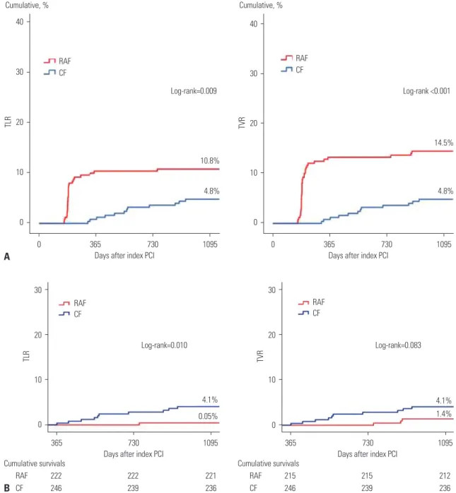
1), our study strictly defined patients who underwent CAG as scheduled (6–9 months after index PCI) absent any of the above conditions as the RAF group. Fig. 4A shows that the total cumu- lative incidences of TLR and TVR in the RAF group were signifi- cantly higher than those in the CF group. Fig. 4B shows that, af- ter the nested control period, the cumulative incidence of TLR
was higher and TVR tended to be higher in the CF group than the RAF group.
We think several possible factors may have influenced these results. As mentioned above, RAF was scheduled at 6–9 months after index PCI. Meanwhile, repeated PCIs that were performed at 10–12 months after index PCI, after the scheduled RAF, were counted as TLRs or TVRs and contributing to the cumulative incidences of TLR or TVR in the RAF group, because these PCIs might be related to RAF strategy. In the RAF group, 96.3% (26 cases/total 27 cases) of TLRs and 91.7% (33 cases/total 36 cas- es) of TVRs occurred days to within one year (6–12 months af- ter index PCI) after RAF. In the CF group, most of TLR and TVR
30
20
10
0
Cumulative survivals
RAF 222 222 221
CF 246 239 236
1095 730
Days after index PCI
4.1%
0.05%
Log-rank=0.010 RAF
CF
365
TLR
B
30
20
10
0
Cumulative survivals
RAF 215 215 212
CF 246 239 236
1095 730
Days after index PCI
4.1%
1.4%
Log-rank=0.083 RAF
CF
365
TVR
40
30
20
10
0 Cumulative, %
1095 730
365
Days after index PCI
10.8%
4.8%
Log-rank=0.009 RAF
CF
0
TLR
A
40
30
20
10
0 Cumulative, %
1095 730
365
Days after index PCI
14.5%
4.8%
Log-rank <0.001 RAF
CF
0
TVR
Fig. 4. Kaplan-Meier curved analysis for TLR and TVR. (A) Total cumulative events curve of TLR and TVR. (B) Cumulative events curve up to 3-year after the nested control period. TLR, target lesion revascularization; TVR, target vessel revascularization; RAF, routine angiographic follow-up; CF, clinical fol- low-up; PCI, percutaneous coronary intervention.
cases occurred after one year (after the nested control period).
The effects thereof are described further in the limitations sec- tion. As shown in Fig. 4B, after the nested control period, the cumulative incidence of TLR and TVR in the CF group was higher than that in the RAF group. After the nested control pe- riod, patients included in the RAF group were relatively stable and not progressive, while patients in the CF group may have been relatively unstable and more likely to be symptomatic due to progression of underlying stenotic CAD. Although present study showed TVR rates were not significantly different be- tween the two groups, if we can follow up for a longer time and investigate much larger-scaled populations, TVR rates may become similar to the cumulative incidence of TLR. According- ly, the authors suggest that longer-term and larger-scaled fol- low-up study is warranted.
Stenotic coronary artery lesions that do not produce ischemic symptoms receive little benefit from revascularization, com- pared with effective optimal medical therapy alone.8 Despite its cost and periprocedural risk, RAF is still performed at select centers to identify angiographically significant stenosis that is not related to ischemic symptoms. The present study showed that total death or MI during the 3 years of follow-up was not reduced in the routine RAF group, compared with the CF group, in AMI patients after index PCI. Although DES can reduce the incidence of clinical and angiographic restenosis rates signifi- cantly, compared with BMS,10 all of the major DES clinical tri- als have required angiographic follow-up.11-13 Protocol-mediat- ed angiographic follow-up in DES may negatively affect safety outcomes,5 and there was substantial controversy regarding the need for and impact of, protocol-mandated angiographic follow-up in PCI trials. Follow-up angiography after PCI has been shown to be associated with accentuated rates of revas- cularization, resulting from the “oculostenotic reflex,” a term describing revascularization with PCI due to anatomic lesion severity, regardless of clinical or physiologic evidence of isch- emia.14 Only a minority (22%) of patients with angiographic re- stenosis show severe (diameter stenosis >70%) stenosis, which is primarily associated with demonstrable myocardial isch- emia.3 In the HORIZONS-AMI study,15 RAF magnified the ben- efit of paclitaxel-eluting stents (PES) over BMS with respect to TVR beyond 1 year, although RAF showed a relatively higher incidence of TLR, compared with the natural event rates before RAF at 13 months after index PCI. A meta-analysis of 11 ran- domized trials16 between DES and BMS in STEMI patients re- vealed a TVR reduction rate of 7.6% in the DES group at 12 months (5.0% vs. 12.6%, p<0.0001). In eight of these 11 studies, RAF was performed before comparison of major clinical out- comes.
In the cases of stable coronary artery disease, TVR was signif- icantly higher at 5 years for patients in whom protocol-mandat- ed angiographic follow-up was planned versus not planned (18.3% vs. 11.1%, p<0.001), although there were no significant dif- ferences in death or reinfarction (8.9% vs. 8.8%, p=0.93).14 These
increased rates of TVR were due to greater treatment of interme- diate stenotic lesions (40–70% stenosis by quantitative angi- ography), not to severe “pre-infarction” stenosis. In addition, intermediate lesions tend to regress over time (2 to 5 years).17,18 In the Clinical Evaluation of the XIENCE VTM Everolimus Elut- ing Coronary Stent System in the Treatment of Subjects With de Novo Native Coronary Artery Lesions (SPIRIT) III trial, there was four times as much TVR in the scheduled angiographic fol- low-up (SAF) group as in the no SAF group, predominantly re- lated to treatment of lesions without documented ischemia (4.5%
vs. 1.0%, p=0.002).17 A substantial proportion of restenosis epi- sodes can present as acute coronary syndrome or MI, and wheth- er RAF or SAF can identify such culprits lesions before they be- come symptomatic, enabling preventive revascularization, is unsettled. Mindrescu, et al.9 also reported that SAF leads to in- creased rates of revascularization without impacting the oc- currences of death, MI, and stent thrombosis. The SPIRIT III trial19 also had indicated that RAF follow-up tended to overes- timate decreases in TVR, compared to routine CF. This suggest that RAF does not appear to adversely affect the long-term safety of patients. At 3 years, rates of death or MI were similar between the RAF and CF groups in this study.
The important causes of recurrent disease at the target lesion site are restenosis and stent thrombosis. Fifty-three patients (21.3%) had revascularization procedure during RAF in our study after PSM analysis (Table 4). Among these patients, 40 pa- tients (75.5%) had revascularization procedures within 2 weeks during a routine follow-up angiography (p<0.001). Thirteen patients (25.5%) had a revascularization procedure thereafter.
When we considered restenosis rates in patients treated with DES in the general population,4 these high incidences of re- vascularization, especially within 2 weeks in RAF group, may include restenotic lesions that do not compromise the lumen area sufficiently enough to cause ischemic symptom. The rates of stent thrombosis were relatively low in our study (1.4% vs.
1.1%, p=NS) during the 3-year follow up period. This may be due to the limitations of a single center study and relatively large proportion of one vessel disease. Moreover, 1st generation DES [Sirolimus-eluting stent (CypherTM)] and PES (TaxusTM) were much more frequently deployed in the unmatched RAF group in our study. This bias regarding stent type, however, was dis- regarded after PSM analysis.
In our study, the CF group was more likely to be elderly, hy- pertensive, and have chronic kidney disease and renal dysfunc- tion. Therefore, we can consider that the CF group faced relative- ly higher angiographic risk (treated vessels, ACC/AHA lesion type, bifurcation type, left main disease, multi-vessel disease, ostial lesion, diffuse long lesion, and small vessel disease). How- ever, revascularization rates and MACE of the CF group were lower than those in the RAF group before and after PSM analy- sis. These results were sustained during the subgroup analysis. The CF group showed favorable results in view of any revasculariza- tion type at 3 years regardless of the subgroup. Subgroup anal-
ysis also showed cardiac death and MI at 3-years to not signif- icantly differ between the two groups (Fig. 3).
This study has some limitations, because it is a non-random- ized design and a single center study. In the aspect of the choice in follow-up modality after index PCI, physician preferences may act as a selection bias, although we feel this better reflects real and routine hospital clinical practice. Although we planned to compare 3-year long-term clinical outcomes between RAF and CF in AMI patients undergoing PCI, those who suffered recurrent events, such as death, any recurrent MI, unplanned revascularization, and unplanned CAG due to typical or atypi- cal chest pain, within 1 year of CF were excluded from our study due to nested control period. It is possible that some propor- tion of MACEs in the CF group could have been included in this nested control period, leading to potential underestimation of MACE in the CF group. Additionally, RAF group patients may have more chances to undergo early detection and early man- agement for their coronary lesions, regardless of the presence of symptoms, than symptom-driven CF patients. In the cases of RAF, the treatment strategies for angiographic stenotic le- sions were left to the judgement of the operators. Unfortunate- ly, in this study, functional studies were done only for a small number of patients (<10%). Practically, we cannot perform rou- tine functional studies including fractional flow reserve (FFR) due to cost issues. In Korea, currently there is no reimburse- ment program for FFR, IVUS, or Cardiac CT and MRI, in addi- tion to CAG. We should depend on angiographic findings and clinical decision in real world clinical practice. Although rela- tively lower rates of functional studies, non-randomized de- sign, and single center study, this study may be meaningful because we tried to reflect “real world” clinical practice. Thus, the patients included in the RAF group underwent CAG as scheduled, regardless of the results of functional study. Also, the patients included in the CF group underwent CAG when- ever angina-like symptoms occurred (symptom-driven CAG), regardless of the results of functional study. Therefore, we think that clinical milieu in terms of function study support in both groups would not be significantly different and would not impact clinical events differently. In the current study, nested control period was defined as 1 year after PCI. However, follow- up CAG was performed 6–9 months after PCI. Therefore, there was a gap of 3 months between “during 1 year after PCI” and the window of the follow-up period. The authors considered that this 3-month gap period may also be related with vulner- able periods, during which procedure-related late complica- tions can happen frequently. During the first 6 months after index PCI, procedure-related acute complications can also occur. Accordingly, we thought 1 year after index PCI was more reasonable as a nested control period than 6 months after in- dex PCI. However, in real-world practice, some patients un- dergo RAF after the scheduled date. Delayed RAF in the pres- ent study was due to personal schedule conflicts and other barrier factors to later than 9 months. Delayed RAF was per-
formed in 16 patients between 10–12 months after index PCI in our study. Exceptionally, in these special cases, these patients were included in the RAF group. However, in these patients, TLR or TVR was not recorded up to 3 years, and they did not contribute to the cumulative incidences of TLR or TVR. In ad- dition, when repeated PCIs were done in these periods (10–12 months after index PCI) after scheduled RAF, these PCIs were counted as TLR or TVR rates and presented in the cumulative incidence of TLR or TVR rates in the RAF group, since these PCIs may be related with RAF strategy. That is to say, these 10–12 months after index PCI included both above exception- ally delayed RAF and the cases of TLR or TVR after RAF in the RAF group. Finally, the index PCI day was considered as the day of enrollment.
Furthermore, like every “real-world” registry, there may have been some under-reporting and/or missing data. Also, we could not obtain exact information concerning the relationship be- tween angina symptom and the degree of angiographic steno- sis due to some missing values. Therefore, we could not de- scribe the differences in TLR rates between the two follow-up groups and intermediate stenotic lesions.
In conclusion, given these limitations, our results do not in- dicate any clinical benefit of RAF in clinical practice. The results of our study confirm that patients who are assigned to RAF undergo more revascularization procedures than CF alone with- out an improvement from death or reinfarction. Therefore, CF seems warranted for asymptomatic patients after PCI for AMI.
REFERENCES
1. Choi YJ, Kim JB, Cho SJ, Cho J, Sohn J, Cho SK, et al. Changes in the practice of coronary revascularization between 2006 and 2010 in the Republic of Korea. Yonsei Med J 2015;56:895-903.
2. Tantawy A, Ahn CM, Shin DH, Kim JS, Kim BK, Ko YG, et al. Nobo- ri-biolimus-eluting stents versus resolute zotarolimus-eluting stents in patients undergoing coronary intervention: a propensity score matching. Yonsei Med J 2017;58:290-5.
3. Garg S, Serruys PW. Coronary stents: current status. J Am Coll Car- diol 2010;56(10 Suppl):S1-42.
4. Kirtane AJ, Gupta A, Iyengar S, Moses JW, Leon MB, Applegate R, et al. Safety and efficacy of drug-eluting and bare metal stents: com- prehensive meta-analysis of randomized trials and observational studies. Circulation 2009;119:3198-206.
5. Cutlip DE, Chauhan MS, Baim DS, Ho KK, Popma JJ, Carrozza JP, et al. Clinical restenosis after coronary stenting: perspectives from multicenter clinical trials. J Am Coll Cardiol 2002;40:2082-9.
6. King SB 3rd, Smith SC Jr, Hirshfeld JW Jr, Jacobs AK, Morrison DA, Williams DO, et al. 2007 focused update of the ACC/AHA/SCAI 2005 guideline update for percutaneous coronary intervention: a report of the American College of Cardiology/American Heart As- sociation Task Force on Practice guidelines. J Am Coll Cardiol 2008;
51:172-209.
7. Silber S, Albertsson P, Avilés FF, Camici PG, Colombo A, Hamm C, et al. Guidelines for percutaneous coronary interventions. The Task Force for Percutaneous Coronary Interventions of the European Society of Cardiology. Eur Heart J 2005;26:804-47.
8. Pijls NH, van Schaardenburgh P, Manoharan G, Boersma E, Bech
JW, van’t Veer M, et al. Percutaneous coronary intervention of func- tionally nonsignificant stenosis: 5-year follow-up of the DEFER Study. J Am Coll Cardiol 2007;49:2105-11.
9. Mindrescu C, Brener SJ, Guerchicoff A, Fahy M, Parise H, Mehran R, et al. Impact of scheduled angiographic follow-up in patients treated with primary percutaneous coronary intervention for ST- segment elevation myocardial infarction. J Interv Cardiol 2013;26:
319-24.
10. Stone GW, Ellis SG, Cox DA, Hermiller J, O’Shaughnessy C, Mann JT, et al. A polymer-based, paclitaxel-eluting stent in patients with coronary artery disease. N Engl J Med 2004;350:221-31.
11. Moses JW, Leon MB, Popma JJ, Fitzgerald PJ, Holmes DR, O’Shaughnessy C, et al. Sirolimus-eluting stents versus standard stents in patients with stenosis in a native coronary artery. N Engl J Med 2003;349:1315-23.
12. Pinto DS, Stone GW, Ellis SG, Cox DA, Hermiller J, O’Shaughnessy C, et al. Impact of routine angiographic follow-up on the clinical ben- efits of paclitaxel-eluting stents: results from the TAXUS-IV trial. J Am Coll Cardiol 2006;48:32-6.
13. Lasala JM, Cox DA, Dobies D, Muhlestein JB, Katopodis JN, Revtyak G, et al. Usage patterns and 2-year outcomes with the TAXUS express stent: results of the US ARRIVE 1 registry. Catheter Cardiovasc In- terv 2008;72:433-45.
14. Uchida T, Popma J, Stone GW, Ellis SG, Turco MA, Ormiston JA, et al. The clinical impact of routine angiographic follow-up in ran- domized trials of drug-eluting stents: a critical assessment of “oc-
ulostenotic” reintervention in patients with intermediate lesions.
JACC Cardiovasc Interv 2010;3:403-11.
15. Stone GW, Parise H, Witzenbichler B, Kirtane A, Guagliumi G, Pe- ruga JZ, et al. Selection criteria for drug-eluting versus bare-metal stents and the impact of routine angiographic follow-up: 2-year insights from the HORIZONS-AMI (Harmonizing Outcomes With Revascularization and Stents in Acute Myocardial Infarction) trial.
J Am Coll Cardiol 2010;56:1597-604.
16. De Luca G, Stone GW, Suryapranata H, Laarman GJ, Menichelli M, Kaiser C, et al. Efficacy and safety of drug-eluting stents in ST-seg- ment elevation myocardial infarction: a meta-analysis of random- ized trials. Int J Cardiol 2009;133:213-22.
17. Asakura M, Ueda Y, Nanto S, Hirayama A, Adachi T, Kitakaze M, et al. Remodeling of in-stent neointima, which became thinner and transparent over 3 years: serial angiographic and angioscopic fol- low-up. Circulation 1998;97:2003-6.
18. Hochman JS, Tamis JE, Thompson TD, Weaver WD, White HD, Van de Werf F, et al. Sex, clinical presentation, and outcome in patients with acute coronary syndromes. Global Use of Strategies to Open Occluded Coronary Arteries in Acute Coronary Syndromes IIb In- vestigators. N Engl J Med 1999;341:226-32.
19. Lansky AJ, Brar SS, Yaqub M, Sood P, Applegate RJ, Lazar D, et al.
Impact of routine angiographic follow-up after percutaneous cor- onary intervention with drug-eluting stents in the SPIRIT III ran- domized trial at three years. Am J Cardiol 2012;110:21-9.