Prognostic Value of Left Atrium Remodeling after Primary
Percutaneous Coronary Intervention in Patients with ST Elevation Acute Myocardial Infarction
The purpose of this study is to assess the relationship between left atrial (LA) size and outcome after acute myocardial infarction (AMI) in patients undergoing primary percutaneous coronary intervention (PCI) and to evaluate dynamic changes in LA size during long-term follow-up. Echocardiographic analyses were performed on 253 AMI patients (174 male and 79 female, 65.4 ± 13.7 yr) undergoing PCI. These subjects were studied at baseline and at 12 months. Clinical follow-up were done at 30.8 ± 7.5 months.
We assessed LA volume index (LAVI) at AMI-onset and at 12-month. Change of LAVI was an independent predictor of new onset of atrial fibrillation or hospitalization for heart failure (P = 0.002). Subjects who survived the 12-month period displayed an increased LAVI mean of 1.86 ± 4.01 mL/m2 (from 26.1 ± 8.6 to 28.0 ± 10.1 mL/m2, P < 0.001). The subject group that displayed an increased LAVI correlated with a low left ventricular ejection fraction, large left ventricle systolic and diastolic dimensions and an enlarged LA size. In conclusion, change of LAVI is useful parameter to predict subsequent adverse cardiac event in AMI patients. Post-AMI echocardiographic evaluation of LAVI provides important prognostic information that is significantly greater than that obtained from clinical and laboratory parameters alone.
Key Words: Echocardiography; Myocardial Infarction; Heart atria; Remodeling; Prognosis Jang Hyun Cho1, Su Hyun Kim1,
Cheol hwan Kim1, Jae Yeong Park1, Seung Choi1, Myung Ho Yun1, Dong Han Kim1, Jae Hyun Mun1, Jun Young Kim1, Hyun Ju Yoon2, Kye Hun Kim2, and Myung Ho Jeong2,3
1Division of Cardiology, Department of Internal Medicine, St. Carollo Hospital, Suncheon;
2The Heart Center, Chonnam National University Hospital, Gwangju; 3The Brain Korea 21 Project, Chonnam National University, Gwangju, Korea Received: 2 July 2011
Accepted: 12 December 2011 Address for Correspondence:
Myung Ho Jeong, MD
Principal Investigator of Korea Acute Myocardial Infarction Registry, Director of Korea Cardiovascular Stent Research Institute of Chonnam National University, Director of Department of Education and Research of Chonnam Natioinal University Hospital, 167 Jaebongro, Dong-gu, Gwangju 501-757, Korea
Tel: +82.62-220-6243, Fax: +82.62-228-7174 E-mail: [email protected]
http://dx.doi.org/10.3346/jkms.2012.27.3.236 • J Korean Med Sci 2012; 27: 236-242
INTRODUCTION
Echocardiographic assessment of the left ventricular (LV) func- tion plays an integral role in the examination of patients with heart failure (HF) undergoing acute myocardial infarction. In addition to the evaluation of myocardial contractility, the pa- rameters of diastolic function may be useful in the providing significant prognostic information and determining therapeu- tic strategies (1-6). Over the past decade, several indicators have been established to predict poor prognosis in patients with chron- ic HF, including age, etiology, left ventricular ejection fraction (LVEF), functional class, exercise capacity, pulmonary arterial pressures, and cardiac output (7-9). Left atrial (LA) size also has been used as an important predictor of prognosis both in the general population and in patients with heart diseases (10), such as LV dysfunction (11), aortic stenosis (12), mitral stenosis (13), mitral regurgitation (14), and atrial fibrillation (AF) (15). In this study, we assessed LAVI in patients who presented with their first acute myocardial infarction (AMI) and were treated with primary percutaneous coronary intervention (PCI). LA volume
has been described as an important predictor of fatal acute myo- cardial infarction (16-19), with smaller LA volume being associ- ated with good prognosis even in patients with depressed sys- tolic function (20). In patients suffered from AMI, the LA volume may serve as a surrogate marker for the measurement of chronic diastolic function and ventricular filling pressure, as it was less affected by acute hemodynamic changes than were transmitral Doppler measurements (21, 22). In this study, we assessed LAVI in patients who presented their first AMI and were treated with primary PCI.
MATERIALS AND METHODS Study group
We evaluated 276 patients with AMI who underwent PCI in our department between January 2007 and December 2008. We per- formed an echocardiography within 1 day to assess the LAVI and evaluated the LA remodeling severity during the 12-month follow-up period after AMI. Our study included 264 consecu- tive patients (174 male and 79 female, 65.4 ± 13.7 yr) with a first
ST-elevation AMI who underwent primary PCI. Twelve patients were excluded before analysis because of underlying heart fail- ure and atrial fibrillation. AMI was identified by clinical symp- toms, new 1-mm ST-segment elevation in 2 contiguous leads, identification of the culprit artery on coronary angiography, and an increase in serum troponin, at least a ‘three-fold-level’ higher than the upper limit of normal as defined in our laboratory (0.4 IU). Inclusion criteria included the patient being 18 yr or more, undergoing primary PCI within 12 hr of hospital admission, the identification of the culprit artery on coronary angiography and the ability to give informed consent. Exclusion criteria were: Non- coronary atherosclerotic etiology of AMI, cardiogenic shock, intra-aortic balloon pump, intravenous inotropic support, pre- vious coronary artery bypass surgery, presence of paced rhythm, the development of complication and known chronic heart fail- ure before the echocardiographic study, inability to assess 3 myo- cardial segments on echocardiogram, and a history of cocaine or drug abuse. Hospital charts were reviewed for age, peak tro- ponin level, lipid profile, electrocardiogram at the initial presen- tation, angiographic findings before and after PCI, in-hospital complications, development of congestive heart failure, the need for urgent revascularization after PCI, and death. Echocardio- graphic examinations were performed at a mean of 3.5 days after the AMI (baseline) and at 12 months after AMI. Clinical follow- up was done for 30.8 ± 7.5 months. The median echocardio- graphic follow-up was 12.5-months (Group I: 12.5 ± 0.7, Group II: 12.4 ± 0.8, P = 0.201). Demographic and clinical characteris- tics of the included subjects are summarized in Table 1.
Ethics statement
The study protocol was reviewed and approved by the institu-
tional review board of the National Evidence-based Healthcare Collaborating Agency (PIRBII-002-1[2]). Informed consent was submitted by all of the subjects.
Echocardiographic analysis
The echocardiographic analysis was performed in the left lateral decubitus position at rest using conventional methods with a commercially available ultrasound system (Siemens, Warsaw, Poland). All echocardiographic studies were analyzed in the main laboratory at the Division of Cardiology of our institute.
During 12-months of follow-up, 11 of 264 patients died (cardiac deaths, 7; non-cardiac deaths, 4). Echocardiographic analyses were collected for the remaining 253 patients at baseline and at 12 months, and clinical follow-up was performed at 30.8 ± 7.5 months through patients visits and telephone interview.
The protocol included calculation of the left ventricular ejec- tion fraction (LVEF) by applying the biplane Simpson Method (with tracing of end-diastolic and end-systolic left ventricular endocardial border from apical 4-chamber and 2-chamber views in 2D echocardiography), assessment of mitral inflow pattern (in an apical 4-chamber view by using the pulse Doppler sam- ple volume at the tips of the opened mitral valve leaflets), pulsed tissue Doppler analysis of the mitral annular motion. Chamber dimensions (both ventricles and left atrium) were measured from 2D-imaging in the parasternal long axis view. Antero-pos- terior dimension of the atrium was measured during end systo- le. Mitral regurgitation (MR) was graded with color flow imag- ing. For wall motion analysis, a 16-segment model was used ac- cording to recommendations by the American Society of Echo- cardiography (23). Analysis of the Echocardiographic image was performed centrally by 2 expert investigators who were unaware of patients’ clinical, electrocardiographic, or angiographic data.
The wall motion score was reached via consensus. A percentage of the wall motion abnormalities were obtained by dividing the number of akinetic, dyskinetic, and aneurysmal segments by the total number of segments evaluated. MR was considered mild when the regurgitant jet area occupied > 5 and < 20% of the LA area, moderate when it occupied > 20 and < 40%, and severe when it occupied more than 40% of the LA area (24). Mitral in- flow Doppler echocardiographic indices were obtained for each patient. We classified diastolic dysfunction according to mitral inflow pattern. An E/A ratio < 1 was accepted as an indication of delayed relaxation profile, E/A > 2 as typical for restriction, and values of E/A between 1 and 2 as either a normal or pseudo- normal pattern. Pulsed tissue Doppler analysis of mitral annular motion was also applied. LA volume was assessed by the biplane area-length method from apical 4-and 2-chamber views at the end-systole from the frame preceding the mitral valve opening.
LAVI was calculated as LA volume to body surface area ratio (mL/m2). LA remodeling was assessed by looking at the change in the LAVI from baseline to 12 months. In all cases, we analyzed Table 1. Baseline characteristics and number of mitral inflow pattern in study popu-
lation (n = 264)
Parameters Findings, No. (%)
Age (yr) 65.2 ± 13.7
Male 182 (68.9)
Body mass index (kg/m2) 24.2 ± 3.0
History of ischemic heart disease 33 (12.5)
Anterior myocardial infarction 131 (49.6)
Hypertension 124 (47.0)
Diabetes mellitus 78 (29.5)
Hypercholesteremia 11 (4.2)
Smoking 149 (56.4)
Killip class ( ≥ III) 32 (12.1)
Heart Rate 77.0 ± 21.0
Systolic blood pressure (mmHg) 130.3 ± 30.0
Diastolic blood pressure (mmHg) 81.3 ± 18.8
Successful PCI 257 (97.3)
Normal profile 83 (31.4)
Delayed relaxation profile 145 (54.9)
Pseudonormal profile 10 (3.8)
Restrictive profile 4 (1.5)
PCI, percutaneous coronary intervention.
medical history and recorded cardiac events such as cause and time of death. After the 12-month follow-up echocardiogram was performed, patients were divided into two groups: decreased LAVI (group I) and increased LAVI (group II) group. Baseline echocardiographic data between these groups were compared.
Relative risk of new onset AF and hospitalization due to exacer- bation of heart failure was calculated and the resulting Kaplan- Meier curves were compared.
Coronary angiography
Selective coronary artery angiography was performed in multi- ple views using Judkin’s technique. It was analyzed by an inde- pendent cardiologist. The infarct-related artery was identified based on a Thrombolysis in the Myocardial Infarction grade 0 to 2 flow, the presence of haziness, suggestive of thrombus, and a 75% narrowing in relation to the reference segment before the PCI. Thrombolysis in the Myocardial Infarction flow grade (0 to 3) after the PCI was also recorded (25). Significant coronary artery disease was defined as a 50% narrowing of the luminal diame- ter of any of the 3 major vessels or a major branch.
Statistical methods
All continuous data were expressed as means ± standard devia- tions and means were compared using Student’s t-tests for un- paired variables. Differences between patients were assessed by un-paired t-testing, and parameters frequencies were exam- ined using the chi-square test. The differences between echocar-
diographic measurements of the 2 groups, changes over time within each group (time effect), and interaction effects were as- sessed by repeated measures analysis of variance. Differences between groups with LAVI change was assessed by the one-way ANOVA and frequency of parameters by chi-square test. Cumu- lative event rate of new atrial fibrillation onset, hospitalization with heart failure was assessed by the Kaplan-Meier method.
The influence of LA remodeling during the 12-month risk of clin- ical outcomes was evaluated by the Cox proportional hazards models. For a multivariable model, we included variables proved to be initial AMI-onset clinical predictors of mortality from the overall study. These included age, creatinine, Killip class, histo- ry of diabetes and echocardiographic parameters (LVEF, E/A ratio, E/E’ ratio), which were found to have a prognostic value in prior studies. We also included the number of repeated revas- cularization. A P value < 0.05 considered statistically significant.
Statistical analysis was performed with SPSS 18.0 for Windows (SPSS Inc., Chicago, IL, USA).
To calculate optimal cutoff values the receiver operating char- acteristic (ROC) Analysis was performed. Sensitivity, specificity, predictive values, and accuracy were calculated by using typical formulas. The Kaplan-Meier survival analysis was applied for determining the parameters differentiating the groups into var- ious prognoses. The multivariate enter logistic regression was performed to identify the independent predictors of adverse outcome. A P value < 0.05 was considered significant.
RESULTS
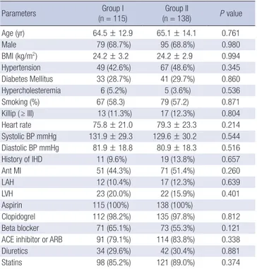
During the mean 30-month follow-up period of 253 patients with AMI, 26 subjects were hospitalized due to heart failure ex- acerbation and 23 subjects developed atrial fibrillation. These subjects were divided into two groups; 115 with decreased LAVI (Group I) and 138 with increased LAVI (Group II). There were no significant differences in baseline clinical characteristics, medi- cation and laboratory findings between the two groups (Tables 2, 3), or in the 12-month persistent rate of medications includ- Table 2. Baseline clinical characteristics and medications between groups
Parameters Group I
(n = 115) Group II
(n = 138) P value
Age (yr) 64.5 ± 12.9 65.1 ± 14.1 0.761
Male 79 (68.7%) 95 (68.8%) 0.980
BMI (kg/m2) 24.2 ± 3.2 24.2 ± 2.9 0.994
Hypertension 49 (42.6%) 67 (48.6%) 0.345
Diabetes Mellitus 33 (28.7%) 41 (29.7%) 0.860 Hypercholesteremia 6 (5.2%) 5 (3.6%) 0.536
Smoking (%) 67 (58.3) 79 (57.2) 0.871
Killip ( ≥ III) 13 (11.3%) 17 (12.3%) 0.804
Heart rate 75.8 ± 21.0 79.3 ± 23.3 0.214
Systolic BP mmHg 131.9 ± 29.3 129.6 ± 30.2 0.544 Diastolic BP mmHg 81.9 ± 18.8 80.9 ± 18.3 0.516
History of IHD 11 (9.6%) 19 (13.8%) 0.657
Ant MI 51 (44.3%) 71 (51.4%) 0.260
LAH 12 (10.4%) 17 (12.3%) 0.639
LVH 23 (20.0%) 22 (15.9%) 0.401
Aspirin 115 (100%) 138 (100%)
Clopidogrel 112 (98.2%) 135 (97.8%) 0.812
Beta blocker 71 (65.1%) 73 (55.3%) 0.121
ACE inhibitor or ARB 91 (79.1%) 114 (83.8%) 0.338
Diuretics 34 (29.6%) 42 (30.4%) 0.881
Statins 98 (85.2%) 121 (89.0%) 0.374
BP, blood pressure; IHD, ischemic heart disease; MI, myocardial infarction; LAH, left atrial hypertrophy; LVH, left ventricular hypertrophy; ACE, angiotensin-converting en- zyme; ARB, angiotensin receptor blocker.
Table 3. Baseline laboratory findings between groups Biochemical
parameters Group I Group II P value
Glucose 179.2 ± 87.9 167.3 ± 70.9 0.234
Creatinine 1.22 ± 1.14 1.31 ± 1.60 0.571
TC 185.0 ± 45.0 187.7 ± 42.3 0.637
LDL-c 117.9 ± 37.5 118.0 ± 39.0 0.973
hs-CRP 0.81 ± 2.15 0.87 ± 2.42 0.882
BNP 404.2 ± 628.9 601.1 ± 782.8 0.032
CPK 1,622.1 ± 1,157.6 1,480.1 ± 1,127.1 0.325
CK-MB 171.4 ± 162.9 181.0 ± 187.9 0.667
TnI 17.6 ± 15.3 17.1 ± 15.4 0.784
TC, total cholesterol; LDL-c, low density lipoprotein cholesterol; hs-CRP, high sensi- tive C-reactive protein; BNP, B-type natriuretic peptide; CPK, creatine phosphokinase;
CK-MB, creatine kinase-MB isoenzyme; TnI, troponin I.
ing beta blockers (Group I; 65 [59.1%], Group II; 82 [64.1%], P = 0.431), renin-angiotensine system (RAS) blockers (Group I; 91 [82.7%], Group II; 107 [84.3%], P = 0.195) and amiodarone (Group I; 0 [0%], Group II; 2 [1.4%], P = 0.195). Table 4 compares the
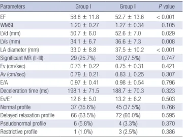
echocardigraphic parameters of the group with decreased LAVI to those of the group with increased LAVI. Regarding the echo- cardiographic parameters, the group with increased LAVI dis- played significantly larger left ventricular systolic and diastolic dimension, lower LVEF values, and larger LA dimension (mea- sured in the long axis view) (P < 0.01, for all parameters) com- pared to the group with decreased LAVI. The classic echocar- Table 4. Comparison of baseline echocardiographic parameters between groups
Parameters Group I Group II P value
EF 58.8 ± 11.8 52.7 ± 13.6 < 0.001
WMSI 1.20 ± 0.27 1.27 ± 0.34 0.105
LVd (mm) 50.7 ± 6.0 52.6 ± 7.0 0.029
LVs (mm) 34.1 ± 6.7 36.6 ± 7.3 0.008
LA diameter (mm) 33.0 ± 8.8 37.5 ± 10.2 < 0.001 Significant MR (II-III) 29 (25.7%) 39 (27.5%) 0.747
Ev (cm/sec) 0.73 ± 0.22 0.75 ± 0.31 0.421
Av (cm/sec) 0.79 ± 0.21 0.83 ± 0.25 0.307
E/A 0.97 ± 0.41 0.98 ± 0.54 0.796
Deceleration time (ms) 198.1 ± 71.5 188.7 ± 70.3 0.323
Ev/E´ 12.6 ± 5.0 13.2 ± 6.2 0.503
Normal profile 37 (35.6%) 45 (37.5%) 0.766
Delayed relaxation profile 66 (63.5%) 72 (60.0%) 0.595
Pseudonormal profile 6 (5.8%) 4 (3.3%) 0.370
Restrictive profile 1 (1.0%) 3 (2.5%) 0.386
EF, ejection fraction; WMSI, wall motion score index; MR, mitral regurgitation; E or A, v, t, dt, early or atrial mitral inflow velocity, time or deceleration time; E´, early diastolic m.a velocity; A´, atrial late diastolic m.a velocity; S´, systolic m.a velocity; Ev/E´, ratio of peak velicity of mitral inflow early phase to peak velocity of early mitral annulus motion.
Table 5. Baseline angiographic characteristics between groups
Angiography findings Group I Group II P value
Infarct-related artery LAD
Non-LAD
Diseased vessel number
47 (43.1%) 62 (56.9%) 1.59 ± 0.71
65 (48.1%) 70 (51.9%) 1.66 ± 0.74
0.433 0.426 ACC/AHA lesion type
B1 B2 C
9 (8.3%) 17 (15.6%) 81 (74.3%)
8 (5.9%) 18 (13.3%) 109 (80.7%)
0.477 0.616 0.229 TIMI flow
0 I II III
59 (54.1%) 7 (6.4%) 20 (18.3%) 23 (21.1%)
72 (53.3%) 10 (7.4%) 26 (19.3%) 27 (20.0%)
0.901 0.764 0.857 0.832
Successful PCI 110 (95.7%) 126 (92.8%) 0.331
LAD, left anterior descending artery; TIMI, Thrombolysis In Myocardial Infarction; PCI, percutaneous coronary intervention; ACC, American College of Cardiology; AHA, American Heart Association.
Table 6. Baseline clinical and echocardiographic characteristics according to changes in left atrial volume index during one year after acute myocardial infarction
Parameters LAVI change (mL/m2)
< -1.4 (n = 65) -1.4 to 1.3 (n = 60) 1.4 to 4.6 (n = 63) > 4.6 (n = 64) P value
Age (yr) 63.8 ± 13.0 64.4 ± 13.0 65.4 ± 13.8 65.8 ± 14.7 0.837
Male, n (%) 43 (66.2%) 43 (71.7%) 45 (71.4%) 42 (65.6%) 0.816
Body mass index (kg/m2) 23.9 ± 3.1 24.6 ± 3.2 24.8 ± 2.8 23.6 ± 2.8 0.094
Systolic B/P (mmHg) 133.7 ± 31.9 130.5 ± 26.3 128.4 ± 28.9 129.7 ± 31.9 0.779
Diastolic B/P (mmHg) 82.9 ± 20.4 80.3 ± 16.2 82.7 ± 17.8 79.1 ± 19.4 0.584
Heart Rate 75.7 ± 20.9 75.2 ± 18.9 79.7 ± 18.8 74.9 ± 22.8 0.056
Killip III-IV 9 (13.8%) 4 (6.7%) 8 (12.7%) 9 (14.1%) 0.547
BNP 452.3 ± 762.1 326.1 ± 375.8 526.0 ± 540.8 735.1 ± 995.5 0.018
Hypertension 27 (41.5%) 27 (45.0%) 28 (44.4%) 34 (53.1%) 0.590
Diabetes 21 (32.3%) 14 (23.3%) 20 (31.7%) 18 (28.1%) 0.674
Previous MI 9 (13.8%) 3 (5.0%) 9 (14.3%) 9 (14.1%) 0.310
Smoking 37 (56.9%) 37 (61.7%) 35 (55.6%) 37 (57.8%) 0.915
Ant. AMI 31 (47.7%) 26 (43.3%) 33 (52.4%) 31 (48.4%) 0.798
Clopidogrel 64 (100%) 58 (96.7%) 62 (98.4%) 62 (96.9%) 0.507
ACE-inhibitor 47 (72.3%) 53 (83.1%) 50 (79.4%) 51 (82.3%) 0.432
Beta blocker 42 (66.7%) 34 (63.0%) 36 (58.1%) 31 (50.8%) 0.311
Statin 59 (90.8%) 47 (79.7%) 58 (93.5%) 54 (84.4%) 0.092
LV-EDV (mL) 100.3 ± 26.0 105.6 ± 26.8 113.0 ± 38.8 114.2 ± 36.8 0.392
LV-ESV (mL) 54.0 ± 22.0 54.4 ± 16.0 60.7 ± 28.9 62.4 ± 32.5 0.575
LV-EF 58.6 ± 11.3 59.5 ± 13.1 51.1 ± 12.6 53.2 ± 13.9 < 0.001
E wave (cm/s) 0.73 ± 0.20 0.71 ± 0.23 0.73 ± 0.23 0.78 ± 0.39 0.583
A wave (cm/s) 0.78 ± 0.22 0.82 ± 0.20 0.82 ± 0.22 0.82 ± 0.29 0.720
E/A wave 1.00 ± 0.42 0.98 ± 0.63 0.94 ± 0.43 0.97 ± 0.44 0.914
Deceleration time (ms) 195.9 ± 75.5 198.9 ± 62.4 184.5 ± 61.6 193.6 ± 82.0 0.736
E/E´ 12.8 ± 5.5 12.5 ± 4.6 13.8 ± 6.5 12.5 ± 5.9 0.633
LAVI, left atrial volume index; B/P, blood pressure; BNP, B-type natriuretic peptide; MI, myocardial infarction; AMI, acute myocardial infarction; ACE, angiotensin-converting enzyme;
LV-EDV, left ventricular end diastolic volume; LV-ESV, left ventricular end systolic volume; LV-EF, left ventricular ejection fraction; E or A, early or atrial mitral inflow velocity; E/E´, ratio of peak velicity of mitral inflow early phase to peak velocity of early mitral annulus motion.
diographic diastolic parameters were not different between the two groups. The comparison of pulsed tissue Doppler variables showed no significant difference for ratio of early wave peak ve- locity of the mitral inflow to peak velocity in early phase of the mitral annulus motion (Ev/E’) (Table 4). The cut-off value of poor outcome was connected with the baseline LAVI at 26.5 mL/m2 (sensitivity = 70.5%, specificity = 65.3%, positve predic- tive value = 29%, negative predictive value = 91.7%).
Left atrial remodeling
In the 253 patients for whom we completed echocardiographic follow-up at the 12-month period, the LAVI increased from 26.1 ± 8.6 at baseline to 28.0 ± 10.1 mL/m2 (P < 0.001) at 12 months. The overall increase in LAVI over the 12-month period
was 1.86 ± 4.01 mL/m2. Increases in the LA volume from base- line to 12 months was observed in 138 patients, while decreases were observed in 115 patients. The increased LAVI group sub- jects had lower LVEF values, larger left ventricular systolic and diastolic dimension, and larger LA dimension, compared to the decreased LAVI group subjects. LA remodeling was not signifi- cantly related to age, sex, hypertension, diabetes, heart rate, base- line Killip class, wall motion score index and location of AMI, and echocardiographic classic diastolic parameters (Tables 2, 4, 5). An increase in LAVI (in the highest compared to lowest quar- tile) was associated with B-type natriuretic peptide (BNP), LVEF (Table 6). Changes in the LAVI were related to change of the LA volume and LVEF, but not significant MR (Table 7).
Left atrial remodeling and clinical outcome
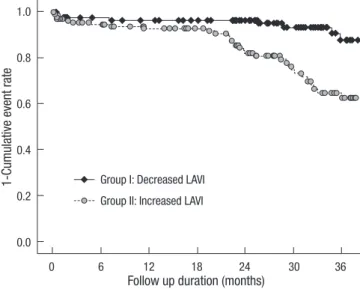
LA-remodeling during the 12 months post-AMI was a predictor of atrial fibrillation, HF hospitalization, even after adjustment for baseling LAVI and covariates (Tables 8, 9). Fig. 1 shows the Kaplan-Meier estimates of adverse cardiac events (new onset atrial fibrillation, hospitalization with heart failure) according Table 7. Comparison of echocardiographic parameters in both groups baseline and 1 yr
Parameters Group I Group II P
(between groups) LAV
Baseline 1 yr
P (within groups)
41.5 ± 9.6 38.5 ± 9.6
< 0.001
45.4 ± 12.3 51.6 ± 12.6
< 0.001
0.004
< 0.001 LAVI
Baseline 1 yr
P (within groups)
24.2 ± 6.6 22.5 ± 6.6
< 0.001
27.9 ± 9.7 32.7 ± 10.4
< 0.001
< 0.001
< 0.001 LV-Ejection fraction
Baseline 1 yr
P (within groups)
58.2 ± 11.6 62.0 ± 9.4
0.029
52.6 ± 13.5 51.8 ± 12.4
0.313
< 0.001
< 0.001 Significant MR (grade II-III)
Baseline 1 yr
P (within groups)
29 (25.7%) 23 (20.4%) 0.109
39 (27.5%) 38 (26.8%) 0.797
0.747 0.234 LAV, left atrial volume; LAVI, left atrial volume index; MR, mitral regurgitation.
Table 8. Major adverse cardiac events between groups
Events Group I Group II P value
Death 1 (0.9%) 2 (1.4%) 0.700
Atrial fibrillation 5 (4.4%) 18 (12.7%) 0.022
Hospitalization with HF 2 (1.8%) 25 (17.6%) < 0.001
Revascularization 3 (2.7%) 3 (2.1%) 0.777
HF, heart failure.
Table 9. Risk of adverse events for each ml/m2 increase in left atrial volume index at 12 months compared with baseline size
Cardiac events HR Lower 95% CI Upper 95% CI P value
Heart failure
hopspitalization Unadjusted Adjusteda Adjustedb
1.209 1.177 1.223
1.094 1.047 1.055
1.337 1.323 1.419
< 0.001 0.006 0.008 Atrial fibrillation Unadjusted
Adjusteda Adjustedb
1.183 1.148 1.193
1.067 1.025 1.054
1.313 1.286 1.350
0.001 0.017 0.005 Heart failure
hopspitalization or Atrial fibrillation
Unadjusted Adjusteda Adjustedb
1.194 1.173 1.216
1.097 1.068 1.091
1.299 1.289 1.354
< 0.001 0.001
< 0.001 Adjusteda for baseline left atrial volume index (LAVI). Adjustedb for baseline LAVI, Age, BMI, heart rate, Killip class, history of ischemic heart disease, Diabetes Mellitus, BNP, re- vascularization, anterior wall myocardial infarction, LV ejection fraction.
Fig. 1. New onset atrial fibrillation or hospitalization with heart failure according to change of LAVI (P value = 0.002).
1-Cumulative event rate
Follow up duration (months) Group I: Decreased LAVI
Group II: Increased LAVI
0 6 12 18 24 30 36
1.0 0.8 0.6 0.4 0.2
0.0
to this classification. The cut-off value of poor outcome that was related to the changes in LAVI was 1.14 mL/m2 (sensitivity = 75%, specificity = 55%, positive predictive value = 26%, nega- tive predictive value = 91.2%).
DISCUSSION
The major finding of this study is that LAVI was proofed as a good prognostic factor for AMI after primary PCI. Noninvasive assessment of LAVI at initial AMI-onset provides superior long- term prognostic information compared with clinical systolic and diastolic echocardiographic variables such as myocardial tissue function assessment by using tissue Doppler imaging. This study demonstrated that change of LAVI is a predictor of a new- onset atrial fibrillation and hospitalization for heart failure after in patients undergoing primary PCI. It was already known that baseline LA size is an independent predictor of death or hospi- talization for HF in patients suffering from an AMI. LA remod- eling begins early after AMI (17) and is influenced by EF, left ven- tricular dimension, wall motion score/16. LA remodeling is pre- dictive of mortality and cardiovascular morbidity.
The LA size is considered to be an expression of the diastolic burden and an increased LA volume usually reflects elevated ventricular filling pressure. During ventricular diastole, the left atrium is directly exposed to LV pressure through the open mi- tral valve. As an adaptation to the decreased ventricular com- pliance following an AMI, the LA pressure rises. This increases the LA wall tension and stretches the atrial myocardium; LA volume reflects the duration and severity of the increased LA pressure (21, 22). In the setting of an AMI, patients with higher chronic LV filling pressure and a previously worsened diastolic dysfunction have lower hemodynamic ‘cardiac reserve’ to help them withstand acute decreases in myocardial contraction (16).
In the early period after AMI, the LA size has shown to provide prognostic information in addition to clinical data and standard echocardiographic predictors (15-17). The present study con- firms and extends these conclusions to a population of patients with AMI and shows that LA size is a predictor of not only of mor- tality but also of cardiovascular morbidity. The increased risk associated with a greater LA size appears to be continuous, and even patients with a mild increase in LA size are at increased risk. We observed that in post-AMI, LA size was a better prog- nostic predictor of outcome than transmitral Doppler indices.
Doppler indices may be quite sensitive to acute changes in the loading conditions secondary to HF and/or to drugs (26, 27).
On the contrary, LA volume is likely to be less affected by acute hemodynamic changes and may represent a more stable indi- cator of the duration and severity of diastolic function and fill- ing pressure over time (22). Likewise, deceleration time, which is a dominant predictor of outcome when in the frankly restric- tive range (28), is less useful when > 140 ms. After an AMI, ven-
tricular structure and functional alteration can lead to ventricu- lar relaxation abnormalities resulting in worsening of diastolic dysfunction and LA remodeling. In our acute MI data, increased LAVI was associated with lower LVEF, atrial fibrillation and LV and LA enlargement in this population. All of these factors are already known to increase LV filling pressure and therefore in- fluence LA volume. LAVI values were not significantly related to gender, diabetes, hypertension, heart rate, Killip class, renal impairment and diastolic echocardiographic parameters. The main finding of our study is that measurement of baseline and changed LAVI values is a strong and useful predictor of adverse events in post-AMI patients undergoing PCI. Interestingly, nei- ther systolic nor diastolic function parameters retained statisti- cal significance in multivariate analysis, although the left ven- tricular EF and the enlarged dimensions of the left ventricle are classic predictors of cardiac mortality after AMI (29), recently published studies emphasize the significance of diastolic param- eters to enable further prognostic stratification, especially in groups with more uniform types of systolic dysfunction (e.g., patients after anterior AMI or below some level of EF impair- ment) (30). Some limitation of the present study should be not- ed. The entry criterion for the study was measurement of change of LAVI. Measurements of LA volume were obtained by multi- ple observers working in a clinical environment, which suggests that the findings can be widely applied. The Doppler assessment may have resulted in a misclassification of diastolic function in several cases (for example, in some patients with normal Dop- pler parameters who had LA enlargement and vice versa).
In patients experiencing AMI followed by primary PCI, the LA size is an independent predictor of both mortality and cardio- vascular morbidity. Moreover, changes in LA size in the months following an AMI predict subsequent adverse outcomes. These data suggest that the assessment of baseline and changing LAVI values is a potential surrogate for LA remodeling in patients un- dergoing PCI after AMI, represents an additions to post-AMI echocardiographic evaluation, and provides more accurate prog- nostic information than that obtained from clinical and labora- tory parameters alone.
REFERENCES
1. Naqvi TZ, Padmanabhan S, Rafii F, Hyuhn HK, Mirocha J. Comparison of usefulness of left ventricular diastolic versus systolic function as a pre- dictor of outcome following primary percutaneous coronary angioplasty for acute myocardial infarction. Am J Cardiol 2006; 97: 160-6.
2. Cho IJ, Pyun WB, Shin GJ . The influence of the left ventricular geometry on the left atrial size and left ventricular filling pressure in hypertensive patients, as assessed by echocardiography. Korean Circ J 2009; 39: 145-50.
3. Ko JS, Jeong MH, Lee MG, Lee SE, Kang WY, Kim SH, Park KH, Shim DS, Yoon NS, Yoon HJ, Hong YJ, Park HW, Kim JH, Ahn YK, Cho JG, Park JC, Kang JC. Left ventricular dyssynchrony after acute myocardial infarction is a powerful indicator of left ventricular remodeling. Korean Circ J 2009;
39: 236-42.
4. Ahn JC, Shim WJ, Park SW, Rha SW, Hwang GS, Song WH, Lim DS, Park CG, Kim YH, Seo HS, Oh DJ, Ro YM. Myocardial reperfusion and long- term change of left ventricular volume after acute anterior wall myocar- dial infarction. Korean Circ J 1997; 27: 1138-46.
5. Kim CM, Kim SR, Youn HJ, Lee MY, Choi KB, Hong SJ. Two-dimension- al echocardiographic predictors of ventricular enlargement after acute myocardial infarction. Korean Circ J 1996; 26: 455-64.
6. Shim WJ, Lee EM, Hwang GS, Ahn JC, Song WH, Lim DS, Park CG, Kim YH, Seo HS, Oh DJ, Ro YM. Microvascular integrity as a predictor of left ventricular remodeling after acute anterior wall myocardial infarction. J Korean Med Sci 1998; 13: 466-72.
7. Packer M. Prolonging life in patients with congestive heart failure: the next frontier. Introduction. Circulation 1987; 75: IV1-3.
8. Gradman A, Deedwania P, Cody R, Massie B, Packer M, Pitt B, Gold- stein S. Predictors of total mortality and sudden death in mild to moder- ate heart failure. J Am Coll Cardiol 1989; 14: 564-70.
9. Cohn JN, Johnson GR, Shabetai R, Loeb H, Tristani F, Rector T, Smith R, Fletcher R. Ejection fraction, peak exercise oxygen consumption, cardio- thoracic ratio, ventricular arrhythmias, and plasma norepinephrine as determinants of prognosis in heart failure. Circulation 1993; 87: S15-6.
10. Benjamin EJ, D’Agostino RB, Belanger AJ, Wolf PA, Levy D. Left atrial size and the risk of stroke and death. The Framingham Heart-Study. Cir- culation 1995; 92: 835-41.
11. Giannuzzi P, Temporelli PL, Bosimini E, Silva P, Imparato A, Corrà U, Galli M, Giordano A. Independent and incremental prognostic value of Doppler-derived mitral deceleration time of early filling in both symp- tomatic and asymptomatic patients with left ventricular dysfunction. J Am Coll Cardiol 1996; 28: 383-90.
12. Rossi A, Tomaino M, Golia G, Santini F, Pentiricci S, Marino P, Zardini P.
Usefulness of left atrial size in predicting postoperative symptomatic im- provement in patients with aortic stenosis. Am J Cardiol 2000; 86: 567-70.
13. Adavane S, Santhosh S, Karthikeyan S, Balachander J, Rajagopal S, Gobu P, Prasath MA, Haddour N, Ederhy S, Cohen A. Decrease in left atrium volume after successful balloon mitral valvuloplasty: an echocardiograph- ic and hemodynamic study. Echocardiography 2011; 28: 154-60.
14. Reed D, Abbot RD, Smucker KL, Kaul S. Prediction of outcome after mi- tral valve replacement in patients with symptomatic chronic mitral re- gurgitation. The importance of left atrial size. Circulation 1991; 84: 23-34.
15. Daccarett M, McGann CJ, Akoum NW, MacLeod RS, Marrouche NF.
MRI of the left atrium: predicting clinical outcomes in patients with atri- al fibrillation. Expert Rev Cardiovasc Ther 2011; 9: 105-11.
16. Moller JE, Hillis GS, Oh JK, Seward JB, Reeder GS, Wright RS, Park SW, Bailey KR, Pellikka PA. Left atrial volume: a powerful predictor of surviv- al after acute myocardial infarction. Circulation 2003; 107: 2207-12.
17. Meris A, Amigoni M, Uno H, Thune JJ, Verma A, Køber L, Bourgoun M, McMurray JJ, Velazquez EJ, Maggioni AP, Ghali J, Arnold JM, Zelenkof- ske S, Pfeffer MA, Solomon SD. Left atrial remodeling in patients with myocardial infarction complicated by heart failure, left ventriclular dys- function, or both: the VALIANT Echo Study. Eur Heart J 2009; 30: 56-65.
18. Popescu BA, Macor F, Antonini-Canterin F, Giannuzzi P, Temporelli PL, Bosimini E, Gentile F, Maggioni AP, Tavazzi L, Piazza R, Ascione L, Stoian
I, Cervesato E, Nicolosi GL; GISSI-3 Echo Substudy Investigators. Left atrium remodeling after acute myocardial infarction (results of the GIS- SI-3 Echo Substudy). Am J Cardiol 2004; 93: 1156-9.
19. Wierzbowska-Drabik K, Krzemińska-Pakula M, Drozdz J, Plewka M, Trzos E, Kurpesa M, Rechciński T, Rózga A, Plońska-Gościniak E, Kasprzak JD. Enlarged left atrium is a simple and strong predictor of poor prognosis in patients after myocardial infarction. Echocardiogra- phy 2008; 25: 27-35.
20. Beinart R, Boyko V, Schwammenthal E, Kuperstein R, Sagie A, Hod H, Matetzky S, Behar S, Eldar M, Feinberg MS. Long-term prognostic sig- nificance of left atrial volume in acute myocardial infarction. J Am Coll Cardiol 2004; 44: 327-34.
21. Hurrell DG, Nishimura RA, Ilstrup DM, Appleton CP. Utility of preload alteration in assessment of left ventricular filling pressure by Doppler echo- cardiography: a simultaneous catheterization and Doppler echocardio- graphic study. J Am Coll Cardiol 1997; 30: 459-67.
22. Appleton CP, Galloway JM, Gonzalez MS, Gaballa M, Basnight MA.
Estimation of left-ventricular filling pressure using two-dimensional and Doppler echocardiography in adult patients with cardiac disease. Addi- tional value of analyzing left atrial size, left atrial ejection fraction and the difference in duration of pulmonary venous and mitral flow velocity at atrial contractioin. J Am Coll Cardiol 1993; 22: 1972-82.
23. Schiller NB, Shah PM, Crawford M, DeMaria A, Devereuix R, Feigen- baum H, Gutgesell H, Reichek N, Sahn D, Schnittger I, Silverman AH, Tajik AJ. Recommendations for quantitation of the left ventricle by two- dimensional echocardiography. American Society of Echocardiography Committee on Standards. Subcommittee on Quantitation of Two-Dimen- sional Echocardiograms. J Am Soc Echocardiogr 1989; 2: 358-67.
24. Helmcke F, Nanda NC, Hsiung MC, Soto B, Adey CK, Goyal RG, Gate- wood RP Jr. Color Doppler assessment of mitral regurgitation with or- thogonal planes. Circulation 1987; 75: 175-83.
25. The TIMI Study Group. The Thrombolysis in Myocardial Infarction (TIMI) trial. Phase 1 findings. N Engl J Med 1985; 312: 932-6.
26. Nishimura RA, Tajik AJ. Evaluation of diastolic filling of left ventricle in health and disease: Doppler echocardiography is the clinician’s Rosetta stone. J Am Coll Cardiol 1997; 30: 8-18.
27. Pritchett AM, Mahoney DW, Jacobsen SJ, Rodeheffer RJ, Karon BL, Redfied MM. Diastolic dysfunction and left atrial volume: a population- based study. J Am Coll Cardiol 2005; 45: 87-92.
28. Temporelli PL, Giannuzzi P, Nicolosi GL, Latini R, Franzosi MG, Gentile F, Tavazzi L, Maggioni AP; GISSI-3 Echo Substudy Investigators. Dop- pler-derived mitral deceleration time as a strong prognostic marker of left ventricular remodeling and survival after acute myocardial infarction:
result of the GISSI-3 echo substudy. J Am Coll Cardiol 2004; 43: 1646-53.
29. Norris RM, Barnaby PF, Brandt PW, Geary GG, Whitlock RM, Wild CJ, Barratt-Boyes BG. Prognosis after recovery from first acute myocardial infarction: determinants of reinfarction and sudden death. Am J Cardiol 1984; 53: 408-13.
30. Møller JE, Egstrup K, Køber L, Poulsen SH, Nyvad O, Torp-Pedersen C.
Prognostic importance of systolic and diastolic function after acute myo- cardial infarction. Am Heart J 2003; 145: 147-53.