557 Copyright © 2013 The Korean Society of Cardiology
Korean Circulation Journal
Introduction
In patients undergoing percutaneous coronary intervention (PCI) for the treatment of acute coronary syndrome, administration of glycoprotein (GP) IIb/IIIa receptor antagonists has been shown to improve their clinical outcomes. However, the associated hemor- rhagic risk may evoke serious complications, such as acute pro- found thrombocytopenia.
1)We present a successfully managed case of a rare complication of acute profound thrombocytopenia af- ter the use abciximab, an intra-aortic balloon pump (IABP) for the treatment of no-reflow phenomenon, and consecutive cardiogenic shock during primary PCI in a patient with ST-segment elevated myo- cardial infarction (STEMI).
Case Report
http://dx.doi.org/10.4070/kcj.2013.43.8.557 Print ISSN 1738-5520 • On-line ISSN 1738-5555
Acute Profound Thrombocytopenia after Using Abciximab
for No-Reflow during Primary Percutaneous Coronary Intervention for ST-Segment Elevation Myocardial Infarction
Soonyoung Park, MD, Jooyoung Lee, MD, Sang Yeub Lee, MD, Jang-Whan Bae, MD, Kyung-Kuk Hwang, MD, Dong-Woon Kim, MD, Myeong-Chan Cho, MD, and Sang Min Kim, MD
Regional Cardiovascular Center, Division of Cardiology, Department of Internal Medicine, Chungbuk National University Hospital, Cheongju, Korea
Glycoprotein IIb/IIIa antagonists are well established for their effectiveness in improving clinical outcomes in acute coronary syndrome patients undergoing percutaneous coronary intervention. Acute profound thrombocytopenia is a rare complication of abciximab. We present a case which was managed successfully for the rare complication of acute profound thrombocytopenia after using abciximab and an intra-aortic balloon pump for the treatment of a no-reflow phenomenon and consecutive cardiogenic shock during primary per- cutaneous coronary intervention. (Korean Circ J 2013;43:557-560)
KEY WORDS: Thrombocytopenia; Abciximab; No-reflow phenomenon.
Received: August 22, 2012
Revision Received: October 31, 2012 Accepted: January 31, 2013
Correspondence: Sang Min Kim, MD, Regional Cardiovascular Center, Di- vision of Cardiology, Department of Internal Medicine, Chungbuk National University Hospital, 776 1sunhwan-ro, Heungdeok-gu, Cheongju 361-711, Korea
Tel: 82-43-269-6027, Fax: 82-43-273-3252 E-mail: sangmin3410@gmail.com
• The authors have no financial conflicts of interest.
This is an Open Access article distributed under the terms of the Creative Commons Attribution Non-Commercial License (http://creativecommons.
org/licenses/by-nc/3.0) which permits unrestricted non-commercial use, distribution, and reproduction in any medium, provided the original work is properly cited.
Case
A 65 year-old man with hypertension visited the local hospital
with resting chest pain for 3 hours. He was transferred to our hospi-
tal due to the abnormal electrocardiogram (ECG) findings showing
an elevated ST-segment in the inferior wall territory with a reciprocal
change, which was suspicious of STEMI. In the emergency room,
blood pressure was 140/80 mm Hg and chest radiography showed
no definite abnormal findings, which was graded as Killip class 1. It
was decided to have the patient undergo primary PCI for the treat-
ment of STEMI. Conventional medical treatments that include a
bolus injection of 5000 IU of heparin, a loading dose of aspirin (300
mg), and clopidogrel (300 mg) was applied before primary PCI. Co-
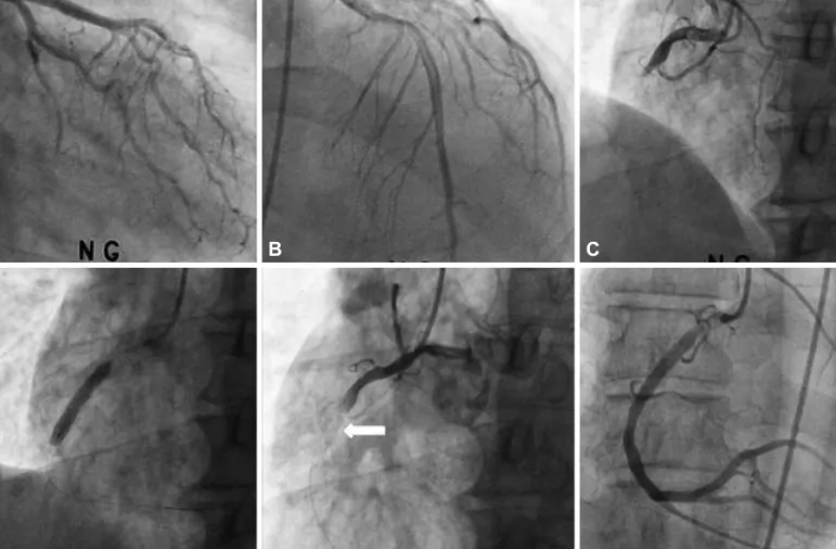
ronary angiography (CAG) showed a totally occluded lesion of the
proximal right coronary artery (RCA) with collateral flow grade 1
from the left anterior descending artery (Fig. 1A, B, and C). After pre-
dilatation with a 2.0×20 mm conventional balloon, a bare metal
stent 5.0×24 mm Liberte® (Boston Scientific, Natick, MA, USA) was
implanted in the proximal RCA lesion (Fig. 1D). After implantation of
a stent, no-reflow phenomenon developed (Fig. 1E) and blood pres-
sure decreased with findings of a complete atrioventricular-block
and re-elevation of the ST-segment on ECG monitoring. The patient
was treated with intracoronary injection of nitrate and adenosine,
and intravenous bolus consecutive injection of abciximab with main-
tenance (intravenous bolus of 0.25 mg/kg, 10 to 60 minutes during
the procedure, followed by 0.125 μg/kg/min infusion for 12 hours).
558 Acute Profound Thrombocytopenia after Administration of Abciximab
http://dx.doi.org/10.4070/kcj.2013.43.8.557 www.e-kcj.org
Although an improvement was observed in the Thrombolysis in Myocardial Infarction flow grade from grade 0 to grade 3 (Fig. 1F) and the patient returned to sinus rhythm, the hypotension was sus- tained. Therefore, an IABP was inserted into the left femoral artery.
The patient was moved to the coronary care unit for intensive moni-
toring with maintenance of abciximab and IABP.
Although the baseline platelet count was 167000/μL before PCI, a routine check determined a complete blood count showing a de- creased platelet count of 6000/μL at 22 hours after bolus administr- ation of abciximab, followed by a platelet count of 3000/μL 4 ho- urs later. It was considered that the patient had acute, profound thrombocytopenia as a complication of abciximab. Heparin induced thrombocytopenia (HIT) was excluded because HIT typically devel- ops after 6 to 10 days of heparin use with no previous exposure to heparin. In this case, the patient had no history of exposure to he- parin. Therefore, the patient was started on clopidogrel 75 mg/day and aspirin 100 mg/day were started to prevent stent thrombosis.
The patient was administered 4 units of platelet concentrates as a precaution. The patient was withdrawn from heparin treatment in consideration of additional hemorrhagic risk, but not HIT. The next followed count of platelet increased to 24000/μL without clinical evidence of stent thrombosis, such as chest pain and a change of ST-segment on ECG. Vital signs were stabilized the next day and A
D
B
E
C
F
Fig. 1. Findings of coronary angiography and primary percutaneous coronary intervention. A and B: no specific stenosis in the left coronary artery. C: a to- tally occluded lesion of the proximal RCA with collateral flow grade 1 from LAD. D: a bare metal stent was implanted in the proximal RCA lesion. E: after implantation of a stent, the coronary angiogram shows no-reflow phenomenon. The arrowindicates the implanted stent. F: improvement of TIMI flow grade from grade 0 to grade 3 after intracoronary injection of nitrate and adenosine, and intravenous injection of abciximab. LAD: left anterior descending artery, RCA: right coronary artery, TIMI: Thrombolysis in Myocardial Infarction.
Fig. 2. The course of platelet count after bolus administration of abciximab decreased, and then gradually increased after cessation of heparin and transfusion of platelet concentration.
200000 150000 100000 50000
0
0 19 hours 22 hours 37 hours 3rd day 4th day 5th day 1 week Transfusion of platelet
concentration (4 pack)
Transfusion of platelet concentration (4 pack) Platelet count/uL