Received: 11 May, 2017 Revised: 26 May, 2017 Accepted: 29 May, 2017
Ⓒ The Korean Society of Mycology
This is an Open Access article distributed under the terms of the Creative Commons Attrib- ution Non-Commercial License (http://creative- commons.org/licenses/by-nc/4.0/) which permits unrestricted non-commercial use, distribution, and reproduction in any medium, provided the original work is properly cited.
Kor. J. Mycol. 2017 June, 45(2): 107-113 https://doi.org/10.4489/KJM.20170013
pISSN : 0253-651X eISSN : 2383-5249
OPEN ACCESS
RESEARCH ARTICLE
Aphanoascus fulvescens: A New Record from Crop Field Soil in Korea
Sun Kumar Gurung
1, Mahesh Adhikari
1, Setu Bazie
1, Hyun Seung Kim
1, Hyun Gu Lee
1, Hyang Burm Lee
2, Youn Su Lee
1*1
Division of Biological Resources Sciences, Kangwon National University, Chuncheon 24341, Korea
2
Division of Food Technology, Biotechnology and Agrochemistry, College of Agriculture and Life Sciences, Chonnam National University, Gwangju 61186, Korea
*Corresponding author: [email protected]
Abstract
Aphanoascus fulvescens KNU16-343 was isolated from crop field soil and identified based on morphological and molecular characteristics. The shape and size of conidia and conidiophores, as well as the internal transcribed spacer region of rDNA, confirmed that the isolate was A.
fulvescens. This is the first report of this fungal species in Korea.
Keywords: Aphanoascus fulvescens, Internal transcribed spacer, Micromorphology
Introduction
The ascomycete genus Aphanoascus consists a large number of species characterized by spherical, lenticular ascospores, pale to dark brown ascomata either discoid or oblate, with or without an equatorial rim, pale to dark brown with a reticulate, pitted or verrucose wall [1]. The genus Aphanoascus was first described in 1890 by Zukal [1, 3]. Aphanoascus fulvescens is mostly found in soil and dung, living as a keratinophilic saprotroph [2].
Although this fungus is a pseudodermatophyte, it is opportunistic and has been reported to cause infection in humans and other mammals [3, 4]. In this study, to assess fungal diversity in Korea, we collected fungal strains from the soil of different crop fields. Among the fungal isolates, we encountered A. fulvescens, which has not been reported before in Korea.
The purpose of the study was to confirm the newly identified isolate as A. fulvescens, by studying its morphology and phylogenetic status, based on rDNA sequence analysis.
Materials and Methods
Soil sampling and isolation of fungi
Soil samples were collected in 2013 from crop fields at various locations in Gyeongnam
city (N 35.081337°, E 128.273832°), Gyeongsangnam-do, Korea. Samples were collected from a depth of 0~15 cm, air-dried, and stored in plastic bags at 4°C. Fungi were isolated using a conventional dilution technique [5] and cultured on potato dextrose agar (PDA;
Difco, Detroit, MI, USA) supplemented with 100 mg/L chloramphenicol (a bacteriostatic agent) for 5~7 days at 25°C until fungal colonies were observed. The pure cultures were maintained on PDA slants at 4°C for further use.
Morphological characterization
Five different types of media were used for the morphological characterization of the study isolate (KNU16-343): PDA, oatmeal agar (OMA), yeast extract sucrose agar (YESA), malt extract agar (MEA), and Czapek yeast extract agar (CYEA). All growth media were prepared according to Samson [6]. The isolate KNU16-343 was cultured in 9 cm petri dishes with three-point inoculation and incubated in the dark at 25°C for 7 days.
Obverse and reverse colony colors, as well as the degree of speculation, were determined.
An HK 3.1 CMOS digital camera (KOPTIC, Seoul, Korea), attached to an Olympus BX50F-3 microscope (Olympus, Tokyo, Japan), was used to capture microscopic images of the fungal isolate. Scanning electron microscopy on a LEO Model 1450VP Variable Pressure Scanning Electron Microscope (Carl Zeiss, Oberkochen, Germany) was used to observe and capture the micro-morphological features of the fungal isolate.
DNA extraction, PCR amplification, sequencing, and data analysis
Genomic DNA was extracted from 1-week-old colonies grown on PDA media, using a DNeasy Plant Mini Kit (Qiagen, Germantown, MD, USA) following the manufacturer’s instructions. The internal transcribed spacer region (ITS) was amplified using ITS1 (5'-TCCGTAGGTGAACCTGCG-3') and ITS4 (5'-TCCTCCGCTTATTGATATGC-3') primers [7]. The amplified PCR products were sequenced on an Applied Biosystems 3730 DNA analyzer (Foster City, CA, USA).
Phylogenetic analysis
Sequences were compared with reference ITS sequences retrieved from GenBank (National Center for Biotechnology Information), using basic local alignment search tool (BLAST) software [8]. This newly identified fungal isolate was deposited in the National Institute of Biological Resources (NIBR) under the deposition number NIBRFG0000499481.
The annotated nucleotide sequence of the KNU16-343 isolate was deposited in GenBank
with the accession number KY906225. Molecular Evolutionary Genetics Analysis (MEGA
6.0) software [9] was used for the alignment of all sequences. The phylogenetic tree was
generated by the neighbor-joining method, utilizing the Kimura 2-parameter model with a
bootstrap analysis of 1,000 replications for each clade.
Results and Discussion
Aphanoascus fulvescens (Cooke) Apinis, Mycopathologia et Mycologia Applicata 35: 99 (1968)
Morphology of the KNU16-343 isolate
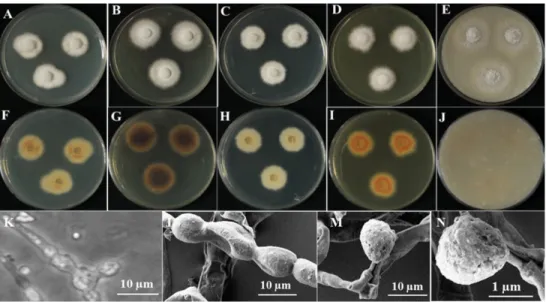
The KNU16-343 isolate attained a diameter of 21~24 mm within 7 days when grown on YESA media at 25°C. Detailed morphological features of the fungal isolate KNU16-343 are shown in Fig. 1. The front side of the mycelium was white, while the rear was black (Fig. 1A, 1F). Sporulation was moderate to dense and the conidia were present in mass, with irregular form and smooth surface. On CYEA media, the isolate attained a diameter of 24~26 mm within 7 days at 25°C. The dorsal side of the mycelium was white, whereas the ventral side was hairy and green in the middle (Fig. 1B, 1G). Sporulation was moderately dense, and the conidia were seen in mass, with irregular form and smooth surface. The KNU16-343 isolate attained a diameter of 18~21 mm within 7 days when grown on PDA media at 25°C. The dorsal side of the mycelium was white, whereas the ventral side was pale brown in the middle (Fig. 1C, 1H). Sporulation was moderately dense, and the conidia were seen in mass, with irregular form and smooth surface. On MEA media, the KNU16-343 isolate attained a diameter of 22~23 mm within 7 days at 25°C. The front and rear sides of the mycelium were black (Fig. 1D, 1I). On OMA media, the isolate attained a diameter of 35~37 mm within 7 days at 25°C. The front and rear sides of the mycelium were dark black
Fig. 1. Morphology of the isolate Aphanoascus fulvescens KNU16-343, grown for 7 days on YESA, CYEA, PDA, MEA, and OMA at 25°C. Obverse colonies (A~E) and reverse colonies (G~J) grown on YESA, PDA, OMA, CYEA and MEA, from left to right. Microscopic images of ascospores (K, L), and scanning electron microscopic images of ascospores (M, N). YESA, yeast extract sucrose agar; CYEA, Czapek yeast extract agar; PDA, potato dextrose agar;
MEA, malt extract agar; OMA, oatmeal agar.
(Fig. 1E, 1J). Sporulation was moderate, and the conidia were present in mass, with irregular form and rough surface.
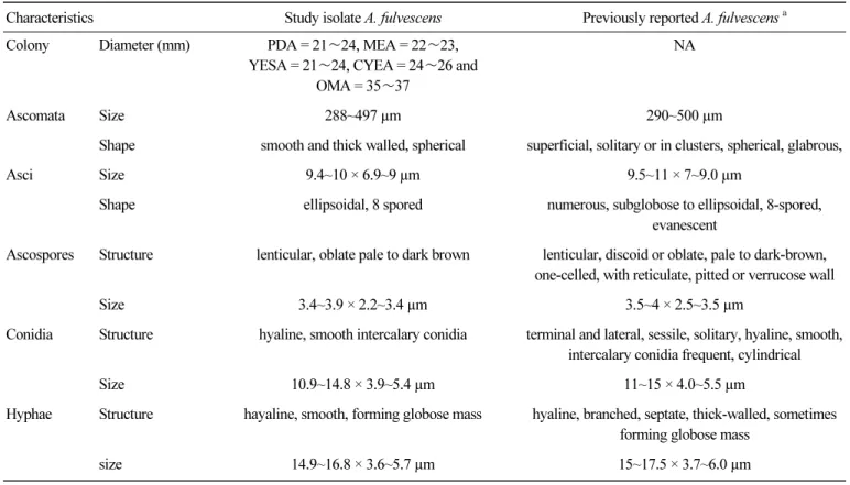
The isolate displayed club-shaped conidia. Hyphae were hyaline, branched, and 1.6~2.7 µm wide. Ascospores were ellipsoidal and lens or disc shaped. Ascomata were smooth and thick walled and asci were ellipsoidal (Fig. 1K, 1N). Detailed comparisons of the morphological characteristics of the KNU16-343 isolate with those previously reported for A. fulvescens [1, 10] are described in Table 1.
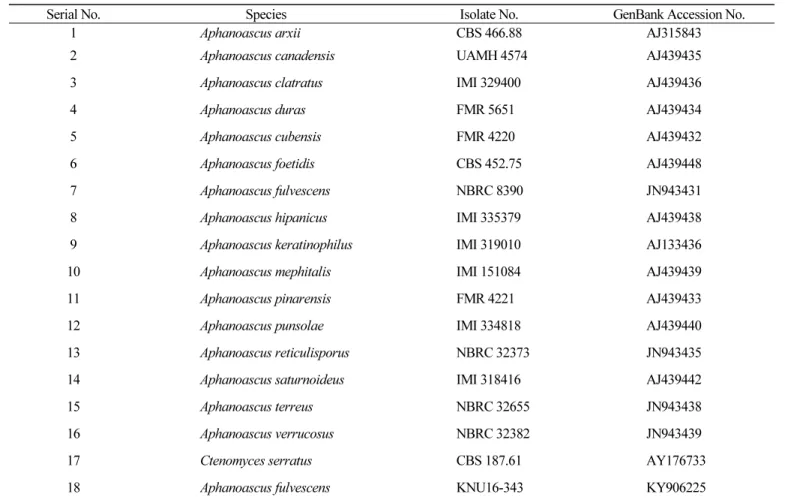
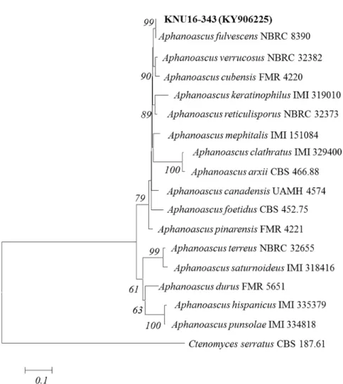
Molecular phylogeny
The KNU16-343 isolate was obtained from the crop field soil of Gyeongnam city, Gyeongsangnam-do, Korea. The isolates used to construct the phylogenetic tree are shown in Table 2, along with their GenBank accession numbers. The KNU16-343 isolate was most closely related to A. fulvescens (NBRC 8390) and formed a monophyletic group, supported by a bootstrap value of 99% (Fig. 2). Phylogenetic analysis revealed that the isolate was A.
fulvescens.
The KNU16-343 isolated from the crop field soil was presumed to be Aphanoascus
Table 1. Morphological comparison between the studied isolate KNU16-343 and a previously described isolate of Aphanoascus fulvescens
Characteristics Study isolate A. fulvescens Previously reported A. fulvescens
aColony Diameter (mm) PDA = 21 ∼24, MEA = 22∼23,
YESA = 21∼24, CYEA = 24∼26 and OMA = 35 ∼37
NA
Ascomata Size 288~497 µm 290~500 µm
Shape smooth and thick walled, spherical superficial, solitary or in clusters, spherical, glabrous,
Asci Size 9.4~10 × 6.9~9 µm 9.5~11 × 7~9.0 µm
Shape ellipsoidal, 8 spored numerous, subglobose to ellipsoidal, 8-spored, evanescent
Ascospores Structure lenticular, oblate pale to dark brown lenticular, discoid or oblate, pale to dark-brown, one-celled, with reticulate, pitted or verrucose wall
Size 3.4~3.9 × 2.2~3.4 µm 3.5~4 × 2.5~3.5 µm
Conidia Structure hyaline, smooth intercalary conidia terminal and lateral, sessile, solitary, hyaline, smooth, intercalary conidia frequent, cylindrical
Size 10.9~14.8 × 3.9~5.4 µm 11~15 × 4.0~5.5 µm
Hyphae Structure hayaline, smooth, forming globose mass hyaline, branched, septate, thick-walled, sometimes forming globose mass
size 14.9~16.8 × 3.6~5.7 µm 15~17.5 × 3.7~6.0 µm
PDA, potato dextrose agar; MEA, malt extract agar; OMA, oatmeal agar.
a