191
A New Record of Neosartorya aureola Isolated from Field Soil in Korea
Mahesh Adhikari
1, Sangwoo Kim
1, Dil Raj Yadav
1, Changmu Kim
2, Hyang Burm Lee
3and Youn Su Lee
1*
1Division of Biological Resource Sciences, Kangwon National University, Chuncheon 24341, Korea
2Biological and Genetic Resources Assessment Division, National Institute of Biological Resources, Incheon 22755, Korea
3Division of Food Technology, Biotechnology & Agrochemistry, College of Agriculture and Life Sciences, Chonnam National University, Gwangju 61186, Korea
ABSTRACT : A new species of Neosartorya was recovered during investigation of the fungal community in soil samples collected from different locations in Korea; Neosartorya aureola KNU14-7 was isolated for the first time from field soil in Korea and identified based on the internal transcribed spacer region of rDNA and morphological characteristics. The species has not been officially reported from Korea and we report it here with description and figures.
KEYWORDS : Molecular identification, Morphology, Neosartorya aureola
Neosartorya is a widespread genus of the Trichocomo- ceae family. It is teloemorph of Aspergillus section Fumi- gati include many species which is important because of its pathogenic or allergenic to human [1]. Most members of the genus Neosartorya Malloch & Cain [2] in the Euro- tiales are of worldwide distribution and are very abun- dant, occurring nearly everywhere in soil, air, dust, food etc. [3-7]. A few Neosartorya species are mycotoxigenic and causal agents of human diseases like aspergillosis, osteomyelitis, endocarditis and mycotic keratitis. [8-16].
Certain species of the genus are used in the production of bioactive metabolites [17]. All of the Neosartyora spe- cies produce heat resistant ascospores that are frequently encountered in different food products and cause the spoilage of processed food products [18, 19]. Thus, there has been considerable recent interest of mycologists in
working with Neosartorya aureola.
During studies on the diversity of fungal communities in field soil of Gangwon-do, Korea, a species of Neosar- torya was encountered. Based on its morphological and molecular characteristics, this species was identified as N.
aureola and named N. aureola KNU14-7. To the best of our knowledge this fungus has not been officially reported in Korea.
Collection of soil samples and fungal isolation
Soil samples were collected from different locations in Samcheok city, Korea in 2014. Soil samples were taken from (0~15 cm) depth, air dried and stored in plastic bags at 4oC until used. The fungi were isolated by con- ventional dilution method and supplemented with 100 µg chloramphenicol per mL potato dextrose agar (PDA;
Difco, Detroit, USA) and grown for 7 days at 28oC until the growth of colonies was observed.
ITS, β-tubulin and calmodulin gene sequencing analysis Genomic DNA of the strain was extracted using DNeasy Plant Mini Kit (QIAGEN, Germantown, MD, USA) follow- ing the manufacturer’s instructions. The internal transcri- bed spacer (ITS) regions, including the 5.8S were ampli- fied with the primers ITS1 and ITS4 [20]. For the sequen- cing of partial β-tubulin and calmodulin genes, the seg- ments of β-tubulin and calmodulin were amplified with the primers BT2a and BT2b and cmd5 and cmd6 [21,
*Corresponding author E-mail: [email protected] Received June 5, 2015 Revised June 13, 2015 Accepted September 8, 2015
This is an Open Access article distributed under the terms of the Creative Commons Attribution Non-Commercial License (http://
creativecommons.org/licenses/by-nc/3.0/) which permits unrestricted non-commercial use, distribution, and reproduction in any medium, provided the original work is properly cited.
Kor. J. Mycol. 2015 September, 43(3): 191-195 http://dx.doi.org/10.4489/KJM.2015.43.3.191 pISSN 0253-651X • eISSN 2383-5249
© The Korean Society of Mycology
22]. The amplified polymerase chain reaction (PCR) pro- duct was purified using QIAquick PCR purification Kit (QIAGEN) following the manufacturer’s instructions. The PCR product was sequenced using ABI Prism 3730 DNA analyzer (Applied Biosystems, Foster city, CA, USA). The sequence was compared with reference ITS1-ITS4 rDNA and β-tubulin and calmodulin rDNA sequences from GenBank using BLAST analysis (http://www.ncbi.nlm.
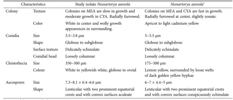
nihgob/blast). The nucleotide sequence was deposited in GenBank and assigned accession number KP966615 for isolate KNU14-7. The sequences of closely related strains were aligned using the MultAlin program. The DNA se- quences were analyzed for phylogenetic relationship using Molecular Evolutionary Genetics Analysis (MEGA 6) soft- ware [23] sequence of present isolate, Neosartorya aureola KNU14-7 was compared with the sequences in the Gen- Characteristics Study isolate Neosartorya aureola Neosartorya aureolaa
Colony Texture Colonies on MEA are slow in growth and moderate growth in CYA. Radially furrowed.
Colonies on MEA and CYA are fast in growth.
Radially furrowed at center, slightly zonate.
Color White in center and wolly growth appearences in surrounding.
Apricot to light cadmium yellow
Conidia Size 3.5~3.8 µm 3~3.3 µm
Shape Globose to subglobose Globose to subglobose
Surface texture Delicately echinulate Delicately echinulate
Conidial head Loosely columnar Loosely columnar
Cleistothecia Size 350~500 µm 175~500 µm
Colour White to yellowish white, globose to ovoid Lemon yellow, surrounded by loose wefts of dark golden yellow hyphae
Ascospores Size 7.3~8.1 × 6.4~6.6 µm 6~7 × 4.4~5 µm
Shape Lenticular with two prominent equatorial crests and with convex surfaces aculeate
Lenticular with two prominent equatorial crests and with convex surfaces conspicuously echinulate
a Source of description [25].
MEA, malt extract agar; CYA, czapek yeast extract agar.
Fig. 1. Neighbor-joining phylogenetic analysis of Neosartorya aureola KNU14-7 partial 18S-ITS1-5.8S-ITS2-28S rDNA region sequence obtained from crop field soil in Korea. Sequence obtained in the study is shown in boldface. Numerical values (> 50) on branches are the bootstrap values as percentage of bootstrap replication from 1,000 replicate analysis. Neosartorya stramania (AB299411) was used as out group.
Bank by using Basic Local Alignment Search Tool (BLAST).
Neighbor-joining tree was constructed by using Kimmura 2-parameter substitution model [24]. The phylogenetic tree was inferred using the maximum-likelihood heuristic search option with the nearest-neighbor interchange.
Bootstrap analysis was performed with 1,000 replications to determine the support for each clade. The isolate was most closely related to N. aureola (EF669954) and formed a monophyletic group with bootstrap value of 98% (Fig.
1), N. aureola (CBS105.55) with bootstrap value of 98%
and 99%, respectively (Fig. 2 and 3). The phylogenetic analysis showed that the isolate is N. aureola.
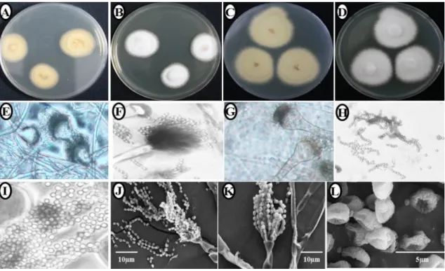
Morphological characteristics and identification Morphological features were observed on malt extract agar (MEA; Fig. 4A and 4B) and czapek yeast extract agar (CYA; Fig. 4C and 4D) by doing three point inoculations in 9 cm petri plates which were incubated in the dark at 28oC for 7 days. The morphological characteristics were examined with the aid of differential interference contrast Fig. 2. Neighbor-joining phylogenetic analysis of Neosartorya aureola KNU14-7 based on β-tubulin sequence data of Aspergillus section Fumigati. Numbers above branches are bootstrap values. Only values above 50% are indicated. The mark (T) indicates the type strain.
Fig. 3. Neighbor-joining phylogenetic analysis of Neosartorya aureola KNU14-7 based on calmodulin sequence data of Aspergillus section Fumigati. Numbers above branches are bootstrap values. Only values above 50% are indicated. The mark (T) indicates the type strain.
microscopy. Photomicrographs were taken with a Kodak 14n digital camera (Rochester, NY, USA) attached to the microscope. Slide material was mounted in water and sometimes with aniline blue staining. Colonies on MEA were slow growing, white, attaining a diameter of 25~30 mm after 7 days at 28oC (Fig. 4A and 4B). Colonies on CYA were moderate growing attaining a diameter 40~45 mm (Fig. 4C and 4D).
Cleistothecia superficial, scattered, white to yellowish white, globose to ovoid, 350~500 µm in diameter (Fig.
4E and 4F), surrounded by a loose covering of yellow to orange, 2~4 µm wide aerial hyphae. Cleistothecial peri- dium hyaline to pale yellowish brown, thin, consisting of irregular, 4~10 µm diam cells. Asci globose to ovoid, 11~
12 × 9.5~11 µm evanescent at maturity (Fig. 4G). Ascos- pores hyaline to pale yellowish brown, broadly lenticular, spore body 4~4.5 × 3.5~4 µm (Fig. 4H~4L), provided with two equatorial crests which are rather appressed up to 1 µm wide and rarely dissected and with the convex surfaces aculeate with spines up to 0.5 µm long.
In conclusion, we identified and reported Neosartorya aureola KNU14-7 as a new record for Korea. The species of Neosartorya are used in the production of bioactive metabolites. Neosartyora species have been described as
causal agents of human diseases including invasive asper- gillosis, osteomyelitis, endocarditis and mycotic keratitis [12]. Thus, in the future, further investigation in this res- pect would be worthwhile.
Acknowledgements
This work was supported by a grant (NIBR2014-01205) from the National Institute of Biological Resources (NIBR), funded by the Ministry of Environment (MOE) of the Republic of Korea for projects on the survey and discov- ery of indigenous Korean fungal species. The authors wish to thank Environmental Chemistry Lab, Kangwon Natio- nal University for providing the soil samples in this study.
REFERENCES
1. Brakhage AA, Langfelder K. Menancing mold: the molecular biology of Aspergillus fumigatus. Annu Rev Microbiol 2002;
56:533-55.
2. Malloch D, Cain RF. The Trichocomataceae: Ascomycetes with Aspergillus, Paecilomyces, and Penicillium imperfect states. Can J Bot 1972;50:2613-28.
3. Domsch KH, Gams W, Anderson TH. Compendium of soil fungi: Vol. 1. London: Academic Press; 1980.
Fig. 4. Morphological characterization of Neosartorya aureola KNU14-7 observed using a compound microscope and scanning electron microscope (SEM). Malt extract agar media: A, Colony in reverse; B, Colony in front. Czapek yeast extract agar media:
C, Colony in reverse D, Colony in front; E~G, Conidiophores; H~I, Simple microscopic picture of ascospores; J~K, SEM of conidiophores; L, SEM of ascospores.
4. Kozakiewicz Z. Aspergillus species on stored products. Wall- ingford: CAB International; 1989.
5. Kozakiewicz Z. CMI descriptions sheets: set 100. Mycopatho- logia 1990;109:183-202.
6. Udagawa S, Yaguchi T. Neosartorya (Eurotiales): taxanomy and significance in applied mycology. In: Deshmukh SK, Rai MK, editors. Biodiversity of fungi: their role in human life. Enfield:
Science Publishers; 2005. p. 341-55.
7. Yaguchi T, Someya A, Udagawa S. A new species of Neosar- torya from Taiwan soil. Mycoscience 1994;35:309-13.
8. Peterson SW. Neosartorya pseudofischeri sp. nov. and its rela- tionship to other species in Aspergillus section Fumigati. My- col Res 1992;96:547-54.
9. De Hoog GS, Guarro J, Gene J, Figueras MJ. Atlas of clinical fungi. 2nd ed. Utrecht: Centraalbureau voor Schimmelcul- tures; 2000.
10. Guarro J, Kallas EG, Godoy P, Karenina A, Gené J, Stchigel A, Colombo AL. Cerebral aspergillosis caused by Neosartorya hiratsukae, Brazil. Emerg Infect Dis 2002;8:989-91.
11. Järv H, Lehtmaa J, Summerbell RC, Hoekstra ES, Samson RA, Naaber P. Isolation of Neosartorya pseudofischeri from blood: first hint of pulmonary Aspergillosis. J Clin Microbiol 2004;42:925-8.
12. Coriglione G, Stella G, Gafa L, Spata G, Oliveri S, Padhye AA, Ajello L. Neosartorya fischeri var fischeri (Wehmer) Malloch and Cain 1972 (anamorph: Aspergillus fischerianus Samson and Gams 1985) as a cause of mycotic keratitis. Eur J Epide- miol 1990;6:382-5.
13. Summerbell RC, de Repentigny L, Chartrand C, St Germain G. Graft-related endocarditis caused by Neosartorya fischeri var spinosa. J Clin Microbiol 1992;30:1580-2.
14. Padhye AA, Godfrey JH, Chandler FW, Peterson SW. Osteo- myelitis caused by Neosartorya pseudofischeri. J Clin Micro- biol 1994;32:2832-6.
15. Lonial S, Williams L, Carrum G, Ostrowski M, McCarthy P Jr. Neosartorya fischeri: an invasive fungal pathogen in an
allogeneic bone marrow transplant patient. Bone Marrow Transplant 1997;19:753-5.
16. Balajee SA, Gribskov JL, Hanley E, Nickle D, Marr KA. As- pergillus lentulus sp. nov., a new sibling species of A. fumi- gatus. Eukaryot Cell 2005;4:625-32.
17. Udagawa S, Tsubouchi H, Horie Y. Neosartorya hiratsukae, a new species of food-borne Ascomycetes. Trans Mycol Soc Jpn 1991;32:23-9.
18. Gómez MM, Pflug IJ, Busta FF. Resistance of Neosartorya fischeri to wet and dry heat. J Pharm Sci Technol 1994;48:16- 23.
19. Kikoku Y, Tagashira N, Gabriel AA, Nakano H. Heat activa- tion of Neosartorya and Talaromyces ascospores and enhance- ment by organic acids. Biocontrol Sci 2009;14:87-95.
20. White TJ, Bruns TD, Lee SB, Taylor JW. Amplification and direct sequencing of fungal ribosomal RNA genes for phylo- genetics. In: Innis MA, Gelfand DH, Sninsky JJ, editors. PCR protocols: a guide to methods and applications. San Diego:
Academic Press; 1990. p. 315-22.
21. Hong SB, Go SJ, Shin HD, Frisvad JC, Samson RA. Polypha- sic taxonomy of Aspergillus fumigatus and related species.
Mycologia 2005;97:1316-29.
22. Glass NL, Donaldson GC. Development of primer sets des- igned for use with the PCR to amplify conserved genes from filamentous ascomycetes. Appl Environ Microbiol 1995;61:
1323-30.
23. Tamura K, Stecher G, Peterson D, Filipski A, Kumar S.
MEGA6: Molecular Evolutionary Genetics Analysis version 6.0. Mol Biol Evol 2013;30:2725-9.
24. Kimmura M. A simple method for estimating evolutionary rates of base substitutions through comparative studies of nu- cleotide sequences. J Mol Evol 1980;16:111-20.
25. Samson RA, Hong S, Peterson SW, Frisvad JC, Varga J. Poly- phasic taxonomy of Aspergillus section Fumigati and its teleo- morph Neosartorya. Stud Mycol 2007;59:147-203.