This is an Open Access article distributed under the terms of the Creative Commons Attribution Non-Commercial License (http: //creativecommons.org/licenses/by- nc/4.0/) which permits unrestricted non-commercial use, distribution, and reproduction in any medium, provided the original work is properly cited.
RESEARCH ARTICLE
First Report of Leptosphaerulina australis Isolated from Soil in Korea
Weilan Li1, Chang-Gi Back2, Seung-Yeol Lee1, Leonid N. Ten1, Hee-Young Jung1,3*
1School of Applied Biosciences, Kyungpook National University, Daegu 41566, Korea
2Horticultural and Herbal Crop Environment Division, National Institute of Horticultural and Herbal Science, Wanju 55365, Korea
3Institute of Plant Medicine, Kyungpook National University, Daegu 41566, Korea
*Corresponding author: [email protected]
ABSTRACT
The fungal strain KNU16-004 was isolated from a field soil sample collected in Seoul. The isolate was identified as Leptosphaerulina australis based on morphological characterization and phylogenetic analysis using the internal transcribed spacer (ITS), large subunit (LSU) rDNA regions, and β-tubulin (Tub2). This is the first report of Leptosphaerulina australis in Korea.
Keywords: Leptosphaerulina australis, Multi-locus phylogeny, Pleosporaceae
INTRODUCTION
The genus Leptosphaerulina was first introduced by McAlpine in 1902, by designating Leptosphaerulinaaustralisasthetypespecies [1]. In1961, GrahamandLuttrell [2] describedthree existingspeciesandthreenewspeciesofLeptosphaerulinafoundonforageplants; thepathogenic Leptosphaerulina arachidicola, L. briosiana, and L. trifolii and the saprobic L. australis, L. argentinensis, andL. americana. However, Irwinand Davis [3] reported thatL. argentinensisis pathogenictoStylosanthesguianensis. Leptosphaerulinaspecieshaveostiolate, papillateascomata, whichareimmersedtoerumpent, andbitunicateascithataredistinctlysaccate. Theascosporesare oblongtocylindrical, generallymuriform, andmostlyhyaline, butbecomebrownatmaturity [2, 4–6]. Thetypespeciesofthegenus, L. australis, wasfoundonapricotleaves (PrunusarmeniacaL.) andhasalsobeenreportedtobeassociatedwithDolichos, Poa, Lolium, andVitisspp. [1, 2]. Inthe UnitedStates, L. australishasbeenshowntocolonizemanyspeciesfromthefollowinggenera: Agrostis, Festuca, Ligustrum, Lolium, Panicum, Poa, Rosa, andTrifolium [2, 7, 8].
Inthepresentstudy, afungalstrain, designatedKNU16-004, whichwasisolatedfromfieldsoil sample, was identifiedas L. australisbasedon morphologicalcharacterization andphylogenetic analysisusingaconcatenateddatasetofthepartialribosomallargesubunit (LSU), internaltranscribed spacer (ITS) region, andβ-tubulin (Tub2) genesequences.
OPEN ACCESS
© 2018 THE KOREAN SOCIETY OF MYCOLOGY.
https://doi.org/10.4489/KJM.20180041
Accepted: November 13, 2018 Revised: November 13, 2018 Received: November 09, 2018 Kor. J. Mycol. 2018 December, 46(4): 369-374 pISSN : 0253-651X
eISSN : 2383-5249
MATERIALS and METHODS
Sampling and fungi isolation
Fungal strain KNU16-004 was isolated from a field soil sample collected in Seoul, Korea (N 37°30'59.5", E127°07'12.6"). Thesoilsamplewascollectedfrom theground, air-dried, andthen storedinaplasticbagat4°Cuntilanalysis. Thefunguswasisolatedaspreviouslydescribed [9]. One gramofthesoilsamplewassuspendedin10mLofsteriledistilledwater, andthesuspensionwas thenvortexedandseriallydiluted. Onehundredmicrolitersofeachdilutionwerespreadontopotato dextroseagar (PDA; Difco, Detroit, MI, USA) plates, whichwereincubatedat25°Cfor3~7daysuntil fungalcolonygrowthwasobserved. Singlecoloniesfromtheseplateswerepurifiedbytransferring themontofreshplatesandincubatingtheplatesunderthesameconditions. Thepurecultureswere routinelyculturedonPDAagarat25°Candmaintainedasaglycerolsuspension (20%, w/v) at −70°C. Formorphologicalanalysis, isolateKNU16-004wasculturedonPDAandincubatedat25°Cinthe darkfor7days. Followingincubation, colonycharacteristics, suchascolour, size, andshape, were recorded. Inaddition, growthwasexaminedonmaltextractagar (MEA), syntheticlow-nutrientagar (SNA), andoatmealagar (OA) plates. Inordertoobserveascomata, ascospores, andasci, thecolonies were exposed toillumination withthe near-UVlight on a12-hrdiurnal cycle. After7 daysof incubation, sampleswerecollecteddailyforoneweekandobservedforthepresenceofascospores usingamodelBX-50lightmicroscope (Olympus, Tokyo, Japan).
DNA extraction, PCR, and sequencing analysis
Forphylogenicanalysis, thegenomicDNAofKNU16-004wasextractedusingtheHiGeneGenomic DNA Prep Kit (BIOFACT, Daejeon, Korea) following the manufacturer’sinstructions. Primers ITS1FandITS4wereusedtoamplifytheITSregionsincludingthe5.8SrRNAregion [10]. Primers LROR [11] andLR5 [12] wereusedforLSUamplificationandprimersBtub2FdandBtub4Rd [13] foramplificationofthepartialTub2generegion. TheamplifiedPCRproductswerepurifiedusing ExoSAP-IT (ThermoFisherScientific, Waltham, MA, USA) andsequenced (SolGent, Daejeon, Korea). Phylogeneticneighborswereidentified, andsimilaritieswithcloselyrelatedspecieswere calculatedusingtheBLASTsearchprogramontheNationalCenterforBiotechnologyInformation (NCBI) website (http://blast.ncbi.nlm.nih.gov/Blast.cgi). The resultingsequences of KNU16-004 havebeendepositedwiththe NCBIGenBank/EMBL/DDBJdatabases, withaccession numbers LC420014forLSUrDNA, LC420015forITSrDNA, andLC420016forTub2.
Phylogenetic analysis
TheLSU, ITS, andTub2genesequenceswerealignedusingtheClustalXcomputerprogram [14].
PhylogenetictreeswereconstructedusingaconcatenateddatasetofpartialLSU, ITS, andTub2gene sequences, assumingthatanalysisusingalongersequencewouldresultinbetterresolution and reliability. NinerelatedtaxawereobtainedfromGenBankandeditedusingtheBioEditprogram [15];
thesestrainsarelistedinTable1. MultiplesequencealignmentswereperformedusingtheClustalX program. Gaps and the5′ and 3′ endsof thealignments weremanually editedin BioEdit [15].
EvolutionarydistancematricesweregeneratedasdescribedbyKimura [16] andphylogenetictrees were constructed using the neighbor-joining [17], maximum-likelihood [18], and maximum- parsimony [19] algorithmsintheMEGA7program [20], withbootstrapvaluescalculatedbasedon 1,000replications.
RESULTS and DISCUSSION
Morphology of isolate KNU16-004
At25°C, isolateKNU16-004formedflatcolonieswithsparseaerialmyceliumandsmoothandlobate
Table 1. Isolates used in this study and their GenBank accession numbers
Taxon Culture accession no. GenBank accession no.
LSU ITS TUB
Leptosphaerulina americana CBS 213.55 GU237981 GU237799 GU237539 Leptosphaerulina arachidicola CBS 275.59 GU237983 GU237820 GU237543 Leptosphaerulina australis CBS 317.83 EU754166 GU237829 GU237540 Leptosphaerulina australis CBS 939.69 EU754167 GU237911 GU237541
Leptosphaeria conoidea CBS 616.75 JF740279 JF740201 KT389804
Leptosphaeria doliolum CBS 505.75T GU301827 JF740205 JF740144
Leptosphaerulina trifolii CBS 235.58 GU237982 GU237806 GU237542
Leptosphaeria maculans CBS 275.63 JF740306 JF740234 KT389841
Herpotrichia juniperi CBS 468.64 DQ384093 GQ203759 GQ203681
Leptosphaerulina australis KNU16-004 LC420014 LC420015 LC420016 CBS, Westerdijk Fungal Biodiversity Institute (formerly CBSKNAW), Utrecht, The Netherlands; T, ex-type strains;
ITS, internal transcribed spacer regions 1 & 2 including the 5.8S nrDNA gene; LSU, 28S large subunit of the nrRNA gene; TUB, β -tubulin.
The newly generated sequences are indicated in bold.
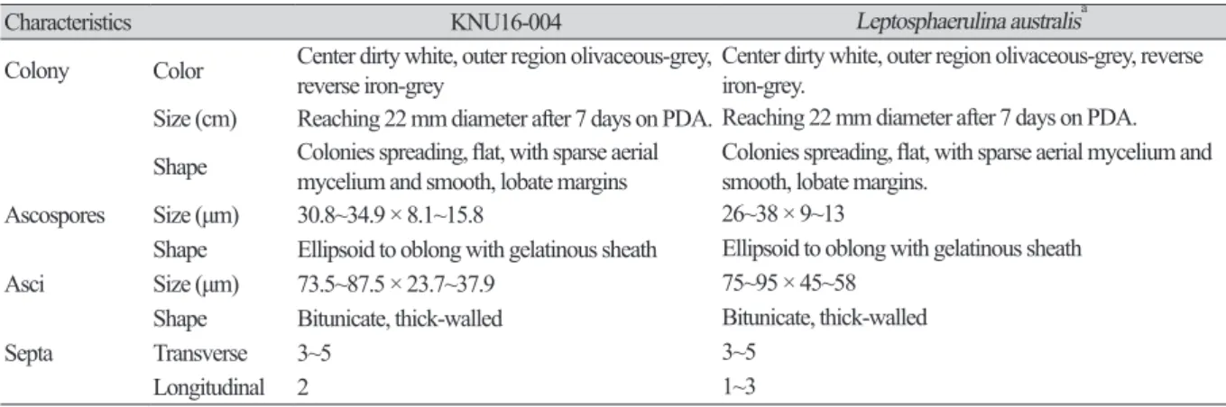
Table 2. Morphological characteristics of KNU16-004 isolated in this study and comparison with previously reported isolates
Characteristics KNU16-004 Leptosphaerulina australisa
Colony Color Center dirty white, outer region olivaceous-grey,
reverse iron-grey Center dirty white, outer region olivaceous-grey, reverse iron-grey.
Size (cm) Reaching 22 mm diameter after 7 days on PDA. Reaching 22 mm diameter after 7 days on PDA.
Shape Colonies spreading, flat, with sparse aerial
mycelium and smooth, lobate margins Colonies spreading, flat, with sparse aerial mycelium and smooth, lobate margins.
Ascospores Size (μm) 30.8~34.9 × 8.1~15.8 26~38 × 9~13
Shape Ellipsoid to oblong with gelatinous sheath Ellipsoid to oblong with gelatinous sheath
Asci Size (μm) 73.5~87.5 × 23.7~37.9 75~95 × 45~58
Shape Bitunicate, thick-walled Bitunicate, thick-walled
Septa Transverse 3~5 3~5
Longitudinal 2 1~3
PDA, potato dextrose agar.
aSource of description [21, 22].
margins. After7daysofincubation, thegrowthdiameterreached22mmonPDA, MEA, andOA agar, butonly12mmonSNAagar (Fig. 1A~1D). Myceliuminitiallywhiteandthenchangedtodeep blackfromthecenterofthecolony. Thesurfaceofthecolonywascoveredbyshort, whiteaerial mycelium, thereversesideofthecolonywasinitiallyblack, andtheendofthecolonyreachedthe edgeofthePDAplatewithin21daysofincubation; ascosporesandasciwereproducedonPDAagar after exposuretothe above-mentioned illuminationand observedunderalightmicroscope. The ascosporeswereellipsoidtooblongwithagelatinoussheath, 30.8~34.9 × 8.1~15.8μminsize, and theasciwerebitunicateandthick-walled, 73.5~87.5 × 23.7~37.9μminsize (Fig. 1F, 1H). Asshown inTable2, thesemorphologicalcharacteristicsofisolateKNU16-004werecompletelyconsistent withthosepreviouslyreportedforL. australis [21, 22].
Molecular phylogeny of isolate KNU16-004
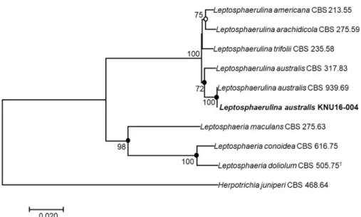
Toidentifytheisolatedfungalstrainatthespecieslevel, thenucleotidesequencesoftheLSU, ITS, andTub2 (890bp, 519bp, and345bp, respectively) wereobtainedandcomparedwiththeGenBank databaseusingBLAST. TheLSUsequenceofKNU16-004exhibitedhighsimilarities (99.8~100%) withvariousL. australisstrains, theITSsequenceshared100% identitywithvariousL. australis strains, andtheTub2sequencewasidentical (100%) toL. australisstrainCBS939.69. Phylogenetic analysis based on aconcatenated alignment of the LSU, ITS, and Tub2 nucleotidesequences performedusingtheneighbor-joining, maximum-likelihood, andmaximum-parsimonymethods, and revealedthatthestrainKNU16-004belongstothegenusLeptosphaerulinaandisgroupedwithtwo L. australisstrainswithahighbootstrapvalueof100% (Fig. 2).
PathogenicitystatusofL. australisisuncertain [23]. However, L. australishasbeendetectedin
Fig. 1. Culture and morphological characteristics of Leptosphaerulina australis KNU16-004. A, Colony on oatmeal agar; B, Colony on potato dextrose agar; C, Colony on malt extract agar; D, Colony on synthetic low-nutrient a gar; E, G, Colony sporulating on ascomata; F, Ascospores; H, Asci (scale bar = 10 µm).
importedagriculturalproduce, namelyonBrassicaoleraceavar. capitateleafspots [21], thus, itcould berelevanttoagriculturalproducttrade. Furtherstudieswillbeneededtoinvestigateitspotential pathogenicityandidentifytherelativesusceptibilityofavailablecultivars. Thisisthefirstreportof LeptosphaerulinaaustralisinKorea.
ACKNOWLEDGEMENTS
ThisresearchwassupportedbyagrantfromtheNationalInstituteofBiologicalResources (NIBR), fundedbytheMinistryofEnvironment (MOE) oftheRepublicofKoreafortheprojectonsurveyand discoveryofindigenousfungalspecies, andBrainPoolProgram (GrantNo. 2018H1D3A2065415) throughthe National Research Foundation (NRF) funded bythe Ministry ofScienceand ICT, RepublicofKorea.
REFERENCES
1. McAlpine D. Fungus diseases of stone fruits in Australia and their treatment. Melbourne: R. S.
Brain, Government Printer; 1902.
2. Graham JH, Luttrell ES. Species of Leptosphaerulina on forage plants. Phytopathology 1961;51:680-93.
Fig. 2. Neighbor-joining phylogenetic tree based on a concatenated alignment of the large subunit, internal transcribed spacer, and β-tubulin sequences. The phylogenic analysis shows the position of Leptosphaerulina australis KNU16-004 among related Leptosphaerulina spp. strains. Bootstrap values (based on 1,000 replications) greater than 50% are shown at the branch points. Filled circles indicate that the corresponding nodes were also recovered in trees generated with the maximum-likelihood and maximum-parsimony algorithms. Open circles indicate that the corresponding nodes were also recovered in the tree generated with the maximum-likelihood algorithm. The tree was rooted using Herpotrichia juniperi CBS 468.64 as an outgroup. Bar, 0.02 substitutions per nucleotide position. CBS, Westerdijk Fungal Biodiversity Institute (formerly CBSKNAW), Utrecht, The Netherlands.
3. Irwin JA, Davis RD. Taxonomy of some Leptosphaerulina spp. on legumes in eastern Australia. Aust J Bot 1985;33:233-7.
4. Crivelli PG. Über die heterogene Ascomycetengattung Pleospora rabh.: Vorschlag für eine Aufteilung [dissertation]. Zurich: ETH Zurich; 1983.
5. Abler SW. Ecology and taxonomy of Leptosphaerulina spp. associated with turfgrasses in the United States [dissertation]. Blacksburg (VA): Virginia Polytechnic Institute and State University; 2003.
6. Zhang Y, Crous PW, Schoch CL, Hyde KD. Pleosporales. Fungal Divers 2012;53:1-221.
7. Couch HB. Diseases of turfgrasses. Malabar: Krieger Publishing Co.; 1995.
8. Wehmeyer LE. The development of the ascocarp in Pseudoplea gaeumannii. Mycologia 1955;47:163-76.
9. Park S, Ten L, Lee SY, Back CG, Lee JJ, Lee HB, Jung HY. New recorded species in three genera of the Sordariomycetes in Korea. Mycobiology 2017;45:64-72.
10. Raja HA, Miller AN, Pearce CJ, Oberlies NH. Fungal identification using molecular tools: a primer for the natural products research community. J Nat Prod 2017;80:756-70.
11. Rehner SA, Samuels GJ. Taxonomy and phylogeny of Gliocladium analysed from nuclear large subunit ribosomal DNA sequences. Mycol Res 1994;98:625-34.
12. Vilgalys R, Hester M. Rapid genetic identification and mapping of enzymatically amplified ribosomal DNA from several Cryptococcus species. J Bacteriol 1990;172:4238-46.
13. Woudenberg JH, Aveskamp MM, de Gruyter J, Spiers AG, Crous PW. Multiple Didymella teleomorphs are linked to the Phoma clematidina morphotype. Persoonia 2009;22:56-62.
14. Thompson JD, Gibson TJ, Plewniak F, Jeanmougin F, Higgins DG. The CLUSTAL_
X windows interface: flexible strategies for multiple sequence alignment aided by quality analysis tools. Nucleic Acids Res 1997;25:4876-82.
15. Hall TA. BioEdit: a user-friendly biological sequence alignment editor and analysis program for Windows 95/98/NT. Nucleic Acids Symp Ser (Oxf) 1999;41:95-8.
16. Kimura M. A simple method for estimating evolutionary rates of base substitutions through comparative studies of nucleotide sequences. J Mol Evol 1980;16:111-20.
17. Saitou N, Nei M. The neighbor-joining method: a new method for reconstructing phylogenetic trees. Mol Biol Evol 1987;4:406-25.
18. Felsenstein J. Evolutionary trees from DNA sequences: a maximum likelihood approach. J Mol Evol 1981;17:368-76.
19. Fitch WM. Toward defining the course of evolution: minimum change for a specific tree topology. Syst Zool 1971;20:406-16.
20. Kumar S, Stecher G, Tamura K. MEGA7: Molecular Evolutionary Genetics Analysis Version 7.0 for Bigger Datasets. Mol Biol Evol 2016;33:1870-4.
21. Hyun IH, Heo NY, Chang SY, Heo JY, Mel’nik V. Identification of three fungi newly intercepted from importing plants in Korea. Mycobiology 2005;33:243-4.
22. Crous PW, Summerell BA, Swart L, Denman S, Taylor JE, Bezuidenhout CM, Palm ME, Marincowitz S, Groenewald JZ. Fungal pathogens of Proteaceae. Persoonia 2011;27:20-45.
23. Mitkowski NA, Browning M. Leptosphaerulina australis associated with intensively managed stands of Poa annua and Agrostis palustris. Can J Plant Pathol 2004;26:193-8.