132
Phialocephala lagerbergii: A New Record from Crop Field Soil in Korea
Mahesh Adhikari
1, Sangwoo Kim
1, Dil Raj Yadav
1, Yong Hyun Um
1, Hyung Seung Kim
1, Hyang Burm Lee
2and Youn Su Lee
1*
1Division of Biological Resource Sciences, Kangwon National University, Chuncheon 24341, Korea
2Division of Food Technology, Biotechnology & Agrochemistry, College of Agriculture and Life Sciences, Chonnam National University, Gwangju 61186, Korea
ABSTRACT : A unrecorded hyphomycete species of Phialocephala was isolated for the first time during the investigation of fungal community in the soil samples collected from different regions of Korea. The fungal isolate was identified as Phialocephala lagerbergii, based on the morphological characteristics and phylogenetic analysis of the ribosomal DNA sequence. In addition, cultural and micro-morphological features were described in detail.
KEYWORDS : Korea, Micromorphology, Phialocephala lagerbergii, rDNA
Introduction
The hyphomycete genus Phialocephala belongs to the family Vibrisseaceae. This fungus is characterized by a pig- mented stipe, a terminal sporogenous head comprising fine hairs of metulae and phialides [1]. Phialocephala spe- cies contribute to a small proportion of the fungal biota, and are closely related to the genus Phialophora. Phialo- cephala species produce conidia in phialides during the peaks of mononematous conidiophores [2]. Conidiophores of Phialocephala are complex with irregular branching and are obconic to globose in shape [3]. Phialocephala species are known to produce a wide variety of extracellular en- zymes such as polyphenyl oxidases, laccases, and protea- ses [4].
Phialocephala lagerbergii covers a wide range of ecolog-
ical niches with diverse morphological characteristics [5].
Color of the colony, pigmentation in the culture, and outgrowths on the stipes are the common morphological features of P. lagerbergii [5]. P. lagerbergii is mainly isola- ted from wood and wood pulp [6]. Moreover, Phialoce- phala species are widely known as endophytes of roots and grow well on agar media [7]. In this study, P. lagerb- ergii was first isolated from the crop field soil during our fungal diversity study in Samcheok city, Korea. The pur- pose of this study was to (i) elucidate the morphological of P. lagerbergii by comparing with the previous descrip- tions of Phialocephala species and (ii) determine its phy- logenetic status, based on ribosomal DNA (rDNA) sequ- ence analysis.
Materials and Methods
Sampling and isolation
Soil samples were collected from various locations in Samcheok city (37º 26' 87.04 N, 128º 90' 04.62 E), Gang- won-do, Korea. Fungal strains were isolated from the soil samples by the conventional dilution technique [8]. After dilution, the isolates were cultured on a potato dextrose agar (PDA; Difco, Detroit, MI, USA), supplemented with 100 μg chloramphenicol (bacteriostat/L PDA) for 5~7 days at 25°C, until the growth of fungal colony was ob- served. Then, a representative isolate (KNU14-11) was preserved at 20°C on PDA slants until further use.
*Corresponding author E-mail: [email protected] Received July 8, 2016 Revised July 23, 2016 Accepted August 21, 2016
This is an Open Access article distributed under the terms of the Creative Commons Attribution Non-Commercial License (http://
creativecommons.org/licenses/by-nc/3.0/) which permits unrestricted non-commercial use, distribution, and reproduction in any medium, provided the original work is properly cited.
Kor. J. Mycol. 2016 September, 44(3): 132-137 http://dx.doi.org/10.4489/KJM.2016.44.3.132 pISSN 0253-651X • eISSN 2383-5249
© The Korean Society of Mycology
First Report of Phialocephala lagerbergii from Crop Field Soil
133
Morphological characterization
Four different types of media were used for the mor- phological characterization of the study isolate (KNU14- 11): potato dextrose agar (PDA), oatmeal agar (OA), yeast extract sucrose agar (YES), and malt extract agar (MEA).
All the growth media were prepared according to Samson [9]. The isolate KNU14-11 was cultured on 9 cm petri dishes with three point inoculation and incubated in dark at 25°C for 7 days. Obverse and reverse colony colors, as well as the degree of speculation, were determined. An HK 3.1 CMOS digital camera (KOPTIC, Seoul, Korea), attached to an Olympus BX50F-3 microscope (Olympus, Tokyo, Japan), was used for capturing microscopic pic- tures of the fungal isolate. Scanning electron microscope (LEO Model 1450VP Variable Pressure Scanning Electron Microscope; Carl Zeiss, Oberkochen, Germany) was used for scanning and capturing the micro-morphological fea- tures of the fungal isolate.
DNA extraction, PCR amplification, sequencing, and data analysis
Genomic DNA was extracted from 1 week old colonies grown on PDA media, using a DNeasy Plant Mini Kit (Qiagen, Germantown, MD, USA) following the manu- facturer’s instructions. The internal transcribed spacer region (ITS) was amplified using ITS1 (5'- TCCGTAGG TGAACCTGCG-3') and ITS4 (5'- TCCTCCGCTTATTG ATATGC-3') primers [10]. The amplified PCR products were sequenced using the Applied Biosystems 3730 DNA analyzer (Foster city, CA, USA). The sequences were compared with the reference ITS sequences, retrieved from GenBank, National Center for Biotechnology Information (NCBI), using the basic local alignment search tool (BLAST) software [11]. The annotated nucleotide sequence of KNU14-11 isolate was deposited in GenBank, with the accession number KP055600. All sequences were aligned using the Molecular Evolutionary Genetics Analysis (MEGA 6.0) software [12]. Phylogenetic tree was genera- ted by the neighbor-joining method, utilizing the Kimura 2-parameter model with a bootstrap analysis of 1,000 replications for each clade.
Results and Discussion
Morphology of the KNU14-11 isolate
Colony morphology: Detailed morphological features of the fungal isolate KNU14-11 are shown in Fig. 1. The KNU14-11 isolate grown on PDA media attained a dia-
meter of 9~12 mm within 7 days at 25°C. The dorsal side of the mycelium was white in color, whereas the ventral side was hairy green in the middle (Fig. 1A and 1E).
Sporulation was moderately dense, and the conidia were seen in mass, having irregular form and smooth surface.
The KNU14-11 isolate grown on OA media attained a diameter of 11~16 mm within 7 days at 25°C. The front and the rear sides of the mycelium were dark black with a circle (Fig. 1B and 1F). Sporulation was moderate, and the conidia were in mass, irregular form and rough sur- face. The KNU14-11 isolate grown on YES media attained a diameter of 8~10 mm within 7 days at 25°C. The front side of the mycelium was white on the margin and black in the center, and the rear was black in color (Fig. 1C and 1G). Sporulation was moderate to dense and the conidia were in mass, having irregular form and smooth surface.
On MEA media, the KNU14-11 isolate attained a diame- ter of 14~17 mm within 7 days at 25°C. The front and rear sides of the mycelium were black in color (Fig. 1D and 1H).
Micromorphology: The conidiophores were macrone- matous, unbranched, erect, septate, smooth, and dark brown in color. Conidiophores were observed to be paler towards the apex. Conidiophores were 40~109 μm high and 3~4.8 μm wide (Fig 1I, 1M, and 1N). Hyphae were hyaline and smooth of about 1.5~2.5 μm wide. Hyphae were observed to be brown near the conidiophores. Coni- dia were catenate, non-septate, smooth, hyaline with rounded apices and truncated bases, tapering modestly to narrower bases, and 2~2.8 × 1~1.5 μm in size (Fig 1L, 1O, and 1P). Phialides were 2~4 in number and 4.8~24 × 1~7.2 μm in size (Fig 1J and 1K). Stipe comprised single to multiseptate and were 4.9~700 μm long. Comparisons of the morphological characteristics of KNU14-11 isolate with those of previously reported species of Phialocephala are described in Table 1.
Molecular phylogeny
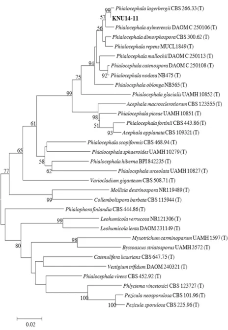
The KNU14-11 isolate was sampled from the crop field soil of Samcheok city, Gangwon-do, Korea. The isolates used for constructing the phylogenetic tree are shown in Table 2, along with their respective GenBank accession numbers. The KNU14-11 isolate was most closely related to P. lagerbergii (CBS 266.336) and formed a monophyle- tic group, supported by a bootstrap value of 99% (Fig.
2). Phylogenetic analysis revealed that the isolate was P.
lagerbergii.
The KNU14-11 isolate belonged to the Phialocephala genus, based on its characteristics (colony, conidia, and conidiophores) that were similar to those of the Phialo- cephala genus. In the KNU14-11 isolate, the micromor- phological structures were similar to those described by Jacobs et al. [13]. Phialocephala species closely resembles the Sporendocladia fumosa in terms of conidial arrange- ment [14]. Moreover, the arrangement of phialides and branching patterns of conidiophores, as well as the struc-
ture of phialides of P. lagerbergii, as described by Day et al. [6], was found to be identical to that of the KNU14- 11 isolate (Fig. 1I-1K). This confirmed that our fungal isolate KNU14-11 was P. lagerbergii. In addition, the ITS sequence of CBS 266.33 was in close proximity (99% sim- ilarity) with that of P. lagerbergii. Based on the ITS se- quence comparison, the present isolate was found to be closely related with P. lagerbergii, which was also strongly supported by their morphological comparison. Produc- Fig. 1. Morphological characteristics of Phialocephala lagerbergii KNU 14-11 isolate grown for 7 days on potato dextrose agar, oatmeal agar, yeast extract sucrose agar, and malt extract agar media at 25°C. A~D, obverse colony from left to right; E~H, reverse colony from left to right; I~K, light microscopic picture of conidiophores; L, light microscopic pictures of conidia; M, N, scanning electronic pictures of conidiophores; O, P, scanning electronic pictures of conidia.
First Report of Phialocephala lagerbergii from Crop Field Soil
135
Table 1. Morphological characteristics of Phialocephala lagerbergii KNU 14-11 isolated in this study Characteristics Study isolate Phialocephala lagerbergii P. lagerbergiia
Colony
Texture Colonies on PDA, OA, YES and MEA
are slow in growth. Colonies on MEA are effuse hairy in the middle.
Color White in front and black in front with
hairy green in the middle Honey to primrose
Conidia
Size 2~2.8 × 1~1.5 µm 2~3 × 1~1.5 µm
Shape Catenate, non-septate, smooth hyaline, round apices and truncate bases
Catenate, non-septate, hyaline, round apices and truncate bases
Surface Tapering modestly Tapering modestly
Conidiophores
Size 40~109 μm high and 3~4.8 μm wide 40~110 μm high and 3~5 μm wide Shape Macronematous, unbranched, erect,
septate, obconic, globose
Macronematous, unbranched, erect and septate, obconic to globose
Phialides
Structure Solitary phialides formed directly on the mycelium Solitary phialides formed directly on the mycelium Size 6.9~9.8 µm long and 3~3.7 µm wide 7~10 µm long and 3~4 µm wide
Shape Thick walled, smooth, lageniform Thick walled, smooth, lageniform
aSource of description [13].
PDA, potato dextrose agar; OA, oatmeal agar; YES, yeast extract sucrose agar; MEA, malt extract agar.
Table 2. Sequences of Phialocephala lagerbergii and allied species used in this study, along with their GenBank accession numbers
S.N. Species Isolate No. GenBank accession No.
1 Phialocephala lagerbergii CBS266.33 NR119426
2 Phialocephala aylmerensis DAOM250106 NR136124
3 Phialocephala dimorphospora CBS 300.62 NR135931
4 Phialocephala repens MUCL1849 EU434847
5 Phialocephala mallochii DAOM250113 NR136123
6 Phialocephala catenospora DAOM250108 NR136122
7 Phialocephala nodosa NB475 NR136121
8 Phialocephala oblonga NB565 KP768317
9 Phialocephala glacialis UAMH 10852 NR136120
10 Acephala macrosclerotiorum CBS 123555 NR121349
11 Phialocephala piceae UAMH 10851 NR111319
12 Phialocephala fortinii CBS 443.86 NR103577
13 Acephala applanata CBS 109321 NR119482
14 Variocladium giganteum CBS 508.71 NR111206
15 Mollisia dextrinospora NR119489 NR119489
16 Leohumicola verrucosa NR121306 NR121306
17 Leohumicola lenta DAOM231149 NR111180
18 Myxotrichum carminoparum UAMH 1597 NR111038
19 Byssoascus striatosporus UAMH 3572 NR111040
20 Phialocephala scopiformis CBS 468.94 NR119460
21 Phlyctema vincetoxici CBS 123727 KJ663857
22 Pezicula neosporulosa CBS 101.96 NR138003
23 Pezicula sporulosa CBS 225.96 KR859262
24 Phialocephala sphaeroides UAMH 10279 NR121302
25 Phialocephala hiberna BPI 842235 NR119465
26 Phialocephala urceolata UAMH 10827 NR111285
27 Collembolispora barbata CBS 115944 NR111443
28 Phialophora finlandia CBS 444.86 AF486119
29 Catenulifera luxurians CBS 647.75 NR121470
30 Vestigium trifidum DAOM240321 NR121556
31 Phialocephala virens CBS 452.92 AF586132
tion of a wide variety of extracellular enzymes such as polyphenyl oxidases, laccases, and proteases by the Phia- locephala species reflects its importance in the field of biotechnology. Further studies on its biotechnological im- portance are worthwhile in the future.
Acknowledgements
This work was supported by a grant from the National Institute of Biological Resources (NIBR), funded by the Ministry of Environment (MOE) of the Republic of Korea
for the project on the survey and discovery of indigenous Korean fungal species.
REFERENCES
1. Kirschner R, Oberwinkkler F. Phialocephala trigonospora, a new hyphomycete species associated with conifericolous bark beetles. Sydowia 1998;50:205-12.
2. Grünig CR, Queloz V, Duò A, Sieber TN. Phylogeny of Pha- emollisia piceae gen. sp. nov.: a dark, septate, conifer-needle endophyte and its relationships to Phialocephala and Aceph- ala. Mycol Res 2009;113:207-21.
Fig. 2. Phylogenetic analysis based on the neighbor-joining method using the partial 18S-ITS1-5.8S-ITS2-28S rDNA region of Phialocephala lagerbergii KNU 14-11 isolate obtained from the crop field soil in Korea. Numerical values (> 50) on branches are the bootstrap values with 1,000 replicates.
First Report of Phialocephala lagerbergii from Crop Field Soil
137
3. Conant NF. The occurrence of a human pathogenic fungus as a saprophytic in nature. Mycologia 1937;29:597-8.
4. Grünig CR. Population biology of the tree-root endophyte Phialocephala fortinii [dissertation]. Zurich (CH): Swiss Fed- eral Institute of Technology; 2003.
5. Jacobs A. The genus Phialocephala: a taxonomic study [dis- sertation]. Pretoria (ZA): University of Pretoria; 2006.
6. Day MJ, Hall JC, Currah RS. Phialide arrangement and cha- racter evolution in the helotialean anamorph genera Cado- phora and Phialocephala. Mycologia 2012;104:371-81.
7. Wang W, McGhee D, Gibas CF, Tsuneda A, Currah RS. My- cologia 2009;101:136-41.
8. Davet P, Rouxel F. Detection and isolation of soil fungi. En- field: Science Publishers; 2000.
9. Samson RA. Food and indoor fungi. Utrecht: CBS-KNAW Fungal Biodiversity Centre; 2010.
10. White TJ, Bruns TD, Lee SB, Taylor JW. Amplification and
direct sequencing of fungal ribosomal RNA genes for phylo- genetics. In: Innis MA, Gelfand DH, Sninsky JJ, editors. PCR protocols: a guide to methods and applications. San Diego:
Academic Press; 1990. p. 315-22.
11. National Center for Biotechnology Information. Basic Local Alignment Search Tool [Internet]. Bethesda (MD): National Center for Biotechnology Information; 2015 [cited 2016 May 25]. Available from: http://www.ncbi.nlm.nih.gov/Blast.cgi.
12. Tamura K, Stecher G, Peterson D, Filipski A, Kumar S.
MEGA6: molecular evolutionary genetics analysis version 6.0.
Mol Biol Evol 2013;30:2725-9.
13. Jacobs A, Coetzee MP, Wingfield BD, Jacobs K, Wingfield MJ.
Phylogenetic relationships among Phialocephala species and other ascomycetes. Mycologia 2003;95:637-45.
14. Crous P, Wingfield MJ. Sporendocladia fumosa and Lauriomy ces bellulus sp. nov. from Castanea cupules in Switzerland. Sy- dowia 1994;46:193-203.