This is an Open Access article distributed under the terms of the Creative Commons Attribution Non-Commercial License (http: //creativecommons.org/licenses/by- nc/4.0/) which permits unrestricted non-commercial
© 2018 THE KOREAN SOCIETY OF MYCOLOGY.
https://doi.org/10.4489/KJM.20180029
RESEARCH ARTICLE
Penicillifer diparietisporus: a New Record from Field Soil in Korea
Kallol Das
1, Chang-Gi Back
2, Seung-Yeol Lee
1, Hee-Young Jung
1*
1
School of Applied Biosciences, College of Agriculture and Life Sciences, Kyungpook National University, Daegu 41566, Korea
2
Horticultural and Herbal Crop Environment Division, National Institute of Horticultural and Herbal Science, Wanju 55365, Korea
*Corresponding author: [email protected]
ABSTRACT
A fungus was isolated from field soil collected from Daegu, Korea. The colony of the isolated fungus showed short, branched, and light to dark yellow pigments with hyaline, yellowish red to orange brown aerial mycelia. In addition, the fungus produced solitary to aggregated perithecia, ovoid to pyriform, short neck, and asci as well as biseriately arranged ascospores.
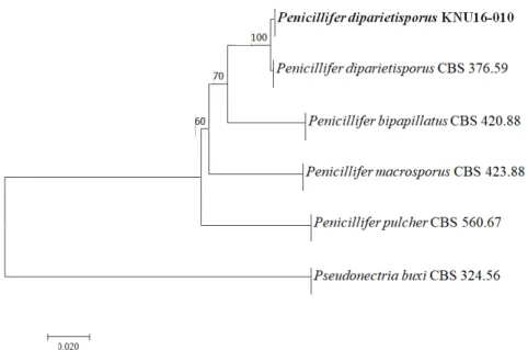
Phylogenetic analysis using the internal transcribed spacer region and translation elongation factor 1-α sequences and morphological characteristics identified the isolated fungus as Penicillifer diparietisporus, which belongs to the family Nectriaceae. To our knowledge, this is the first report of Penicillifer diparietisporus in Korea.
Keywords: Nectriaceae, Penicillifer diparietisporus , Soil fungi
INTRODUCTION
Approximately 2,700 fungal species from 240 genera are currently recognized in eight families in the order Hypocreales [1, 2]. The species produce light- to bright-colored, ostiolate, perithecial ascomata containing unitunicate asci with hyaline ascospores. The asexual morphs found in nature are, most frequently, moniliaceous and phialidic [3-7]. The fungal species are globally found in various environments; they are of great importance in agriculture and medicine and extensively exploited for industrial and commercial applications [5]. Moreover, several species have been reported as important opportunistic human pathogens [8-10], whereas others produce mycotoxins that are of medical concern [5].
The purpose of this study was to screen for unreported fungal species in field soil in Korea.
This study provides information for further studies on the use of such organisms in agriculture and medicine as well as for industrial applications. On the basis of morphological and molecular characteristics, the strain KNU16-010, identified as Penicillifer diparietisporus , was reported
Accepted: August 18, 2018 Revised: August 18, 2018 Received: August 11, 2018
Kor. J. Mycol. 2018 September, 46(3): 227-233 OPEN ACCESS
pISSN : 0253-651X eISSN : 2383-5249
MATERIALS AND METHODS
Collection of soil samples
In 2016, samples were collected from field soil during the screening of fungal species in Daegu, Korea (35°54′11.3″N, 128°36′56.1″E). A sterile trowel and spatula were used to collect the soil samples from a depth of 15~30 cm. The collected soil samples were placed in polythene zipper bags and transferred to the laboratory. Then, the soil samples were prepared for serial dilutions, and 1 g of soil was diluted with 10 mL of sterile distilled water. The serial dilutions were performed until a concentration of 10
-3was achieved, and, then, 100 µL of each sample was spread on potato dextrose agar (PDA; Difco, Detroit, MI, USA) plates and incubated for 2~3 days at 25°C. Single germinating fungal colonies were transferred to fresh PDA plates and incubated under the same conditions. The strain KNU16-10 was selected for further morphological and molecular phylogenetic analyses.
Morphological characterization
To study the cultural and morphological characteristics, PDA, oatmeal agar (OA; Difco), and malt extract agar (MEA; Difco) were used; the incubation period was 10 days at 25°C. Then, cultural characteristics such as colony color, fungal growth, and texture were observed and recorded, and the morphological characteristics were observed under a light microscope (BX- 50; Olympus, Tokyo, Japan).
Genomic DNA extraction, PCR amplification, and sequencing
Genomic DNA was extracted from 7-day-old colonies of the strain KNU16-010 grown on PDA by using the HiGene Genomic DNA Prep kit (BIOFACT, Daejeon, Korea), according to the manufacturer’s instructions. Partial gene sequences were amplified by the internal transcribed spacer (ITS) region, ITS1/ITS4, and translation elongation factor (TEF) 1-α gene, EF1-728F/EF2, as described previously [11-13]. Then, the amplified PCR products were purified with EXOSAP-IT (Thermo Fisher Scientific, Waltham, MA, USA) and sequenced by Solgent (Daejeon, Korea). The similarities of the sequences were analyzed using BLAST of NCBI. The sequences obtained from KNU16-010 were deposited in NCBI GenBank (accession numbers LC387549 and LC387601 for the ITS region and TEF gene, respectively).
Phylogenetic analysis
The consensus sequences were compared with other sequences in the NCBI database by using
BLAST to determine the percentage of shared sequence identity with other sequences of
fungal species. The alignments were performed using MEGA 7.0 [14] with 1,000 bootstrap
replicates, and the evolutionary distance matrices were generated based on Kimura’s neighbor-
joining algorithm model [15].
RESULTS AND DISCUSSION
Morphology of the strain KNU16-010
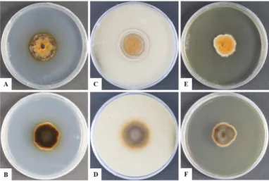
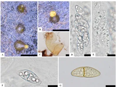
To observe the morphological characteristics, the strain KNU16-010 was cultured on PDA, OA, and MEA at 25°C for 10 days, and the diameter of the colonies was 28.7~29.7 mm, 32.0~32.7 mm, and 31.9~32.5 mm, respectively (Fig. 1). The morphology of the strain KNU16- 010 was compared with previous descriptions of Penicillifer diparietisporus [16] and Pseudonectria diparietospora [17] (Table 1). Pseudonectria diparietospora produced reddish brown colonies on PDA [17]. Moreover, Neocosmospora arxii (basionym: P seudonectria diparietispora ) colonies are composed of hyaline or sometimes yellow-pigmented, branched, septate, smooth-walled, hyphae that are 2.5~5 μm wide, and they produce semi-immersed to immersed perithecia, scattered or aggregated in small groups, yellowish orange to yellowish brown, broadly ovoid to somewhat pyriform, 220~340 × 160~270 μm in diameter, almost glabrous, conical shaped with short neck [18]. However, our strain formed orange brown to yellowish red colonies in all the media, and PDA was used to observe the mycological characteristics (Fig. 1). The fungal colonies showed wavy mycelia at the edges and rough wart- like structures. They also formed septate, irregular, and short hyphae, and the average width of the hyphae was 2.42 μm. They produced submerged perithecia, solitary to aggregated, yellowish orange to yellowish brown, ovoid to pyriform, smooth, conical shaped with short neck (Fig. 2A, 2C). Asci clavate, 70.9~81.1 × 16.3~19.7 μm, thin wall, and 8-spored (Fig. 2D, 2E). Ascospores were oval, septate, guttulate, 2-celled, distichous, double wall, biseriate, tubular, hyaline and pale green, smooth and blunt at both ends with a diameter (n = 30) of 17.9~30.7 × 6.7~10.9 μm and no conidia (Fig. 2F, 2G). The ascospores were distichous, elliptical, continuous, hyaline, with thick, double wall, smooth 18~24 × 12~14 μm and no
Fig. 1. Cultural characteristics of KNU16-010. A, B, Front and reverse sides of the colony on potato
Fig. 2. Morphological characteristics of KNU16-010. A, B, Mature perithecia on potato dextrose agar;
C, Perithecium; D, E, Asci; F, G, Ascospores (scale bars: A, B = 500 μm, C = 50 μm, D~G = 10 μm).
Table 1. Morphological characteristics of the strain KNU16-010 with reference to Penicillifer diparietisporus
Characteristics KNU16-010
aPenicillifer diparietisporus
bPseudonectria diparietospora
cColony Color Yellowish to orange and dark brown in reverse on PDA. Light yellow in center, white edge and brown color in reverse on MEA.
Reddish white edge, changing to greenish yellow on OA and yellowish brown to brown, light brownish orange to dark brown on PDA. Dark brown color in reverse both media.
Reddish-brown color on PDA and colonies composed of tangled masses of conidial chains.
Size (Diam.) PDA: 28.7~29.7 mm; OA: 32.0~32.7 mm and MEA: 31.9~32.5 mm in 10 days at 25°C.
PDA: 22mm; OA:30 mm in 10 days at
25°C; MEA: N/A N/A
Shape Edge margin irregular, wavy and
inadequate aerial mycelium. Edges submerged into irregular margin
and scant of aerial mycelium. Lightly floccose or plane and appressed and thin.
Asci Size (Diam.) and
Shape Clavate, 70.9~81.1 × 16.3~19.7 μm,
thin wall and 8-spored. Clavate, 60~85 × 12~25 μm,
unitunicate and 8-spored. Broadly clavate to spindle shaped with very thin wall, short stalked, 60~80 × 19~22 μm and 8-spored.
Ascosspores Size (Diam.) 17.9~30.7 × 6.7~10.9 μm. 21~25 × 12~15 μm. 18~24 × 12~14 μm.
Shape and Color Oval, septate, distichous, double wall, tubular, hyaline to pale green, smooth and blunt in both ends.
Biseriate, thick walled, broadly ellipsoidal, surrounded by a thin walled sheath collapsing at maturity and sometimes giving the ascospores roughened appearance.
Distichous, elliptical, continuous, hyaline, with thick, double wall, smooth.
Perithecia Submerged, solitary to aggregated, yellowish orange to yellowish brown, ovoid to pyriform,smooth, conical shaped and short neck.
Solitary to densely aggregated, superficial, globose, ovoid to pyriform, 270~300 × 240~270 μm diameter, brown, red-orange to orange.
Superficial, transparent wall, globose- conoid with very short neck, smooth, yellowish-red, 400~500 × 300~400 μm.
PDA, potato dextrose agar; MEA, malt extract agar; OA, oatmeal agar; Diam., diameter; N/A, not available in previous references.
a
Fungal strain studied in this paper.
b
Sources of the descriptions [16].
c