145
A New Record of Pseudeurotium bakeri from Crop Field Soil in Korea
Mahesh Adhikari
1, Sangwoo Kim
1, Dil Raj Yadav
1, Yong Hyun Um
1, Hyun Seung Kim
1, Hyang Burm Lee
2and Youn Su Lee
1*
1Division of Biological Resource Sciences, Kangwon National University, Chuncheon 24341, Korea
2Division of Applied Bioscience and Biotechnology, College of Agriculture and Life Sciences, Chonnam National University, Gwangju 61186, Korea
ABSTRACT : Pseudeurotium bakeri KNU14-21 isolated from crop field soil is a previously unreported fungal species in Korea. This fungal species was identified through morphological and molecular characteristics. Based on the shape and size of conidia and conidiophores as well as the internal transcribed spacer region of rDNA, the isolate was confirmed to be Pseudeurotium bakeri.
This species has not been previously reported from Korea, and this is the first report of its incidence in Korea.
KEYWORDS : Characteristics, Morphology, Pseudeurotium bakeri, Species
Introduction
Pseudeurotium bakeri is an ascomycetous fungus belong- ing to the Pseudeurotiaceae family. The general features of this fungal species include having hyaline or brown ascospores in a thin walled ascus [1]. Pseudeurotium ba- keri has been reported as an endophytic fungus having a cosmopolitan distribution, and can survive in high con- centrations of diesel fuel in soil [2]. The conidial phase of P. bakeri has been noted as sporothrix, beauvaria, or acremonium-like structures [3].
Soil fungi play vital ecological roles as decomposers, pa- thogens, and symbionts [4]. Fungi have a cosmopolitan distribution and grow in a vast area of habitats. Despite their ecological importance, little is known about their distribution and biodiversity. Therefore, we collected fun-
gal strains to discover the diversity of fungi in the crop field soil from Suncheon-si, Jeollanam-do, Korea. Among the fungal isolates, Pseudeurotium bakeri, which has not been reported officially from Korea, was encountered. The purpose of this study was to describe the isolated fungal species of Pseudeurotium by morphologically comparing it with previously described Pseudeurotium spp.
Materials and Methods
Sampling of soil and fungal isolation
Soil samples were obtained from different areas of Sun- cheon-si, Jeollanam-do, Korea. Each soil sample was taken at a depth of 1~15 cm. All the collected soil samples were stored in plastic bags and stored at 4°C. The fungi were isolated by the conventional dilution method [5] and cul- tured on potato dextrose agar (PDA; Difco, Detroit, MI, USA) supplemented with 100 μg chloramphenicol (bac- teriostatic agent) per liter of PDA for 5~7 days at 26°C.
For further use, pure cultures of PDA slants were main- tained at 4°C at the plant microbiology and biotechnology lab of Kangwon National University.
Morphological examination
Morphological features of the isolate were observed in PDA by performing one point inoculation in 9 cm petri plates. Inoculated plates were incubated in the dark at 26°C. The morphological characteristics were observed
*Corresponding author E-mail: [email protected] Received August 25, 2016 Revised September 12, 2016 Accepted September 21, 2016
This is an Open Access article distributed under the terms of the Creative Commons Attribution Non-Commercial License (http://
creativecommons.org/licenses/by-nc/3.0/) which permits unrestricted non-commercial use, distribution, and reproduction in any medium, provided the original work is properly cited.
Kor. J. Mycol. 2016 September, 44(3): 145-149 http://dx.doi.org/10.4489/KJM.2016.44.3.145 pISSN 0253-651X • eISSN 2383-5249
© The Korean Society of Mycology
by using a differential contrast microscope. The color, size, and shape of the microstructures of the isolate were ob- served by using an HK3.1 CMOS digital camera (KOP- TIC, Seoul, Korea) attached to an Olympus BX50F-3 microscope (Olympus, Tokyo, Japan). Scanning electronic microscopy images of the conidia and conidiophores were taken by a LEO Model 1450VP Variable Pressure (SEM;
Carl Zeiss, Oberkochen, Germany).
DNA extraction, PCR amplification, sequencing and data analysis
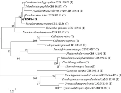
Genomic DNA was extracted from fully grown mycelia on PDA media. DNA was extracted using a DNeasy Plant Mini Kit (Qiagen, Germantown, MD, USA) following the manufacturer’s instructions. For amplification of the inter- nal transcribed spacer (ITS) gene region, primers ITS1 (5'-TCCGTAGGTGAACCTGCG-3') and ITS4 (5'-TCCT CCGCTTATTGATATGC-3') [6] were used. The amplified PCR products were sequenced using an ABI Prism 3730 DNA analyzer (Applied Biosystems, Foster City, CA, USA). The sequences were compared with reference ITS sequences deposited in GenBank at the National Center for Biotechnology Information (NCBI) using the basic local alignment search tool (BLAST) [7]. The nucleotide sequence obtained from the isolate KNU14-21 was depo- sited in GenBank and was assigned an accession number KP055601. The isolates used for the construction of the phylogenetic tree are shown in Table 2. The neighbor- joining relationship was analyzed using MEGA 6.0 [8]. A neighbor-joining tree was constructed using the Kimura 2-parameter substitution model [9]. The branch robust- ness was tested by 1,000 replicates of bootstrap analysis.
Results and Discussion
Morphological features of the isolate KNU14-21 assessed by optical and scanning electron microscopes are shown in Fig. 1. The colony grew moderately, attaining a diame- ter of 25~32 mm. The isolate had a smooth colony surf- ace. The colony was white in color at the backside and light yellow on the front side. The center of the isolate was slightly powdery (Fig. 1A, 1B). The conidiophores were produced from the aerial or substrate mycelium (Fig. 1F). Short side-branches of the conidiophores were observed (23~57 μm in length) (Fig. 1G). Conidia were subglobose or ellipsoid (Fig. 1E). The size of the conidia was 4.8~5.5 × 3.0~3.2 μm (Table 1). Conidia were present in a chain-like structure (Fig. 1H). The conidial head was loosely columnar. The terminal conidiogenous cells were 9~21 μm in size and lageniform. The morphological des- cription of our study isolate reasonably fits with the des- cription of P. bakeri [10]. Formation of conidia and coni- diophores by the isolate is similar to that of the Teberdinia state of P. bakeri reported by Sogonov et al. [10].
To confirm the identification, molecular analysis was carried out. ITS gene region was used for determination of the phylogenetic relationship with the previously des- cribed Pseudeurotium species. Our results showed that the isolate is most closely related to P. bakeri (CBS878.71), with a bootstrap value of 99% (Fig. 2). Additional sequence similarity analysis of the ITS region using BLAST search also supported that KNU14-21 is P. bakeri (Table 3).
Although this is a common fungal species with a cosmo- politan distribution, this is the first report of its incid- ence in Korea.
Since Pseudeurotium bakeri has been reported as a
Characters Present isolate Pseudeurotium bakeri P. bakeria
Colony
color White in back and light yellow in front side NA growth pattern Colonies on PDA are moderate in growth,
irregular NA
Conidia
size 4.8~5.5 × 3.0~3.2 µm 5.0~5.7 × 3.2~3.5 µm
shape Subglobose or ellipsoid Glabrous,subglobose, ellipsoid or obvoid
conidial head Loosely columnar Loosely columnar
Conidiophores structure Aerial or substrate mycelium Aerial or substrate mycelium Terminal
conidiogenous cells 9~21 μm, lageniform 10~22 μm, lageniform
NA, Not Available; PDA, potato dextrose agar.
a Source of description [10].
Fig. 1. Morphological characteristics of Pseudeurotium bakeri KNU14-21 grown for 7 days on potato dextrose agar. A, reverse view; B, obverse view; C, D, Simple microscopy picture of conidiophores; E, Simple microscopy picture of conidia; F, G, Scan- ning electronic microscopic pictures of conidiophores; H, Scanning electronic microscopic pictures of conidia.
Table 2. Sequences of Pseudeurotium and allied species used in this study with their GenBank accession numbers
Serial No. Species Isolate No. GenBank accession No.
1 Pseudeurotium hygrophilum CBS 102670 NR111128
2 Teberdinia hygrophila CBS 102671 AY129292
3 Pseudeurotium ovale var. ovale CBS 389.54 AY129289
4 Pseudeurotium bakeri CBS 878.71 AY129287
5 Pseudeurotium zonatum CBS 329.36 NR111127
6 Thelebolus globosus CBS 113940 NR138367
7 Pseudeurotium desertorum CBS 986.72 AY129288
8 Collophora rubra CBS120873 NR119747
9 Collophora capensis CBS120879 NR137726
10 Collophora hispanica CBS 128568 NR111680
11 Pseudofabraea citricarpa CBS 130297 KR859279
12 Phialocephala virens CBS 452.92 NR103564
13 Phacidium pseudophacidioides CBS 590.69 KJ663853
14 Phacidium grevilleae CBS139892 NR137977
15 Allantophomopsis lunata CBS137781 NR132922
16 Geomyces auratus CBS 108.14 NR11187
17 Pseudogymnoascus destructans ATCC MYA-4855 NR111838
18 Pseudogymnoascus appendiculatus UAMH 10509 NR137875
19 Gymnostellatospora frigida UAMH 9304 NR111200
20 Gymnostellatospora alpina UAMH 9430 NR111201
fungal species resistant to biodiesel contaminated soil [2], this finding has important biotechnological implications. However, the main purpose of our present study was limited to identify and describe the isolate morphologically and molecularly. Further studies on its use and implications in the field of biotechnology are thus needed.
Acknowledgements
This work was supported by a grant from the National Institute of Biological Resources (NIBR), funded by the Ministry of Environment (MOE) of the Republic of Korea for the project on survey and discovery of indigenous fungal species.
REFERENCES
1. Cannon PF, Kirk PM. Fungal families of the world. Walling- ford: CAB International. 2007.
2. Flewelling AJ, Johnson JA, Gray CA. Isolation and bioassay screening of fungal endophytes from North Atlantic marine macroalgae. Bot Mar 2013:56:287-97.
3. Ferrari BC, Zhang C, van Dorst J. Recovering greater fungal diversity from pristine and diesel fuel contaminated sub-Ant- arctic soil through cultivation using both a high and a low nu- trient media approach. Front Microbiol 2011;2:217.
4. Swift MJ, Heal OW, Anderson JM. Decomposition in terrestrial ecosystems. Berkeley: University of California Press; 1979.
5. Davet P, Rouxel F. Detection and isolation of soil fungi. En- field: Science Publishers; 2000.
6. White TJ, Bruns TD, Lee SB, Taylor JW. Amplification and direct sequencing of fungal ribosomal RNA genes for phylo- genetics. In: Innis MA, Gelfand DH, Sninsky JJ, editors. PCR Fig. 2. Neighbor-joining phylogenetic analysis of the partial 18S-ITS1-5.8S-ITS2-28S rDNA sequences of Pseudeurotium bakeri KNU14-21 obtained from crop field soil in Korea. The phylogenetic tree was constructed using the MEGA 6 program. The sequence obtained in the study is shown in boldface. The mark (T) indicates type strain. Numerical values (> 50) on branches are the bootstrap values as percentage of bootstrap replication from a 1,000 replicate analysis. The scale bar represents the number of substitutions per site.
Table 3. Identification of fungal isolate to species level with reference species based on the analyses of internal transcribed spacer gene sequence
Gene NIBR No. GenBank accession No. GenBank library strain Sequence similarity (%) ITS rDNA NIBRFG0000142402 KP055601 Pseudeurotium bakeri (the present isolate)
CBS878.71 AY129288 Pseudeurotium bakeri (the closest isolate) 98 ITS, internal transcribed spacer.
protocols: a guide to methods and applications. San Diego:
Academic Press; 1990. p. 315-22.
7. National Center for Biotechnology Information. Basic Local Alignment Search Tool [Internet]. Bethesda (MD): National Library of Medicine; 2015 [cited 2016 June 15]. Available from: https://blast.ncbi.nlm.nih.gov/Blast.cgi.
8. Tamura K, Stecher G, Peterson D, Filipski A, Kumar S.
MEGA6: molecular evolutionary genetics analysis version 6.0.
Mol Biol Evol 2013;30:2725-9.
9. Kimura MA. A simple method for estimating evolutionary rates of base substitutions through comparative studies of nuc- leotide sequences. J Mol Evol 1980;16:111-20.
10. Sogonov MV, Schroers HJ, Gams W, Dijksterhuis J, Summer- bell RC. The hyphomycete Teberdinia hygrophila gen. nov., sp.
nov. and related anamorphs of Pseudeurotium species. Myco- logia 2005;97:695-709.