This is an Open Access article distributed under the terms of the Creative Commons Attribution Non-Commercial License (http: //creativecommons.org/licenses/by- nc/4.0/) which permits unrestricted non-commercial
© 2019 THE KOREAN SOCIETY OF MYCOLOGY.
RESEARCH ARTICLE
A new record of Scleroconidioma sphagnicola isolated from soil in Korea
Benjamin Yaw Ayim1, Gihwan Sung1, In-Kyu Kang1, Seung-Yeol Lee1,2, Hee-Young Jung1,2*
1College of Agriculture and Life Sciences, Kyungpook National University, Daegu 41566, Korea
2Institute of Plant Medicine, Kyungpook National University, Daegu 41566, Korea
*Corresponding author: [email protected]
ABSTRACT
A fungal isolate, designated KNU-JJ-1824, was isolated from a soil sample collected from a field on Jeju, Korea. Colonies of the isolates cultured on PDA, MEA and CMAD for 14 days grew to diameters of 49~51 mm, 60~63 mm, and 47~50 mm, respectively. The colonies of this fungal isolate were dark green in color on both sides, and had irregular margins on the PDA media. Mycelia submerged and exhibited clear lines from the center to the edge on MEA media. On CMAD media, they were dark brown to greenish-brown, with narrow white margins.
The shape of the conidia was guttulate, fusiform or clavate, and 1.9~3.4 × 6.5~10.2 μm in size. A molecular phylogenetic tree was constructed using dataset from small subunit (SSU) rDNA and the internal transcribed spacer (ITS) regions, and the isolate was found to cluster with Scleroconidioma sphagnicola UAMH 9731. Based on the results of the phylogenetic tree analysis and the cultural and morphological characteristics, the isolate was identified as Scleroconidioma sphagnicola. We report S. sphagnicola for the first time in Korea.
Keywords: Morphological characteristics, Phylogenetic analysis, Scleroconidioma sphagnicola
INTRODUCTION
The genus Scleroconidioma Tsuneda, Currah & Thormann was identified for the first time in diseased leaves of Sphagnum fuscum in Canada, and later reported from Sweden and the Czech Republic [1-3]. It is similar to members of the genera Phaeotheca and Phaeosclera, but shows differences in conidiogenesis and cultural characteristics [4,5]. Scleroconidioma sphagnicola, the type and sole species of the genus Scleroconidioma, has distinctive characteristics. It is a strongly melanized fungus, allied to the Dothidiales, and represents a group of fungi found on plant litter [6,7]. It has the ability to adapt to stressful environmental conditions [8]. S. sphagnicola is an anamorphic ascomycete, and a pathogen of peat bogs (Sphagnum fuscum) as has been reported from Canada [6,9]. It was isolated in Sweden and the Czech Republic, where it was observed that the fungus has saprotrophically colonized wood and coniferous litter [2,3]. It has also been reported that S. fuscum cell walls are highly resistant to biological degradation due to a high content of phenolic compounds [10]. During surveying unreported fungal species in Korea, a fungal
Accepted: June 19, 2019 Revised: June 18, 2019 Received: May 17, 2019 https://doi.org/10.4489/KJM.20190016 Kor. J. Mycol. 2019 June, 47(2): 125-30
OPEN ACCESS pISSN : 0253-651X eISSN : 2383-5249
Hee-Young Jung
https://orcid.org/0000-0002-4254-3367
based on morphological characteristics and phylogenetic analysis result. In this study, we firstly report and describe the characteristics of S. sphagnicola KNU-JJ-1824 which has not been reported in Korea.
MATERIALS AND METHODS
Soil sample collection and fungal isolation
Fungal isolate KNU-JJ-1824 was isolated from a soil sample collected on Jeju, Korea (N 33°25'20.3'', E 126°33'04.0'') in 2018. The sample was collected from the ground, air-dried, and stored in a plastic bag at 4°C until analysis. One gram of the soil sample was suspended in 10 mL of sterile distilled water. The suspension was vortexed, diluted, and a defined volume was spread on potato dextrose agar (PDA; Difco, Detroit, MI, USA). Individual colonies that developed on the agar were purified by subculture on fresh PDA and incubated at 25°C until mycelium development. The pure cultures were preserved on PDA slants at 4°C.
Morphological characterization of cultures
For morphological analysis, isolate KNU-JJ-1824 was grown on potato dextrose agar (PDA), malt extract agar (MEA), and corn meal agar with 1% dextrose (CMAD) for 14 days [1]. After incubation, colony characteristics such as color, size, and shape were recorded. Samples were observed using a model BX-50 light microscope (Olympus, Tokyo, Japan).
Genomic DNA extraction, PCR amplification and data analysis
Genomic DNA was extracted from strain KNU-JJ-1824 using HiGene Genomic DNA Prep Kits (BIOFACT, Daejeon, Korea), following the manufacturer�s protocol. Small subunit ribosomal rDNA (SSU) and the internal transcribed spacer region (ITS) rDNA were amplified using the primers NS1/ITS4, while NS4, NS6, NS8 and ITS1F primers were used for inner sequence analysis [11,12]. The amplified PCR products were purified using ExoSAP-IT (Thermo Fisher Scientific, Waltham, MA, USA) and sequenced by Macrogen (Daejeon, Korea). Similarities between the sequences were analyzed using NCBI BLAST and GENETYX-WIN (ver. 3.2). A partial sequence of the SSU and ITS regions of allied species of S.
sphagnicola were retrieved from NCBI to generate a phylogenetic analysis (Table 1). The sequences from KNU-JJ-1824 were deposited in NCBI GenBank under the accession number MK880096.
Phylogenetic analysis
The isolates used to construct the phylogenetic tree are summarized in Table 1, with their strain and GenBank accession numbers. Phylogenetic trees were constructed based on concatenated SSU and ITS region sequences using the maximum likelihood method in the MEGA6 software program with a bootstrap analysis of 1,000 replications [13].
RESULTS AND DISCUSSION
Morphology of KNU-JJ-1824
The isolated KNU-JJ-1824 was cultured on three different media; PDA, MEA, and CMAD for 14 days, and incubated at 25°C. The diameters of the resulting colonies were 49~51 mm, 60~63 mm, and 47~50 mm, respectively. Both sides of the colonies were black and carbonaceous on PDA and had irregular margins and a solid texture. On MEA, mycelia submerged into the medium and made clear lines from the center to the edge. Colonies were dark brown to greenish brown with narrow white margins on CMAD (Fig. 1). Morphological characteristics were observed for colonies on the CMAD. Stromata with conidia, masses of sclerotic cells and clusters of shiny white tufts were observed (Fig. 2A, B and E). Conidia were pigmented, guttulate, variable in shape, but mostly fusiform or clavate, 1.9~3.4 × 6.5~10.2 μm in size (Fig. 2C). Bacilliform conidia were aseptate, hyaline, and 0.9~1.2 × 2.2~3.3 μm in size (Fig. 2D).
Sympodial conidia arising directly from hyphae were observed (Fig. 2F). The morphological characteristics of KNU-JJ-1824 were similar to those previously reported for S. sphagnicola [1] (Table 2). Based on the morphological characteristics, the fungal isolate KNU-JJ-1824 was identified as S. sphagnicola.
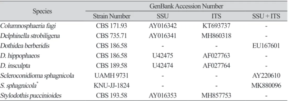
Table 1. List of the sequences used in this study.
Species GenBank Accession Number
Strain Number SSU ITS SSU + ITS
Columnosphaeria fagi CBS 171.93 AY016342 KT693737 -
Delphinella strobiligena CBS 735.71 AY016341 MH860318 -
Dothidea berberidis CBS 186.58 - - EU167601
D. hippophaeos CBS 186.58 U42475 AF027763 -
D. insculpta CBS 189.58 U42474 AF027764 -
Scleroconidioma sphagnicola UAMH 9731 - - AY220610
S. sphagnicola* KNU-JJ-1824 - - MK880096
Stylodothis puccinioides CBS 193.58 AY016353 MH857753 -
* The fungal isolated used in this study.
Table 2. Comparison of the morphological characteristics of an isolate KNU-JJ-1824 and the reference strain Scleroconidioma sphagnicola Characteristics Scleroconidiom a sphagnicola KNU-JJ-1824a Scleroconidiom a sphagnicola UAMH 9731b Colony Shape and color On CMAD, dark green to brown, irregular with white
margin, smooth and becoming floccose with age On CMAD, dark brown to greenish brown with a narrow white margin, smooth when young, becoming somewhat flocculose
with age
Size (mm) CMAD: 47~50, PDA: 49~51, MEA: 60~63 CMAD: 49~51, PDA: N/A, MEA: N/A Conidia Shape and position produced on conidiomata: guttulate, mostly fusiform or
clavate, 1.9~3.4 × 6.5~10.2 μm Conidia (bacilliform): aseptate, hyaline,
0.9~1.2 × 2.2~3.3 μm
produced on conidiomata: degree of pigmentation, commonly fusiform, clavate or spathulate, 1~3 × 6.5~9 μm
Conidia (bacilliform): hyaline, one-celled, 0.9~1 × 3~5.5(7.5) μm
Sclerotic cell Abundant Abundant
a The fungal isolate used in this study
Fig. 1. Cultural characteristics Scleroconidioma sphagnicola KNU-JJ-1824 after 14 days in 25°C. A, B, Colonies on potato dextrose agar; C and D, Colonies on malt extract agar; E and F, Colonies on CMAD.
Front (A, C, E) and reverse (B, D, F) sides.
Fig. 2. Morphological characteristics of Scleroconidioma sphagnicola KNU-JJ-1824. A, Stromata with conidia; B, Mass of sclerotic cells; C, Conidia; D, Conidia (Bacilliform); E, Shiny white tuft clusters; F, Sympodial conidia directly arising from hyphae (arrowhead). White scale bar = 10 μm, Black scale bar
= 200 μm).
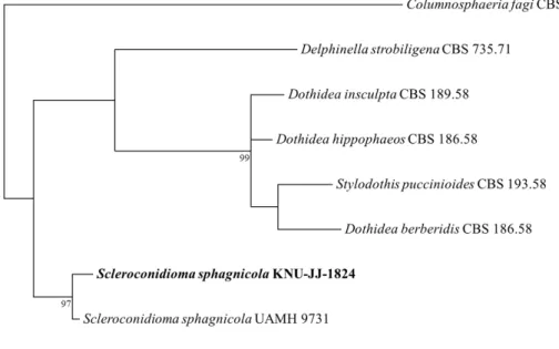
Molecular phylogeny of the isolate KNU-JJ-1824
A phylogenetic tree for isolate KNU-JJ-1824 was constructed to examine its phylogenetic relationship with allied species. Sequencing produced 2,240 bp, covering the small subunit (SSU) and the internal transcribed spacer (ITS) regions. BLAST search results revealed that strain KNU-JJ-1824 has 99% similarity with S. sphagnicola UAMH 9731 (AY220610), previously reported from Canada. A phylogenetic tree was constructed from partial sequences of the SSU and ITS regions using the neighbor-joining method. This tree indicated that strain KNU-JJ-1824 clustered with S. sphagnicola UAMH 9731 with 97% bootstrap value (Fig. 3). Furthermore, it has been reported that only S. sphagnicola UAMH 9731 is the sole species of the genus Scleroconidioma, it was confirmed that KNU-JJ-1824 was closely related with S. sphagnicola. Thus, S.
sphagnicola KNU-JJ-1824 was confirmed using morphological and molecular phylogenetic characteristics and was deposited in the National Institute of Biological Resources (accession no. NIBRFGC000502240).
S. sphagnicola was described as an anamorphic necrotrophic parasite on Sphagnum fuscum in Canada [1]. In Korea, it has been reported that Sphagnum spp. grow naturally in Jeju island [14]. In this reason, it is expected that S. sphagnicola inhabiting Sphagnum spp. can be found on Jeju Island. According to our result, this is the first report of the presence of genus Scleroconidioma and S. sphagnicola in Korea.
Fig. 3. Maximum likelihood phylogenetic tree based on a concatenated dataset of SSU and ITS gene sequences, showing the phylogenetic position of Scleroconidioma sphagnicola. The strain isolated in this study is shown in boldface. Bootstrap values based on 1,000 replications are shown at the branch points.
Columnosphaeria fagi CBS 171.93 was used as an outgroup. Bar is 0.01 substitutions per nucleotide position.
ACKNOWLEDGEMENTS
This research was supported by the project on the survey and excavation of Korean indigenous species of the National Institute of Biological Resources (NIBR 201801105) under the Ministry of Environment, Republic of Korea.
REFERENCES
1. Tsuneda A, Thormann MN, Currah RS. Scleroconidioma, a new genus of dematiaceous Hyphomycetes. Can J Bot 2000;78:1294-8.
2. Vasiliauskas R, Lygis V, Larsson KH, Stenlid J. Airborne fungal colonization of coarse woody debris in North temperate Picea abies forest: Impact of season and local spatial scale. Mycol Res 2005;109:487-96.
3. Koukol O, Kovářová M. Autecology of Scleroconidioma sphagnicola particularly in Šumava National Park (Czech Republic). Czech Mycol 2007;59:111-23.
4. Sigler L, Tsuneda A, Carmichael JW. Phaeotheca and Phaeosclera, two new genera of dematiaceous Hyphomycetes and a redescription of Sarcinomyces Lindner. Mycotaxon 1981;12:449-67.
5. Tsuneda A, Murakami S. Endoconidium development and release in the Hyphomycete Phaeotheca fissurella. Mycologia 1985;77:433-40.
6. Tsuneda A, Chen MH, Currah RS. Conidiomatal morphogenesis and pleomorphic conidiogenesis in Scleroconidioma sphagnicola. Mycologia 2001;93:1164-73.
7. Soderstrom BE. Vertical distribution of microfungi in a spruce forest soil in the south of Sweden. Trans Br Mycol Soc 1975;65:419-25.
8. Sterflinger K, de Hoog GS, Haase G. Phylogeny and ecology of meristematic ascomycetes.
Stud Mycol 1999;43:5-22.
9. Hambleton S, Tsuneda A, Currah RS. Comparative morphology and phylogenetic placement of two microsclerotial black fungi from Sphagnum. Mycologia 2003;95:959-75.
10. Tsuneda A, Thormann MN. Currah RS. Modes of cell-wall degradation of Sphagnum fuscum by Acremonium cf. curvulum and Oidiodendron maius. Can J Bot 2001;79:93-100.
11. White TJ, Bruns T, Lee S, Taylor JW. Amplification and direct sequencing of fungal ribosomal RNA genes for phylogenetics. In: Innis MA, Gelfand DH, Sninsky JJ, White TJ, editors. PCR Protocols: a guide to methods and applications. San Diego: Academic Press; 1990. p. 315-22.
12. Gardes M, Bruns TD. ITS primers with enhanced specificity for basidiomycetes - application to the identification of mycorrhizae and rusts. Mol Ecol 1993;2:113-8.
13. Tamura K, Stecher G, Peterson D, Filipski A, Kumar S. MEGA 6: Molecular evolutionary genetics analysis version 6.0. Mol Biol Evol 2013;30:2725-9.
14. Kim JW, Lee GY, Kim YH, Eom BC. A synecological description of Ohmi moor with Sphagnum islet in Jeju, Korea. Korean J Ecol Environ 2018;51:174-83.