• 검색 결과가 없습니다.

관동맥

N/A
N/A
Protected

Academic year: 2022

Share "관동맥"

Copied!
8
0
0

로드 중.... (전체 텍스트 보기)

전체 글

(1)

Original Articles Korean Circulation J 2000;;;;30((((1))))::::23-30

논문접수일:1999년 6월 28일 심사완료일:1999년 12월 22일

교신저자:박승정, 138-736 서울 송파구 풍납동 388-1 울산대학교 의과대학 서울중앙병원 내과학교실 전화:(02) 2224-4818・전송:(02) 475-6898

E-mail:[email protected]

관동맥 스텐트 시술 후 혈관조영상 재협착의 예측 인자에 대한 연구:혈관내 초음파를 이용한 스텐트 단면적과 길이에 따른 재협착율에 대한 연구

울산대학교 의과대학 서울중앙병원 내과학교실

이내희·홍명기·박성욱·이철환·김영학·조구영 나득영·강덕현·송재관·김재중·박승정

Predictors of Angiographic Restenosis after Intracoronary Stenting according to Stent Lumen Cross Sectional Area and Stent Length in Native Coronary Artery Lesions::An Intravascular Ultrasound Study

Nae-Hee Lee, MD, Myeong-Ki Hong, MD, PhD, Seong-Wook Park, MD, PhD, FACC, Cheol Whan Lee, MD, Young-Hak Kim, MD, Goo-Young Cho, MD, Deuk-Young Nah, MD, Duk-Hyun Kang, MD, PhD,

Jae-Kwan Song, MD, PhD, Jae-Joong Kim, MD, PhD and Seung-Jung Park, MD, PhD, FACC Department of Internal Medicine, College of Medicine, University of Ulsan, Cardiovascular Center, Asan Medical Center, Seoul, Korea

ABSTRACT

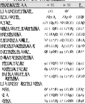
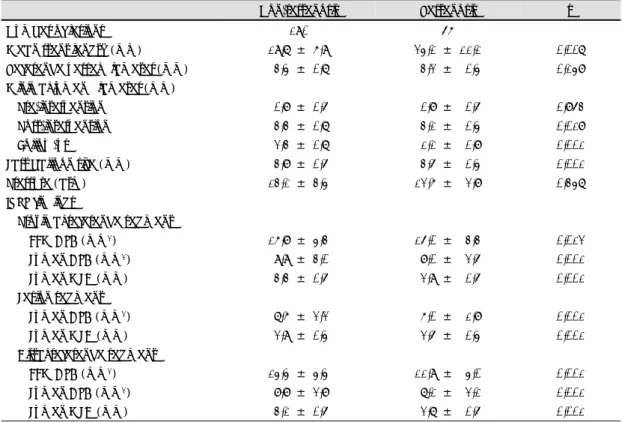
Background:The adequate intravascular ultrasound (IVUS) criteria for stent optimization have not been determined in long coronary stenting. We evaluated the predictors of angiographic restenosis and compared that according to stent lumen cross-sectional area (CSA) and stent length between short (stent length <20 mm) and long (≥20 mm) coronary stenting. Methods:IVUS-guided coronary stenting was successfully performed in 285 consecutive patients with 304 native coronary lesions. Six-month follow-up angiogram was performed in 236 patients (82.8%) with 246 lesions (80.9%). Results were evaluated using conventional (clinical, angiographic, and IVUS) methodology. Results:The overall angiographic restenosis rate was 22.8% (56/246)(short stent 17.6% vs long stent 32.2%, p=0.009). Using multivariate logistic regression analysis, the independent predictors of angiographic restenosis were the IVUS stent lumen CSA (odd ratio=

1.51, 95% CI 1.18-1.92, p=0.001) and stent length (odd ratio=0.95, 95% CI 0.91-1.00, p=0.039). The angiographic restenosis rate was 54.8% in stent lumen CSA <5.0 mm

2

(short stent 37.5% vs long stent 73.3%, p=0.049), 27.4% between 5.0 and 7.0 mm

2

(short stent 24.1% vs long stent 31.7%, p=0.409), 10.5%

between 7.0 and 9.0 mm

2

(short stent 10.0% vs long stent 12.5%, p=0.772), and 11.4% in stent lumen CSA

≥9.0 mm

2

(short stent 10.4% vs long stent 13.3%, p=0.767)(p=0.001). Conclusions:Compared with short

coronary stenting, long coronary stenting is effective treatment modality to cover long lesions with com-

parable long-term clinical outcomes in cases of stent lumen CSA ≥7.0 mm

2

. Regardless of the stent length,

(2)

the most important factor determining the angiographic restenosis was the IVUS stent lumen CSA in relatively large coronary artery lesions. ((((Korean Circulation J 2000;30((((1)))):23-30))))

KEY WORDS:Stent·Coronary artery disease·Intravascular ultrasound.

서 론

비교적 큰 관동맥의 국소 병변에서의 스텐트 시술의 유용성은 잘 알려져 있지만,

1)2)

미만성 병변의 경우는 긴 스텐트나 여러 개의 스텐트를 시술한 경우에 재협착 율이 높다고 보고되고 있다.

3-5)

즉 스텐트 길이 자체가 스텐트 시술후의 재협착을 초래하는 요인의 하나로 인 식되어 왔다. 하지만 다발 스텐트 시술(스텐트 수 ≥3) 도 2개 이하 스텐트 시술과 비교하여 병소재개율(tar- get lesion revascularization) 및 심 합병증 발생율 등 이 비슷하다는 보고도 있다.

6)

따라서 미만성 병변의 치 료로서 풍선성형술과 스텐트 시술 중 어느 방법이 우수 한 가에 대해서도 이견이 많은 상태이다.

7-9)

스텐트 시술의 적정화(stent optimization)에 대한 혈관내 초음파(intravascular ultrasound, 이하 IVUS) 상의 기준은 대부분의 연구에서 짧은 단일 스텐트에 관 한 것이고

10-12)

긴 스텐트 시술에 대해서는 잘 알려져 있지 않다. 일부연구에서는 스텐트 재협착을 예견하는 데 절대적인 혈관 내강의 크기가 스텐트의 상대적 확장 정도(스텐트 내경면적 / 참조혈관 내경면적의 비) 보다 더 중요하다는 연구결과도 있다.

13-15)

그래서 본 연구는 1) 혈관조영상 재협착의 예측 인자를 찾기 위해 임상적, 정량적 혈관조영상(quantitative coronary angiography, QCA), IVUS상의 예측인자를 분석하고, 2) 짧은 스텐 트(<20 mm)와 긴 스텐트(≥20 mm) 시술 군의 스텐 트 단면적(cross sectional area, 이하 CSA)과 길이에 따른 혈관조영상 재협착율을 비교하고자 하였다.

방 법

대상 환자

IVUS를 이용한 스텐트 시술을 성공적으로 시행한 285 명의 연속된 환자 304병변을 대상으로 하였다. 모든 환자는 시술후 1, 3, 6개월에 외래에서 임상적 추적 관 찰을 하였으며 6개월 시점에 추적 혈관 조영술을 시행

하였다. 6개월 추적 혈관조영술은 236명(82.8%)의 환 자 246병변(80.9%)에서 시행되었다. 대상 병변이 좌 주간지에 존재하거나 관동맥 우회술을 받은 경우는 본 연구에서 제외되었다. 추적 혈관조영술을 시행하지 못 한 49명의 환자도 본 연구에서 제외되었다. 대상 환자 들은 짧은 스텐트(<20 mm)를 시술한 151명의 환자, 159병변 군과 긴 스텐트(≥20 mm)를 시술한 85명의 환자, 87병변 군으로 분류하였다. 대상 환자는 객관적 인 심허혈의 증거가 있고 혈관조영상 70% 이상의 내 강 협착이 있는 경우로 하였다. 모든 환자는 본 시술에 동의한 경우로 하였다.

스텐트 시술

이전의 스텐트 시술에 대한 연구보고와 같은 방법으

로 시행되었다.

1)2)16)17)

시술중 활성 응고 시간(acti-

vated clotting time)이 300초가 넘도록 정맥내로 헤

파린을 투여하였다. 짧은 스텐트 시술로 GFX 스텐트

79병변, CrossFlex 스텐트 32병변, NIR 스텐트 25병

변, tantalum Cordis 스텐트 10병변, Wiktor 스텐트 5

병변, Palmaz-Schatz 스텐트 4병변 및 Multilink 스

텐트를 4병변에 삽입하였다. 긴 스텐트 시술로는 GFX

스텐트 27병변(24 mm 14병변, 30 mm 8병변 및 40

mm 5병변), 25 mm CrossFlex 스텐트 12병변, Cr-

own 스텐트 12병변(22 mm 1병변 및 30 mm 11병

변), Wallstent(평균길이 40.4 mm) 10병변, Giantur-

co-Roubin Ⅱ스텐트 9병변(20 mm 7병변 및 40 mm

2병변) 및 25 mm Multilink 스텐트를 1병변에 삽입하

였다. GFX 스텐트는 9기압 이상으로 풍선을 확장하여

삽입하였다. 추가적인 풍선 확장은 스텐트가 불충분하

게 확장된 경우에 시행되었다. Tantalum Cordis와 Cr-

oss-Flex 스텐트는 스텐트가 풍선에 premount된 채

로 병변까지 이송하였고, 시술시 12~14기압으로 확장

하였다. 이 두 스텐트는 스텐트 삽입후 다른 풍선을 이

용한 2차 풍선 확장술은 필요하지 않았다.

18)19)

GFX,

tantalum Cordis 및 CrossFlex 스텐트를 제외하고는

병변 부위에서 풍선을 확장하여 스텐트를 삽입하였다.

(3)

스텐트 삽입후 혈관조영상 적정화(angiographic stent optimization)를 위해 16기압까지 추가적인 고압력 풍 선 확장술을 시행하였다. IVUS는 혈관조영상 스텐트 시술의 적정화가 된 후 시행하였다. IVUS상 적정 스텐 트 확장(optimum stent expansion) 기준에 만족할 때 스텐트 시술을 종료하였다. 적정 스텐트 확장 기준은 스텐트 CSA가 가장 작은 곳이 근위부 및 원위부 참조 혈관의 평균 CSA의 80% 이상이고, 스텐트와 혈관이 완전히 밀착된 경우로 하였다. 이 기준에 적합하지 않 으면 추가적인 풍선 확장술을 시행하였다. 모든 환자에 게 항혈전제로 아스피린과 티클로피딘을 투여하였다.

티클로피딘은 시술 전 적어도 3일 전부터 시술 후 1개 월까지 250 mg씩 하루 두 번 투약되었고 아스피린은 영구적으로 투약되었다. 스텐트 삽입 후 헤파린은 즉시 중단 하였다.

정량적 혈관조영 분석

관동맥 내로 니트로글리세린 0.2 mg을 주입 후 혈관 조영을 시행하였다. 유도 도관을 내경 측정의 표준으로 하여 혈관조영 분석(ANCOR V2.0, Siemens, Germ- any)을 하였고 퍼센트 내경 협착(percent diameter stenosis), 최소 혈관 내경(minimal luminal diameter, 이하 MLD), 참조 혈관 내경(reference vessel dia- meter)을 측정하였다. 이 수치는 혈관의 내강이 가장 작게 관찰되고, 시술 전,후 및 추적 관찰시 확장기에 일 치하는 영상에서 측정하였다. 혈관조영상 재협착은 내 경 협착이 50% 이상인 경우로 하였다.

IVUS 시술

관동맥 내로 니트로글리세린 0.2 mg을 주입 후 IVUS (CVIS system, SCIMED life system INC, Boston Scientific Corporation, USA)를 시행하였다. 초음파 도관을 대상 병변의 원위부 10 mm 이상까지 삽입 후 대동맥과 관동맥 개구부 교차점까지 촬영하였다. 영상은 자동 당김 장치(automatic pullback device)에 의해 0.5 mm/sec의 속도로 후진하면서 촬영하였다. IVUS 도관은 3.2 F의 영상 도관 내에 1,800 rpm으로 회전 가능하고 유연한 몸체 끝에 트랜서듀서가 장치된 것이다. 초음파 영상은 시술 후 연구를 위해 고화질 s-VHS 테이프에 저 장하였다.

정량적 IVUS 분석

경화반(plaque)의 성분 분석, 외층 탄성막(external elastic membrane, 이하 EEM), 내강(lumen) 및 경 화반+중막(plaque+media)의 CSA 측정에 IVUS의 유용성은 이미 알려져 있다.

20)21)

EEM CSA는 혈관전 체의 CSA를 의미하며 외막(adventitia)의 leading edge를 추적하여 측정하였다. 시술 후 대상 병변, 근위 부 및 원위부 참조 분절(reference segment)의 최소, 최대 내경 및 CSA를 측정하였다. 병변 위치(lesion site) 는 IVUS 영상상 내강의 CSA가 가장 작은 곳으로 하였 다. 참조 분절은 대상 병변의 근위부 및 원위부 10 mm 이내에서 단면이 가장 정상적으로 보이는 부분으로 하 였다. 스텐트 CSA와 스텐트 길이에 따른 혈관조영상 재협착율을 구하였다. 이상의 결과들은 스텐트의 길이에 따라 짧은 스텐트 시술군과 긴 스텐트 시술군, 두 군으 로 구분하여 비교 하였다.

통계 분석

SPSS 통계 프로그램을 이용해 분석하였다. 범주 변 수는 빈도로 표시하고 연속 변수는 평균 ±1 표준편차 로 표시하였다. 비교 분석은 Student t 분석, χ

2

분석 이나 Fisher’s exact법을 사용하였다. 혈관 조영상 재 협착 예측 인자를 찾기 위해 단계별 로지스틱 회귀 분 석을 사용하였다. 통계적으로 의의 있는 경우는 p값이 0.05 이하인 경우로 하였다.

결 과

짧은 스텐트 시술 군에서 긴 스텐트 시술군 보다 고 혈압(p=0.007)이 많았으나 다른 임상적 차이는 없었 다(Table 1). 혈관조영상 및 시술상 특성은 Table 2와 같다. 긴 스텐트는 모두 B2나 C형 병변에서 사용되었 다(p=0.01). 평균 스텐트 길이는 짧은 스텐트 군이 15.8 mm 였으며 긴 스텐트 군이 29.7 mm 였다(p=

0.001). 두 군간에 QCA 상 참조 혈관 내경 및 시술 전후 MLD의 차이는 없었다. 긴 스텐트 군에서 최종 풍선 확장 압력이 높았으며(p=0.001), 짧은 스텐트 군에서 큰 풍선을 사용한 경향이 있었다(p=0.190).

스텐트 시술 후 IVUS 소견은 Table 3과 같다. 윈위

부 참조 분절의 EEM, 내강 CSA 및 내강 MLD는 긴

스텐트 사용 군에서 작았다. 병변 분절(lesion segm-

(4)

ent)의 내강의 CSA 및 MLD도 긴 스텐트 사용 군에 서 작았다.

혈관조영상 재협착율은 총 22.8%(56/246)였으며, 짧은 스텐트 사용 군이 17.6%(28/159)로 긴 스텐트 사용 군의 32.2%(28/87) 보다 낮았다(p=0.009). 6 개월 추적 관찰시 재협착이 발생한 군과 발생하지 않은 군간의 혈관 조영상, IVUS소견상의 비교는 Table 4와 같다. 재협착이 발생한 군에서 평균 스텐트 길이가 길 었으며(24.1 대 19.7 mm, p=0.007), 시술 후 QCA 상 MLD도 작았고(p=0.001), 시술시 작은 풍선이 사 용되었다(p=0.001). 또한 재협착 군에서 근위부 및 원위부 참조분절의 EEM, 내강 CSA, 내강 MLD가 의 미있게 작았다. 병변 분절의 내강의 CSA와 MLD도 재 협착군에서 작았다.

단변수 분석에서 p<0.2인 경우의 변수만을 이용하여 다변량 분석을 시행하였다. 다변량 로지스틱 회귀 분석 에서 IVUS상 스텐트 CSA(odd ratio=1.51, 95% CI

=1.18~1.92, p=0.001) 및 스텐트 길이(odd ratio=

0.95, 95% CI=0.91~1.00, p=0.039)가 혈관조영상 재협착의 독립적 예측 인자였다. 스텐트 CSA와 길이 에 따른 혈관조영상 재협착율은 Table 5와 같다.

고 안

미만성 관동맥 병변은 시술 성공율이 낮고 시술에 따 른 초기 합병증 및 후기 재협착이 잘 발생한다고 알려 져 있다. 또한 당뇨, 다혈관 질환 등과 같은 임상적 특 징과 동반해서 잘 나타나고 심한 경우에는 관동맥 우회 Table 1. Baseline clinical characteristics (%)

Stent length, mm < 20 ≥ 20 p Number of patients 151 85

Age (years) 57±9 59±8 0.073 Males 112 (74.2) 68 (80.0) 0.198 Prior myocardial infarction 11 ( 7.3) 11 (12.9) 0.116 Hypertension 60 (39.7) 20 (23.5) 0.007 Diabetes mellitus 25 (16.6) 17 (20.0) 0.319 Hypercholesterolemia 18 (11.9) 9 (10.6) 0.468 Cigarette smoking 73 (48.3) 44 (51.8) 0.356 Clinical presentation

Stable angina 41 (27.2) 25 (29.4) 0.411 Unstable angina 84 (55.6) 46 (54.1) 0.465 Acute myocardial

infarction 26 (17.2) 14 (16.5) 0.518 Number of disease vessel

One 85 (56.3) 44 (51.8) 0.296 Two 42 (27.8) 25 (29.4) 0.454 Three 24 (15.9) 16 (18.8) 0.343

Table 2. Baseline angiographic characteristics and pr- ocedural results (%)

Stent lengt h, mm < 20 ≥ 20 p Number of lesions 159 87 Coronary artery dilated

Left anterior descending 89 (56.0) 52 (59.8) 0.330 Left circumflex 22 (13.8) 8 ( 9.2) 0.196 Right coronary 48 (30.2) 27 (31.0) 0.501 Restenotic lesions 4 ( 2.5) 1 ( 1.2) 0.420 Infarct-related artery 19 (11.9) 11 (12.6) 0.512 Debulking before stenting 12 ( 7.5) 10 (11.5) 0.209 Lesion morphology 0.001

A 7 ( 4.4)

B1 45 (28.3)

B2 82 (51.6) 11 (12.6) C 25 (15.7) 76 (87.4) Mean stent length (mm) 15.8±2.3 29.7±7.5 0.001 Reference vessel

diameter (mm) 3.3±0.7 3.3±0.4 0.715 Minimal lumen diameter

(mm)

Pre-intervention 0.8±0.5 0.7±0.4 0.370 Post-intervention 3.3±0.7 3.2±0.4 0.139 Follow-up 2.1±0.8 1.8±0.8 0.048 Last balloon size (mm) 3.8±0.5 3.7±0.5 0.190 Pressure (atm) 12.4±3.3 14.0±3.1 0.001

Table 3. Post-intervention IVUS findings

Stent length, mm < 20 ≥ 20 p Proximal reference

segment

EEM CSA (mm

2

) 16.0±4.0 17.1±4.4 0.078 Lumen CSA (mm

2

) 9.5±2.9 9.5±3.3 0.861 Lumen MLD (mm) 3.2±0.5 3.2±0.6 0.918 Lesion segment

Lumen CSA (mm

2

) 7.5±2.3 6.7±2.0 0.009 Lumen MLD (mm) 2.8±0.5 2.7±0.4 0.023 Distal reference segment

EEM CSA (mm

2

) 14.5±4.6 12.7±3.9 0.003

Lumen CSA (mm

2

) 8.8±2.9 7.6±2.3 0.001

Lumen MLD (mm) 3.1±0.5 2.9±0.5 0.001

(5)

술을 시행하기 어려운 형태를 한 경우도 있어 경피적 치료가 불가피한 경우가 있을 수가 있다. 그러나 국소 병변에 비해 미만성 병변에서의 경피적 풍선성형술은 재협착율이 높다고 보고되고 있다.

22-24)

단일병원에서 시행한 연구결과에 의하면 20 mm 이상 길이의 병변에 서의 풍선성형술에 따른 재협착율이 63%로 높아 스텐 트 시술이 유용하리라는 연구 결과가 있었지만 대상 환 자군이 작고 초록 형식의 발표였다.

24)

하지만 지금까지 여러 연구에서는 미만성 병변에서 스텐트 및 절제 기구 (ablative device) 등의 새로운 치료 방법이 풍선성형 술보다 우수하다는 증거는 아직 없다.

25-27)

즉, 이처럼 미만성 관동맥 병변의 치료에는 아직 이견이 많은 상태

이다.

본 연구에서 스텐트 시술 후 혈관조영상 재협착의 독 립적 예측 인자는 IVUS상 스텐트 내강 CSA와 스텐트 길이였고, 스텐트 CSA가 가장 중요한 결정 인자였다.

이 결과는 스텐트 CSA가 임상적, 혈관조영상 재협착 과 밀접한 관계가 있다는 이전의 여러 연구결과와 일치 하는 결과이다.

10-15)

본 연구에서 짧은 스텐트 시술 시 스텐트 CSA와 재협착율의 관계는 스텐트 CSA가 5.0 mm

2

미만인 경우는 37.5%였으며, 5.0 mm

2

이상 7.0 mm

2

미만인 경우는 24.1%, 7.0 mm

2

이상 9.0 mm

2

미만인 경우는 10.0%, 9.0 mm

2

이상인 경우는 10.4%

였다(p=0.025). 이상의 스텐트 CSA에 따른 혈관조영 Table 4. Angiographic and IVUS findings between restenosis and non-restenosis (%)

Non-restenosis Restenosis p

Number of lesions 190 56

Mean stent length (mm) 19.7 ± 6.9 24.1 ± 11.1 0.007 Reference vessel diameter (mm) 3.4 ± 0.7 3.2 ± 0.4 0.148 Minimal lumen diameter (mm)

Pre-intervention 0.8 ± 0.5 0.8 ± 0.5 0.853 Post-intervention 3.3 ± 0.7 3.1 ± 0.4 0.018 Follow-up 2.3 ± 0.7 1.1 ± 0.8 0.001 Last balloon size (mm) 3.8 ± 0.5 3.5 ± 0.4 0.001 Pressure (atm) 13.1 ± 3.4 12.6 ± 2.8 0.347 IVUS findings

Proximal reference segment

EEM CSA (mm

2

) 16.8 ± 4.3 15.0 ± 3.3 0.012 Lumen CSA (mm

2

) 9.9 ± 3.0 8.0 ± 2.5 0.001 Lumen MLD (mm) 3.3 ± 0.5 2.9 ± 0.5 0.001 Lesion segment

Lumen CSA (mm

2

) 7.6 ± 2.2 6.0 ± 1.8 0.001 Lumen MLD (mm) 2.9 ± 0.4 2.5 ± 0.4 0.001 Distal reference segment

EEM CSA (mm

2

) 14.4 ± 4.4 11.9 ± 4.0 0.001 Lumen CSA (mm

2

) 8.8 ± 2.8 7.1 ± 2.1 0.001 Lumen MLD (mm) 3.1 ± 0.5 2.7 ± 0.5 0.001

Table 5. Angiographic restenosis rate (%) according to postintervention lumen CSA and stent length Stent length, mm

Lumen CSA, mm

2

Total < 20 ≥ 20 p

< 5.0 17/31 (54.8) 6/16 (37.5) 11/15 (73.3) 0.049 5.0≤ <7.0 26/95 (27.4) 13/54 (24.1) 13/41 (31.7) 0.409 7.0≤ <9.0 8/76 (10.5) 6/60 (10.0) 2/16 (12.5) 0.772

≥ 9.0 5/44 (11.4) 3/29 (10.4) 2/15 (13.3) 0.767

P 0.001 0.025 0.001

(6)

상 재협착률에 대한 본 연구의 결과는 짧은 길이의 스 텐트를 사용한 이전의 다른 연구와도 유사한 결과이다.

시술에 사용된 스텐트의 갯수도 재협착 예측 인자 중 하나로 알려져 있는데,

3-5)

본 연구도 유사한 결과를 나 타내었다.

스텐트 시술 후 우수한 임상 경과를 나타내는 IVUS 상 여러 지표들이 제안되었는데,

10-12)

이 지표들을 짧 은 스텐트 시술시에 적용하면 재협착 감소에 기여한다 고 알려져 있다.

13-15)

그러나 긴 스텐트 시술에서는 IVUS상 재협착 예측 인자에 대해 잘 알려져 있지 않 다. 본 연구에서는 긴 스텐트를 시술한 경우에도 짧은 길이의 스텐트를 대상으로한 다른 연구 결과처럼

13-15)

혈관조영상 재협착의 가장 중요한 예측 인자는 절대적 인 내강의 크기(absolute lumen dimension)이었다. 본 연구에서 긴 스텐트 시술시에 스텐트 CSA와 재협착율 의 관계는 CSA가 5.0 mm

2

미만인 경우는 73.3%, 5.0 mm

2

이상 7.0 mm

2

미만인 경우는 31.7%, 7.0 mm

2

이상 9.0 mm

2

미만인 경우는 12.5%, 9.0 mm

2

이상인 경우는 13.3%였다(p=0.001).

긴 관동맥 스텐트 시술시에는 긴 길이의 병변자체의 특성상 대개 근위부 및 윈위부 참조 혈관 사이에 혈관 의 크기에 차이가 있다. 긴 스텐트를 적절하게 삽입하 였는 경우에 근위부 참조 혈관이 큰 경우에도 원위부 참조 혈관은 아주 작은 경우도 흔히 관찰할 수도 있다.

이 경우 스텐트의 중간부위가 놓이게 되는 초기병변의 가장 좁았던 부위는 크게 유지되나 스텐트의 원위부 끝 은 내경이 좁아 이 부분이 최소 내경 CSA 및 MLD 부 위가 된다. 이전의 대부분의 긴 관동맥 병변을 대상으 로 한 연구들은 작은 윈위부 혈관과 큰 근위부 혈관을 갖는 병변을 일부에서 대상으로 포함 하였을 가능성을 배제할 수 없다. 본 연구에서 혈관의 크기가 작은 경우 (내강 CSA <5.0 mm

2

)에는 긴 스텐트 시술이 짧은 스 텐트 시술에 비해 혈관조영상 재협착율이 현저히 높았 다. 이상의 결과는 긴 스텐트 시술시 원위부 혈관의 크 기가 재협착의 중요한 예측 인자라는 연구결과와

27)

일 맥상통한다고 하겠다. 그러나 혈관이 비교적 큰 경우 (내강 CSA ≥7.0 mm

2

)에는 긴 스텐트 시술도 짧은 스텐트 시술과 비교하여 재협착율의 차이가 없었다. 이 는 스텐트 내강의 CSA가 7.0 mm

2

이상으로 큰 경우 에는 긴 스텐트 시술도 짧은 스텐트 시술 못지않은 우 수한 결과를 낼 수 있다는 것을 의미하는 결과이다. 즉

혈관의 크기에 관계없이 긴 스텐트 시술은 재협착율이 높아 풍선 성형술보다 우수한 점이 없다는 이전의 생각 은 재고되어야 한다고 사료된다. 결국 비교적 큰 혈관 에서는 스텐트의 길이에 관계없이 IVUS상 스텐트 CSA가 가장 중요한 혈관조영상 재협착을 결정하는 가 장 중요한 인자였다.

본 연구는 몇 가지 점에서 한계점이 있다. 첫째로 후 향적 연구였기 때문에 모든 미만성 병변에서 긴 스텐트 시술이 반드시 재협착을 감소시킨다는 것을 의미하는 것은 아니다. 이 점은 추후 전향적 연구로 증명을 필요 로 한다고 하겠다. 두 번째로 스텐트 시술시 IVUS를 사용한 선택적 환자만를 대상으로 하였으므로 IVUS를 시행하지 않은 다른 환자에서 이 결과를 적용시키기에 는 어려움이 있다. 세 번째로 스텐트 시술 후 IVUS를 시행하였으므로 스텐트 시술전의 IVUS에 대한 영향은 평가할 수 없었다. 네 번째로 본 연구에서 사용한 스텐 트의 평균 길이는 30 mm 정도로 40 mm 이상의 긴 스텐트에 대해서는 같은 연구결과를 적용하기에는 어 렵다고 하겠다. 다섯 번째로 감적술(debulking pro- cedure)을 시행한 경우가 적어서 재협착을 줄이기 위 한 감적술에 대한 영향은 평가하기 어려웠다. 마지막으 로 여러 종류의 스텐트를 사용하였으므로 스텐트 종류 에 따른 재협착율의 차이는 비교할 수 없었다.

요 약

연구배경:

긴 스텐트 시술에서 시술의 적정화(stent optimiza- tion)를 위한 혈관내 초음파(IVUS)의 기준은 아직 명 확하지 않다. 본 연구에서는 짧은 스텐트(<20 mm)와 긴 스텐트(≥20 mm) 시술 후 스텐트 내강 단면적(CSA)과 스텐트 길이에 따른 혈관조영상 재협착율을 차이를 비 교하고자 하였다.

방 법:

IVUS를 이용하여 성공적으로 관동맥 스텐트를 시술 한 연속된 285명의 환자 304병변을 대상으로 하였다.

6개월 추적 혈관조영술은 236명(82.8%)의 환자 246 병변(80.9%)에서 시행되었다. 연구 대상에 대한 임상 적, 혈관조영상 및 IVUS 결과를 분석하였다.

결 과:

혈관조영상 재협착율은 22.8%(56/246)였으며 짧은

(7)

스텐트 시술 군(17.6%)이 긴 스텐트 시술 군(32.2%) 보다 낮았다(p=0.009). 로지스틱 다변량 분석하였을 때 재협착의 예측 인자는 IVUS상 스텐트 내강 CSA (odd ratio=1.51, 95% CI=1.18~1.92, p=0.001)와 스텐트의 길이(odd ratio=0.95, 95% CI=0.91~1.00, p=0.039)였다. 스텐트 내강 CSA에 따른 재협착율(짧 은 스텐트:긴 스텐트)은 CSA <5.0 mm

2

인 경우 54.8%

( 37.5%:73.3%, p=0.049), 5.0 mm

2

≤CSA <7.0 mm

2

인 경우 27.4%(24.1%:31.7%, p=0.409), 7.0 mm

2

≤CSA <9.0 mm

2

인 경우 10.5%(10.0%:12.5%, p=0.772) 및 CSA ≥9.0 mm

2

인 경우가 11.4% (10.4%:

13.3%, p=0.767)였다(p=0.001).

결 론:

긴 관동맥 병변에서 스텐트 CSA가 7.0 mm

2

이상으 로 큰 경우에는 긴 스텐트 시술도 장기 임상 경과가 짧 은 스텐트시술과 같이 우수하였다. 상대적으로 큰 관동 맥 병변에서는 스텐트 길이에 관계없이 IVUS상 스텐 트 내강 CSA가 혈관 조영상 재협착의 가장 중요한 결 정 인자였다.

중심 단어 :스텐트・재협착・혈관내 초음파.

REFERENCES

1)

Serruys PW, Jaegere PD, Kiemeneij F, Macaya C, Rut- sch W, Heyndrickx G, et al. A comparison of balloon

expandable stent implantation with balloon angioplasty in patients with coronary artery disease. N Engl J Med 1994;331:489-95.

2)

Fischman DL, Leon MB, Baim DS, Schatz RA, Savage MP, Penn I, et al. A randomized comparison of coronary-

stent placement and balloon angioplasty in the treatment of coronary artery disease. N Engl J Med 1994;331:496-501.

3)

Kastrati A, Schomig A, Elezi S, Schuhlen H, Dirschin- ger J, Hadamitzky M, et al. Predictive factors of resteno-

sis after coronary stent placement. J Am Coll Cardiol 1997;

30:1428-36.

4)

Ellis SG, Savage M, Fischman D, Baim DS, Leon MB, Goldberg S, et al. Restenosis after placement of Palmaz-

Schatz stents in native coronary arteries. Circulation 1992;

86:1836-44.

5)

Haude M, Erbel R, Straub U, Dietz U, Meyer J. Short

and long term results after intracoronary stenting in hu- man coronary arteries: Monocentre experience with balloon- expandable Palmaz-Schatz stent. Br Heart J 1991; 6:337-45.

6)

Kornowski R, Mehran R, Hong MK, Satler LF, Pichard AD, Kent KM, et al. Procedural results and late clinical

outcomes after placement of three or more stents in sin- gle coronary lesions. Circulation 1998;97:1355-61.

7)

Hamasaki N, Nosaka H, Kimura T, Nakagawa Y, Yokoi H, Tamura T, et al. Influence of lesion length on late an-

giographic outcome and restenotic process after success- ful stent implantation. J Am Coll Cardiol 1997;29(suppl A):239A, (abstract).

8)

Moussa I, Di Mario C, Moses J, Reimers B, Blengino S, Colombo A. Single versus multiple Palmaz-Schatz stent

implantation. J Am Coll Cardiol 1997;29(suppl A):276A,

(abstract).

9)

Gaxiola E, Vlietstra R, Browne K, Ebersole DG, Brenn-er AS, Weeks TT, et al. Six-month follow-up of

patients with multiple stents in a single coronary artery. J Am Coll Cardiol 1997;29(suppl A):276A, (abstract).

10)

Hoffmann R, Mintz GS, Mehran R, Pichard AD, Kent KM, Satler LF, et al. Intravascular ultrasound predictors

of angiographic restenosis in lesions treated with Pal- maz-Schatz stents. J Am Coll Cardiol 1998;31:43-9.

11)

Mudra H, Regar E, Klauss V, Werner F, Henneke KH, Sbarouni E, et al. Serial follow-up after optimized ultra-

sound-guided deployment of Palmaz-Schatz stents. Circu- lation 1997;95:363-70.

12)

Hong MK, Park SW, Lee CW, Kang DH, Song JK, Kim JJ, et al. Intravascular ultrasound findings in stenting of

unprotected left main coronary artery stenosis. Am J Car- diol 1998;82:670-3.

13)

Hayase M, Oshima A, Cleman MW, Pepine CJ, Bailey SR, Shani J, et al. Relation between target vessel revas-

cularization and minimum stent area by intravascular ultrasound (CRUISE Trial). Comparison of ultrasound vs angiographic guidance for stenting in the CRUISE study.

J Am Coll Cardiol 1998;31:386A, (abstract).

14)

Ziada KM, Tuzcu EM, De Franco AC, Franco I, Potts WJ, Magyar WA, et al. Absolute, not relative, post-stent

lumen area is a better predictor of clinical outcome. Cir- culation 1996;94:I-453, (abstract).

15)

Moussa I, Di Mario C, Moses J, Reimers B, Blengino S, Colombo A. The predictive value of different intravascu-

lar ultrasound criteria for restenosis after coronary stenting.

J Am Coll Cardiol 1997;29:60A, (abstract).

16)

Schomig A, Neumann FJ, Kastrati A, Schuhlen H, Bl- asini R, Hadamitzky M, et al. A randomized comparison

of antiplatelet and anticoagulant therapy after the place- ment of coronary artery stents. N Engl J Med 1996;334:

1084-9.

17)

Colombo A, Hall P, Nakamura S, Almagor Y, Maiello L, Martini G, et al. Intracoronary stenting without antico-

agulation accomplished with intravascular ultrasound guidance. Circulation 1995;91:1676-88.

18)

Park SJ, Park SW, Lee CW, Hong MK, Kim JJ, Hong MK, et al. Immediate results and late clinical outcomes

after new CrossFlex coronary stent implantation. Am J Cardiol 1999;83:502-6.

19)

Park SJ, Park SW, Hong MK, Cheong SS, Lee CW, Kim JJ, et al. Late clinical outcomes of Cordis tantalum cor-

onary stenting without anticoagulation. Am J Cardiol 1997; 80:943-7.

20)

Tobis JM, Mallery J, Mahon D, Lehmann K, Zalesky P, Griffith J, et al. Intravascular ultrasound imaging of hu-

man coronary arteries in vivo: Analysis of tissue charact- eristics with comparison to in vivo histologic specimens.

Circulation 1991;83:913-26.

21)

Nishimura RA, Edwards WD, Warnes CA, Reeder GS, Holmes DR, Tajik AJ, et al. Intravascular ultrasound

(8)

imaging: In vitro validation and pathologic correlation. J Am Coll Cardiol 1990;16:145-54.

22)

Hirshfeld JW, Schwartz JS, Jugo R, MacDonald RG, Goldberg S, Savage MP, et al. Restenosis after coronary

angioplasty: a multivariate statistical model relate lesion and procedure variables to restenosis. J Am Coll Cardiol 1991;18:647-56.

23)

Rensing BJ, Hermans WR, Vos J, Tijssen JG, Rutch W, Danchin N, et al. Luminal narrowing after percutaneous

transluminal coronary angioplasty. A study of clinical, procedural, and lesional factors related to long-term an- giographic outcome. Coronary Artery Restenosis Preven- tion on Repeated Thromboxane Antagonism (CARPORT) Study Group. Circulation 1993;88:975-85.

24)

Nobuyoshi M, Yokoi H, Narins CR. Long lesions and

diffuse disease. In: Topol EJ, ed. Textbook of interventio- nal cardiology. Philadelphia, Pennsylvania: W.B. Saunders

company;1999. p.335-45.

25)

Reifart N, Vandormael M, Krajcar M, Gohring S, Pr- eusler W, Schwarz F, et al. Randomized comparison of

angioplasty of complex coronary lesions at a single center:

Excimer laser, rotational atherectomy, and balloon an- gioplasty comparison (ERBAC) study. Circulation 1997;96:

91-8.

26)

Appelman YEA, Piek JJ, Strikwerda S, Tijssen JGD, De Feyter PJ, David GK, et al. Randomized trial of

excimer laser angioplasty versus balloon angioplasty for treatment of obstructive coronary artery disease. Lancet 1996;347:79-84.

27)

Kerr AJ, Stewart RA, Low CJ, Restieaux NJ, Wilkins GT. Long stenting in native coronary arteries: Relation

between vessel size and outcome. Cathet Cardiovasc Di-

agn 1998;44:170-4.

수치

Table 3. Post-intervention IVUS findings
Table 5. Angiographic restenosis rate (%) according to postintervention lumen CSA and stent length  Stent length, mm  Lumen CSA, mm 2 Total  &lt; 20  ≥ 20  p  &lt; 5.0  17/31 (54.8)   6/16  (37.5) 11/15  (73.3) 0.049  5.0≤  &lt;7.0 26/95  (27.4) 13/54  (24

참조

관련 문서

In conclusion, emergent balloon angioplasty and bailout self- expandable stent placement may be a safe and effective method to achieve successful recanalization in acute large

Comparison of a Nitinol Stent versus Balloon Angioplasty for Treatment of a Dysfunctional Arteriovenous Graft 1.. 인조혈관 동정맥루의 기능부전시 니티놀

In conclusion, sequential self-expandable stent fitting into the CCA lumen upon balloon-expandable stent-graft fitting of the ECA lumen is a useful troubleshooting protocol for the

Coronary angiograms showing:(A) class Ⅲ coro- nary artery perforation by stent implantation:and (B) final result after deployment of the PTFE-coated stent with no further

coronary angioplasty and predictors of recurrent reste- nosis. Coronary intervention in the diabetic patients: improved outcome following stent implantation versus

We describe the removal of the proximal segment of a balloon catheter that had become disconnected from the distal shaft of the stent balloon catheter and entrapped following

We report on our experience with balloon-expandable stent implantation for native coarctation of the aorta in a 23-year-old man with congenital mitral stenosis.. Aortogram showed

13) Benestent Study Group:A comparison of balloon-exp- andable-stent implantation with balloon angioplasty in patients with coronary artery disease. N Engl J Med 331 : 489-495,