원 저 Korean Circulation J 2003;33(4):277-283
경피적 관동맥 중재술에 따른 관동맥 천공의 임상적 고찰
성균관대학교 의과대학 내과학교실 삼성서울병원 심혈관센터
김병진·권현철·홍종서·성지동·이상철·박승우·김준수 전은석·김덕경·이상훈·홍경표·박정의·서정돈
Clinical Aspects of Coronary Artery Perforation during Percutaneous Coronary Intervention
Byung Jin Kim, MD, Hyeon-Cheol Gwon, MD, Jong Seo Hong, MD, Ji Dong Sung, MD, Sang-Chol Lee, MD, Seung Woo Park, MD, June Soo Kim, MD,
Eun-Seok Jeon, MD, Duk Kyung Kim, MD, Sang Hoon Lee, MD, Kyung Pyo Hong, MD, Jeong Euy Park, MD and Jung Don Seo, MD
Department of Medicine, Cardiac and Vascular Center, Sungkyunkwan University School of Medicine, Samsung Medical Center, Seoul, Korea
ABSTRACT
Background and Objectives:Coronary artery perforation is a rare, but potentially deadly, complication of per- cutaneous coronary intervention (PCI). The purpose of this study was to analyze the clinical characteristics, and outcome, of coronary artery perforation. Subjects and Methods:We retrospectively reviewed 3,782 consecut- ive PCIs, performed between January, 1994 and May, 2002 at the Samsung Medical Center, from the database re- cords. The medical records and angiograms of the patients were also reviewed. The coronary artery perforations were classified according to Ellis’ classification. Results:A coronary artery perforation was noted in 24 patients (0.6%). It was most commonly observed during PCI of the right coronary artery (46%) and a chronic total occlu- sion intervention (42%). The number of the patients with Ellis’ classes I, II and III were 11, 8 and 5, respectively.
The most frequent causes of the perforation were guidewire, followed by balloon (11 and 8 cases, respectively).
The interventional modality with the highest risk of perforation in this study was rotational atherectomy, (4 out of 157, 2.6%). Five patients had cardiac tamponade, of which four occurred during a rotablator procedure. Pericardi- ocentesis was performed in 5 patients, while 3 patients with class III perforations received emergent coronary artery bypass surgery. There were no in-hospital mortalities, although the duration of the hospital stay for the class III patients was longer than those with classes I or II perforations. Conclusion:A coronary artery perforation during percutaneous coronary intervention is a potentially serious complication. However, the immediate and adequate management results in a fairly good prognosis. (Korean Circulation J 2003;33(4):277-283)
KEY WORDS:Coronary disease;Angioplasty, transluminal, percutaneous, coronary;Atherectomy, coronary.
논문접수일:2003년 1월 2일 심사완료일:2003년 2월 6일
교신저자:권현철, 135-710 서울 강남구 일원동 50 성균관대학교 의과대학 내과학교실 삼성서울병원 심혈관센터 전화:(02) 3410-3995·전송:(02) 3410-3849·E-mail:[email protected]
서 론
관동맥 천공은 경피적 관동맥 중재술의 매우 드문 합 병증이지만 임상적으로 심각한 문제를 초래하는 합병증 중 하나이다. 이러한 관동맥 천공은 경피적 관동맥 중재 술을 받는 환자들 중 0.1%에서, 많게는 3%까지 발생 할 수 있다고 보고되고 있으며 최근 보다 나은 시술 결 과를 위해 platelet GP IIb/IIIa 차단제의 사용과 죽상 반 절제술과 같은 새로운 기구들이 등장함에 따라 관동 맥 천공의 빈도가 다소 증가하고 있다.1-10) 저자들은 경 피적 관동맥 중재술을 받은 허혈성 심질환 환자들에서 시술 후 관동맥 천공의 발생빈도와 임상적, 혈관 조영 술상의 특징 및 이들의 치료방침과 임상경과에 대해 알 아보고자 한다.
대상 및 방법
1994년 1월부터 2002년 5월까지 본원에서 경피적 관동맥 중재술을 시술 받은 환자는 3,782명이었고 경피 적 관동맥 풍선 확장술은 1,176명, 스텐트 삽입술은 2,344명, 종축 죽상반 절제술 14명, 회전 죽상반 절제 술 153명, 그리고 엑시머 레이저 시술은 39명에서 시 행 되었다. 이 중 관동맥 천공이 있었던 환자는 모두
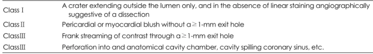
24명(0.6%)이었다. 본 연구에서는 관동맥 천공이 있었 던 24명의 환자들을 대상으로 본원 심혈관 센터 데이 터베이스와 의무기록을 분석하고 관동맥 조영술을 검토 하여 이들의 임상적, 혈관 조영술상의 특징 및 임상경 과에 대해 고찰하였다. 관동맥 천공은 Ellis 분류를 이 용하여 각각 ClassⅠ, Class Ⅱ, Class Ⅲ로 분류하였다 (Table 1)(Fig. 1).6)
결 과
관동맥 천공이 있었던 24명의 환자의 평균연령은 64±
11세였고, 남자가 16명(0.6%), 여자가 8명(0.7%)이 었다. 고혈압이 15명에서, 당뇨병이 6명에서, 흡연력이 6명에서, 그리고 고지혈증이 8명에서 관찰되었다. 안정 형 협심증 환자가 10명, 불안정형 협심증 환자가 6명, 그리고 급성 심근경색증 환자가 5명이었다. 첫 번째 관 동맥 중재술인 경우가 92%였고, 다혈관 질환은 62%에 서 관찰되었다(Table 2).
관동맥 천공이 있었던 혈관은 우 관동맥이 46%로 가 장 많았다. 병변 형태는 American College of Cardiol- ogy/American Heart Association(ACC/AHA)병변 분 류로 type B1가 4%, typeB2가 21%, type C가 75%로 복합 병변이 많았다. 완전 폐쇄 병변이 10명(42%), 심 Table 1. Ellis classification of coronary perforation
ClassⅠ A crater extending outside the lumen only, and in the absence of linear staining angiographically suggestive of a dissection
ClassⅡ Pericardial or myocardial blush without a≥1-mm exit hole ClassⅢ Frank streaming of contrast through a≥1-mm exit hole
ClassⅢ Perforation into and anatomical cavity chamber, cavity spilling coronary sinus, etc.
Fig. 1. Coronary angiograms demonstrating of coronary perforation:classⅠ(A), classⅡ(B) and class Ⅲ(C). Arrows show the site of perforation on coronary angiograms.
A B C
한 석회화 침착 병변이 10명(42%), 미만성 병변이 5명 (21%), 분지부 병변이 4명(17%), 굴곡이 심한 병변이 3명(13%)에서 관찰되었다 (Table 3).
관동맥 천공의 형태는 ClassⅠ이 11명, Class Ⅱ가 8명, Class Ⅲ가 5명이었다. Abciximab은 ClassⅠ천 공을 보였던 1예에서만 투여하고 있었다. 관동맥 천공
은 24명의 환자들 중 23명에서 관동맥 중재술 동안 확 인이 되었고, 나머지 1명은 관동맥 중재술 후 14시간 뒤에 확인되었으며 Class Ⅱ 관동맥 천공을 보였다.
관동맥 천공의 원인으로는 유도철선에 의한 경우가 11명(46%)으로 가장 높은 빈도를 보였으며, 풍선에 의 한 경우가 8명(33%), 회전 죽상반 절제술에 의한 경우 가 4명(17%), 스텐트에 의한 경우가 1명(4%)이었다.
전체 환자에서 시술별 관동맥 천공의 발생률은, 회전 죽 상반 절제술에 의한 경우가 153명 중 4명으로(2.6%) 가장 많았으며, 유도철선에 의한 경우가 3,782명 중 11명 (0.3%), 풍선에 의한 경우 3,653명 중 8명(0.2%), 그 리고 스텐트에 의한 경우가 2,344명 중 1명(0.04%)이 었다.
유도철선에 의한 관동맥 천공은 환자 11명 중 8명 (74%)에서 ClassⅠ천공이었고, 7명(63%)에서 완전 폐 쇄 병변의 시술도중 발생하였다. 시술에 쓰인 유도철선 의 종류는 floppy 유도철선이 7명(74%), intermediate 유도철선이 3명(27%), standard 유도철선이 1명(9%) 에서 사용되었고, floppy 유도철선을 사용한 경우는 5명 에서 친수성으로 코팅된 유도철선(hydrophilic coated guidewire)을 사용한 경우였다.
풍선에 의한 관동맥 천공 환자 8명에서 풍선 대 혈 관의 비(balloon to artery ratio)는 0.83~1.17이었고, 5명의 환자에서 16기압이상의 높은 압력으로 풍선 확 장을 시행한 후 발생하였다.
회전 죽상반 절제술에 의한 관동맥 천공은 기구 대 혈관의 비(burr to artery ratio)는 0.5~0.72이었고, 주 로 원위부 혈관에서, 심한 석회화 침착이 동반된 굴곡이 있는 병변에서 발생하였다. 또한 4명의 환자 중 3명에 서 Class Ⅲ 천공을 보였다(Table 4).
심낭 압전은 5명에서 관찰되었고, Class Ⅱ 천공이 2명, Class Ⅲ 천공이 3명이었다. 관동맥 천공의 원인 별로는 회전 죽상반 절제술 시술에 의한 경우가 3명, 풍 선 확장술에 의한 경우가 2명이었다. 심낭 압전을 보였 던 5명의 환자 모두에서 즉각적으로 성공적인 심낭천자 가 시행 되었고 이들 중 Class Ⅲ 천공을 보였던 3명의 환자들은 모두 응급 관동맥 우회로술을 시행 받았다 (Table 5). Class Ⅱ 천공을 보였던 2명의 환자들은 심 낭 천자 후 낮은 압력에서 10분간 풍선 확장술을 시행 한 후 혈관 조영술상 천공이 관찰되지 않았다. 심낭 압 전이 있었던 5명의 환자 모두에서 심낭 압전과 관련된 Table 2. Baseline clinical characteristics (n=24)
Age (yrs) 64±11 (range 38-83)
Male/Female 16 (67%)/8 (33%)
Diabetes mellitus 06 (25%)
Hypertension 15 (63%)
Smoking 06 (25%)
Hyperlipidemia 08 (33%) Clinical diagnosis
Acute myocardial infarction 05 (21%) Stable angina 10 (42%) Unstable angina 06 (25%) Non Q myocardial infarction 03 (13%) Prior myocardial infarction 06 (25%) Prior PCI* (any vessels) 02 (08%) Prior PCI* (target vessels) 02 (08%) Ejection fraction≤50% 05 (21%)
*:percutaneous coronary intervention
Table 3. Baseline angiographic characteristics (n=24) Lesion morphology
ACC/AHA lesion class
Type A 00 (00%)
Type B1 01 (04%)
Type B2 05 (21%)
Type C 18 (75%)
Total occlusion 10 (42%)
Calcification 10 (42%)
Diffuse lesion 05 (21%)
Ostial lesion 01 (04%)
Bifurcation 04 (17%)
Thrombus 02 (08%)
Acute angulation 03 (13%)
Target coronary artery
Left anterior descending artery 08 (33%) Left circumflex artery 05 (21%) Right coronary artery 11 (46%) AHA/ACC:American college of cardiology/American heart association
사망은 없었다.
관동맥 천공의 치료로는 24명 환자들 모든 환자에서 먼저 2~6기압으로 10분 이상 풍선 확장술을 시행하였다.
10분 후 추적 관동맥 조영술 결과 21명(88%)에서 관 동맥 천공의 소견이 더 이상 관찰되지 않았고, 이 환자 들은 시술 종료 후 중환자실로 옮겨 흉통 유무를 주기 적으로 확인하였고, 심전도 모니터링, 추적 혈청 심장 효소 검사 및 심 초음파 검사를 시행하였다. 환자들 중 스텐트에 의한 Class Ⅲ 관동맥 천공을 보였던 1명은 시술 종료 후 3시간 뒤 흉통이 지속되어 추적 관동맥 조영술을 시행한 결과, 조영제의 누출이 확인되어 PTFE (polytetrafluoroethylene) covered stent를 삽입하였 고 이후 관동맥 천공의 소견은 관찰되지 않았다(Fig. 2).
PTFE covered stent는 2예에서 시도되었으나 1예에 서는 근위부의 굴곡과 석회화로 인하여 통과에 실패 하여 응급 관동맥 우회로술을 시행 받았다. 회전 죽상 반 절제술 시술에 의한 Class Ⅲ 천공이 있었던 환자 3
명에서는 성공적인 심낭 천자에도 불구하고 심낭 압전 및 저혈압이 지속 또는 재발되어 응급 관동맥 우회로 술을 시행하였다.
관동맥 천공이 있었던 환자들에서 입원 기간 중 사망은 없었으며, 입원일수는 평균 13.9±12.7일이었고, Class Ⅲ 천공이 있었던 환자들이 ClassⅠ과 Class Ⅱ 천공이 있 었던 환자들보다 입원 일수는 의미 있게 길었다(ClassⅠ 28.4±22.3, Class Ⅲ 10.1±4.9, p<0.01).
고 찰
본 연구에서 경피적 관동맥 중재술에 따른 관동맥 천공 의 발생빈도는 0.6%였고, 관동맥 천공의 형태는 ClassⅠ 이 11명, Class Ⅱ가 8명, Class Ⅲ가 5명이었다. 천공 의 원인으로는 유도 철선에 의한 경우가 가장 높은 빈 도를 보였지만, 시술별 관동맥 천공의 발생률은 회전 죽 상반 절제술에 의한 경우가 가장 높았다. 또한 심낭 압 전이 있었던 환자 5명은 즉각적인 심낭 천자가 시행되 었고 24명의 관동맥 천공 환자들의 입원 기간 동안 사 망은 없었다.
관동맥 천공에 대한 국내의 보고는 유도철선에 의해 유발된 원위부 좌회선지 천공 환자에서 관동맥 내 poly- vinyl alcohol 주입에 의한 색전으로 치유된 1예와11) 관 동맥 천공 환자에서 autologous vein graft-coated st- ent로 성공적인 치료를 보인 1예12)의 보고 외에는 아직 없는 실정이다. 이에 본 연구는 경피적 관동맥 중재술을 받은 허혈성 심장질환 환자들에서 시술에 따른 관동맥 천공의 발생빈도와 임상적, 혈관 조영술상의 특징 및 이 들의 치료방침과 임상경과에 대한 체계적인 첫 번째 국 내 연구로서 의미가 있을 것으로 생각된다.
Table 4. Incidence and classification of coronary artery perforation by interventional modality
Classification Cause Incidence
ClassⅠ ClassⅡ ClassⅢ Guidewire 11/3782 (0.3%) 8 3
Balloon 08/3653 (0.2%) 3 4 1
Stent 01/2344 (0.04%) 1
Rotablator 04/0153 (2.6%) 1 3 ELCA 00/0039 (0%)
DCA 00/0014 (0%)
ELCA:excimer laser coronary angioplasty, DCA:dire- ctional coronary atherectomy
Table 5. Treatment and in-hospital outcomes of 24 pati- ents with coronary artery perforation
Class of perforation ClassⅠ ClassⅡ Class Ⅲ Pericardiocentesis,
n (%) 00 (0%) 02 (4%) 03 (13%) PTFE-covered stent,
n (%) 00 (0%) 00 (0%) 01 (04%) Urgent CABG,
n (%) 00 (0%) 00 (0%) 03 (13%) Death,
n (%) 00 (0%) 00 (0%) 00 (00%) In-hospital stay, days
(mean±SD) 10.6±6.1 10.5±5.2 26±25 PTFE:polytetrafluoroethylene, CABG:coronary artery bypass graft
Fig. 2. Coronary angiograms showing:(A) class Ⅲ coro- nary artery perforation by stent implantation:and (B) final result after deployment of the PTFE-coated stent with no further leakage.
A B
과거 문헌들에 의하면, 경피적 관동맥 풍선 확장술을 시행 받은 환자들에서 관동맥 천공의 빈도는 대략 0.1%
정도이며, 최근 회전 죽상반 절제술이나 종축 죽상반 절 제술 등과 같이 조직을 깎거나 제거하는 시술 중 관동 맥 천공의 발생빈도는 0.5~3%정도로 그 빈도가 풍선 확장술보다 다소 높은 편이라고 보고하고 있다.1-10) 본 연구에서의 경피적 관동맥 중재술 동안 발생한 관동맥 천공의 빈도는 0.6% 로서 여러 문헌들과 유사한 빈도 를 보인다.
풍선 확장술이나 스텐트 삽입술 중 관동맥 천공은 대 개 유도철선이나 풍선을 진행하는 도중 혈관 벽의 손상 으로 인하여 발생하거나 풍선의 파열 및 높은 압력으로 풍선을 확장 시킨 경우, 그리고 혈관의 직경보다 큰 풍 선을 사용한 경우에 발생한다.13-17) 본 연구에서 유도철 선에 의한 천공의 경우 친수성 코팅 유도철선(hydrophi- lic coated guidewire)에 의한 빈도가 높아서 시술 중 코팅 된 유도철선의 진행시 주의가 필요할 것으로 사 료된다. 그러나 Dixon 등은 유도철선에 의한 관동맥 천 공은 glycoprotein IIb/IIIa blocker를 투여하지 않는 한 대개는 심낭 압전과 같은 심각한 합병증은 발생되지 않 는다고 보고하고 있다.17) 본 연구 결과 역시 유도철선에 의한 관동맥 천공은 10분에서 20분 정도의 풍선 확장 술로 모두 좋은 결과를 보였다.
Cohen 등18)은 특히 회전 죽상반 절제술에 의한 관 동맥 천공은 관동맥 혈관 조영술상 병변 형태가 편심성 이거나, 병변의 길이가 10 mm 이상인 경우, 또는 혈관 의 굴곡이 있는 경우 더 잘 발생한다고 보고하고 있다.
또한 기구 대 혈관비(device to artery ratio)가 0.8이 상일 경우 천공의 빈도가 증가한다고 보고하고 있다.4) 본 연구에서의 회전 죽상반 절제술에 의한 관동맥 천공 의 경우도 4명 모두에서 병변의 길이가 10 mm 이상의 심한 석회화를 동반한 편심성 병변이었다. 또한 회전 죽 상반 절제술에 의한 관동맥 천공이 있었던 4명의 환자 들 중 3명(75%)에서 심낭 압전과 같은 중대한 합병증 의 발생을 초래하는 Class Ⅲ 천공이었고, 3명 모두 응 급 관동맥 우회술을 시행 받았다. 그러므로 회전 죽상반 절제술과 같은 조직을 깎거나 제거하는 시술 동안의 관 동맥 천공은 임상적으로 심각한 합병증을 초래할 수 있 으므로 혈관 병변의 형태 및 기구 대 혈관의 크기 비 등 을 세밀히 검토하여 시술에 임해야 할 것으로 사료된다.
관동맥 천공의 치료는 원인에 관계없이 우선적으로 비
수술적으로 천공된 부위를 막고 혈역동학적으로 환자를 안정화 시켜야 한다. 비수술적 치료로는 풍선을 이용하 는 방법,4)5)19)20) 스텐트를 삽입하는 방법,21-24) 심낭천 자,2) coil 색전술,21)25)26) 경도관 알코올 주입(transca- theter injection of polyvinyl alcohol form)11) 등이 있 으며, 그 외에도 protamine, 혈소판 수혈2) 등이 있다.
풍선을 이용할 때는, 혈관 밖으로 조영제가 분출되는 부위에 즉시 풍선을 위치시키고 2~6기압 정도의 낮은 압력으로 적어도 10분 이상 풍선을 확장해야 하며, 환 자가 장시간의 혈관 폐색을 견디지 못하거나 10분간의 풍선 확장에도 불구하고 천공부위가 불완전하게 막혔을 경우에는 관류형 풍선 카테타를 이용하여 15분에서 45 분 동안 다시 낮은 압력으로 풍선을 부풀리는 것이 필 요하다.2) 과거 문헌들은 풍선을 이용한 방법으로 관동 맥 천공 환자의 60~70%는 수술적 치료가 필요하지 않다고 보고하고 있는데4)6)20)21) 본 연구에서도 24명 의 환자들 중 21명(88%)에서 수술적 치료가 필요하 지 않았으며 다른 문헌들의 보고보다 높은 비수술적 치 료의 결과는 유도 철선에 의한 ClassⅠ과 같은 경한 관 동맥 천공이 많음에 기인하는 것으로 사료된다.
스텐트를 이용하는 방법에는 stent-vein allograft 나12)22) PTFE-covered stent가23)24) 이용되지만, 전 자는 기술적으로 시간을 요하므로 특히 혈역동학적으로 불안정한 환자들에게 이용하기는 부적절할 것으로 생각 된다. Class Ⅲ 관동맥 천공이 있었던 본원의 2예에서 PTFE-covered stent의 삽입을 시도하였고, 1예에서 는 다른 심각한 합병증 없이 성공적으로 천공된 부위를 막을 수 있었다. 그러나 다른 1예에서는 천공 부위가 석 회화와 굴곡이 심한 혈관의 원위부에 위치하여 스텐트 통과에 실패하여 응급 관동맥 우회로술을 시행하였다.
심낭 천자는 천공된 부위에 풍선을 위치시킨 후 심낭 내 출혈 및 심낭 압전 소견이 관찰되면 즉시 시행되어 져야 하며, 6~24시간 동안 배액을 시키면서 심낭 삼출이 지속되는지를 6~12시간마다 심 초음파를 이용하여 확 인하여야 한다. 만약 출혈이 지속되면 응급 관동맥 우회 로술을 고려해야 한다.2)
응급 관동맥 우회로술은 심한 허혈성 병변과 동반된 천공의 크기가 큰 경우나, 비수술적 치료를 해도 천공이 지속적으로 관찰되거나 혈역동학적으로 불안정한 경우에 시행되어야 한다. 대개 관동맥 천공 환자의 30~40%에 서 응급 관동맥 우회로술이 필요하였지만4)5)18) 최근에
는 PTFE covered stent의 등장으로 응급 관동맥 우회 로술의 역할이 감소되고 있는 추세이다.
유도철선에 의한 원위부 관동맥 천공에 있어서 특히, 혈관크기가 작거나 원위부에 위치하는 경우, 천공된 혈 관이 심근의 국한된 부위인 경우, 만성 완전 폐쇄 병변 이었던 경우 등과 같은 수술적 치료가 어려운 환자들에 서 최근에는 gelfoam을 이용한 coil 색전술이 시행되어 지고 있다.17)25)26)
본 연구는 후향성 연구로 관동맥 천공 환자들의 치료 에 있어서 체계적인 연구계획에 따른 것이 아니라는 제 한점을 가지고 있다. 또한 본 연구에서 종축 죽상반 절 제술에 의한 관동맥 천공은 관찰되지 않았는데, 이는 시 술 받은 환자 수가 많지 않았기 때문으로, 본 연구의 결 과가 종축 죽상반 절제술에 의한 관동맥 천공의 빈도를 대변할 수는 없을 것으로 사료된다.
결론적으로 경피적 관동맥 중재술에 따른 관동맥 천공 은 임상적으로 심각한 합병증을 초래하지만 심낭 천자 등과 같은 즉각적이고 적절한 치료가 행해진다면, 심지 어 Class Ⅲ 천공이나 심낭 압진이 생긴 경우라도 좋은 예후를 기대할 수 있을 것으로 사료된다.
요 약
배경 및 목적:
관동맥 천공은 경피적 관동맥 중재술의 드물지만 임 상적으로 심각한 문제를 초래하는 합병증 중 하나이다.
저자들은 경피적 관동맥 중재술을 받은 허혈성 심장질 환 환자들에서 시술에 따른 관동맥 천공의 발생빈도와 임상적, 혈관 조영술상의 특징 및 임상경과에 대해 알아 보고자 하였다.
방 법:
경피적 관동맥 중재술을 시술 받은 환자 3,782명 중 시술에 따른 관동맥 천공이 있었던 환자 24명(평균나 이:64(11세, 남:여=16:8)의 의무기록과 관동맥 혈 관 조영술 결과를 고찰하였다. 관동맥 천공은 Ellis의 분 류에 따라 분류하였다.
결 과:
경피적 관동맥 중재술에 따른 관동맥 천공은 환자 3,782 명 중 24명에서 발생하였으며, 발생빈도는 0.6%였다. 관 동맥 천공의 형태는 ClassⅠ이 11명, Class Ⅱ가 8명, Class Ⅲ가 5명이었다. 관동맥 천공은 우 관동맥 병변
의 시술(46%)과 만성 완전 폐쇄 병변의 시술(42%)동 안 가장 많이 발생하였다. 관동맥 천공의 원인으로는 유 도 철선에 의한 것이 11명(46%)으로 가장 높은 빈도 를 보였고 풍선에 의한 것이 8명으로 두 번째 높은 빈 도를 보였다. 시술별 관동맥 천공의 발생률은 회전 죽상 반 절제술에 의한 것이 157명 중 4명(2,6%)으로 가장 많았다. 심낭 압전은 5명의 환자에서 관찰되었고 회전 죽상반 절제술에 의한 경우가 4명으로 가장 많았다. 5 명의 심낭 압전을 보였던 환자 모두에서 심낭 천자가 시 행되었고 이들 중 Class Ⅲ 천공을 보였던 3명의 환자들 은 응급 관동맥 우회로술을 시행 받았다. 관동맥 천공이 있었던 환자들에서 입원 기간 중 사망은 없었으며 입원 일수는 Class Ⅲ 천공이 있었던 환자들에서 ClassⅠ과 Class Ⅱ 환자들보다 의미 있게 길었다.
결 론:
경피적 관동맥 중재술에 따른 관동맥 천공은 임상적 으로 심각한 합병증을 초래한다. 하지만 즉각적이고 적 절한 치료가 행해진다면, 심지어 Class Ⅲ 천공이나 심 낭 압진이 생긴 경우라도 좋은 예후를 기대할 수 있을 것으로 사료된다.
중심 단어:관동맥 질환;경피적 관동맥 혈관 확장술;관 동맥 죽상반 절제술.
REFERENCES
1) Gruberg L, Pinnow E, Flood R, Bonnet Y, Tebeica M, Wak- sman R, Satler LF, Pichard AD, Kent KM, Leon MB, Lindsay J Jr. Incidence, management, and outcome of coronary artery perforation during percutaneous coronary intervention. Am J Cardiol 2000;86:680-2.
2) Safian RD, Freed MS. The manual of interventional cardiol- ogy. 2nd ed. Birmingham: Physicians’ Press; 2001. p.423-30.
3) Dippel EJ, Kereiakes DJ, Tramuta DA, Broderick TM, Shi- mshak TM, Roth EM, Hattemer CR, Runyon JP, Whang DD, Schneider JF, Abbottsmith CW. Coronary perforation during percutaneous coronary intervention in the era of abciximab platelet glycoprotein IIb/IIIa blockade: an algorithm for pe- rcutaneous management. Catheter Cardiovasc Interv 2001;
52:279-86.
4) von Sohsten R, Kopistansky C, Cohen M, Kussmaul WG 3rd.
Cardiac tamponade in the “new device” era: evaluation of 6999 consecutive percutaneous coronary interventions. Am Heart J 2000;140:279-83.
5) Ajluni SC, Glazier S, Blankenship L, O’Neill WW, Safian RD.
Perforation after percutaneous coronary interventions: clin- ical, angiographic, and therapeutic observations. Cathet Ca- rdiovasc Diagn 1994;32:206-12.
6) Ellis SG, Ajluni S, Arnold AZ, Popma JJ, Bittl JA, Eigler NL, Cowley MJ, Raymond RE, Safian RD, Whitlow PL. Increa-
sed coronary perforation in the new device era: incidence, classification, management, and outcome. Circulation 1994;
90:2725-30.
7) Friedrich SP, Berman AD, Baim DS, Diver DJ. Myocardial perforation in the cardiac catheterization laboratory. Cathet Cardiovasc Diagn 1994;32:99-107.
8) van Suylen RJ, Serruys PW, Simpson JB, de Feyter PJ, Stra- uss BH, Zondervan PE. Delayed rupture of right coronary artery after directional atherectomy for bail-out. Am Heart J 1991;121:914-6.
9) Bittl JA, Ryan TJ Jr, Keaney JF Jr, Tcheng JE, Ellis SG, Isner JM, Sanborn TA. Coronary artery perforation during exci- mer laser coronary angioplasty. J Am Coll Cardiol 1993;
21:1158-65.
10) Saffitz JE, Rose TE, Oaks JB, Roberts WC. Coronary arter- ial rupture during coronary angioplasty. Am J Cardiol 1983;
51:902-4.
11) Yoo BS, Yoon J, Lee SH, Kim JY, Lee HH, Ko JY, Lee BK, Hwang SO, Choe KH. Guidewire-induced coronary artery perforation treated with transcatheter injection of polyvinyl alcohol form. Catheter Cardiovasc Interv 2001;52:231-4.
12) Chae JK, Park SW, Kim YH, Hong MK, Park SJ. Successful treatment of coronary artery perforation during angioplasty using autologous vein graft-coated stent. Eur Heart J 1997;
18:1030-2.
13) Kimbiris D, Iskandrian AS, Goel I, Bemis CE, Gehl L, Ow- ens J, Segal BL. Transluminal coronary angioplasty compli- cated by coronary artery perforation. Cathet Cardiovasc Di- agn 1982;8:481-7.
14) Cherry S, Vandormael M. Rupture of a coronary artery and hemorrhage into the ventricular cavity during coronary an- gioplasty. Am Heart J 1987;113:386-8.
15) Meier B. Benign coronary artery perforation during percut- aneous transluminal coronary angioplasty. Br Heart J 1985;
54:33-5.
16) Grollier G, Bories H, Commeau P, Foucault JP, Potier JC.
Coronary artery perforation during coronary angioplasty. Clin Cardiol 1986;9:27-9.
17) Dixon SR, Webster MW, Ormiston JA, Wattie WJ, Hammett CJ. Gelfoam embolization of a distal coronary artery guide- wire perforation. Catheter Cardiovasc Interv 2000;49:214-7.
18) Cohen BM, Weber VJ, Relsman M, Casale A, Dorros G. Co- ronary perforation complicating rotational ablation: the U S multicenter experience. Cathet Cardiovasc Diagn 1996;
(Suppl 3):55-9.
19) Holmes DR Jr, Reeder GS, Ghazzal ZM, Bresnahan JF, King SB 3rd, Leon MB, Litvack F. Coronary perforation after ex- cimer laser coronary angioplasty: the excimer laser coronary angioplasty registry experience. J Am Coll Cardiol 1994;
23:330-5.
20) Cowley MJ, Dorros G, Kelsey SF, van Raden M, Detre KM.
Acute coronary events associated with percutaneous transl- uminal coronary angioplasty. Am J Cardiol 1984;53:12C-6C.
21) Dorros G, Jain A, Kumar K. Management of coronary artery rupture: covered stent or microcoil embolization. Cathet Ca- rdiovasc Diagn 1995;36:148-54.
22) Caputo RP, Amin N, Marvasti M, Wagner S, Levy C, Giam- bartolomei A. Successful treatment of a saphenous vein graft perforation with an autologous vein-covered stent. Catheter Cardiovasc Interv 1999;48:382-6.
23) Ramsdale DR, Mushahwar SS, Mooris JL. Repair of coron- ary artery perforation after rotastenting by implantation of the Jostent covered stent. Cathet Cardiovasc Diagn 1998;45:
310-3.
24) Casella G, Werner F, Klauss V, Mudra H. Successful treatm- ent of coronary artery perforation during angioplasty using a new membrane-covered stent. J Invasive Cardiol 1999;11:
622-6.
25) Gaxiola E, Browne KF. Coronary artery perforation repair using microcoil embolization. Cathet Cardiovasc Diagn 1998;
43:474-6.
26) Thomas WJ, Moskowitz WB, Freedman A, Vetrovec GW, Goudreau E. Therapeutic embolization for unusual latroge- nic complications related to coronary revascularization. Ca- theter Cardiovasc Interv 1999;46:457-62.