submit.radiology.or.kr J Korean Soc Radiol 2011;65(6):569-572
569 INTRODUCTION
The carotid stump is a blind, pouch-like remnant of the oc- cluded internal carotid artery (ICA) at its proximal aspect.
When ischemic symptoms persist in a properly compensated ICA occlusion, the carotid stump is a potential source of mi- croemboli (1). The turbulence in the stump contributes to progressive atherosclerosis and thrombogenesis, and a subse- quent microembolism may occur through the anastomotic channel between the external carotid artery (ECA) and the distal ICA. Carotid stump syndrome has been reported in 7.5% of carotid endarterectomy series, which were treated by conventional methods, with surgical excision of the stump or
oversewing of the ICA origin (2). More recently, either the stump was excluded from the vascular lumen by placing a self-expandable stent graft (Wallgraft endoprosthesis; Boston Scientific, Natick, MA, USA; 3-5), or acutely near-occluded ICA was reopened anterogradely under proximal balloon pro- tection (6, 7). In our patient, our attempt to place a balloon- expandable stent graft for the exclusion of the stump resulted in some issues; a stent graft mounted on a balloon with a di- ameter of more than 7 mm is usually compatible with a shut- tle sheath of at least 7 Fr., was difficult to advance along the acute angle between the aortic arch and the carotid artery (8).
Furthermore, the considerable difference in diameter between the ECA and the common carotid artery (CCA) carries a risk
Case Report
pISSN 1738-2637
J Korean Soc Radiol 2011;65(6):569-572
Received July 1, 2011; Accepted August 27, 2011 Corresponding author: Ho Kyun Kim, MD
Department of Radiology, Catholic University of Daegu School of Medicine, 3056-6 Daemyeong 4-dong, Nam- gu, Daegu 705-718, Korea.
Tel. 82-53-650-4309 Fax. 82-52-650-4339 E-mail: [email protected]
This work was supported by research grants from the Catholic university of Daegu in 2011. Otherwise, the authors have no conflicts of interests and/or disclosure.
Copyrights © 2011 The Korean Society of Radiology
Carotid stump, the blind remnant of an occluded internal carotid artery, can be a potential source of microemboli, and warrants its exclusion from the vascular lu- men to prevent the recurrence of a microembolism. In a 69-year-old male with a symptomatic carotid stump and acute angle between left common carotid artery and aortic arch, a 7-Fr. shuttle sheath was scarcely placed into the left carotid ar- tery but the 7-mm-diameter stent-graft-loading balloon could not be inserted into the 7-Fr. shuttle sheath. With the mounting a stent graft over a 5-mm balloon, the balloon-expandable stent graft was unfolded. The self-expandable stent was placed over the stent graft, and an 8-mm balloon was subsequently expanded. Self-ex- panding stenting can be useful for troubleshooting in a case of device incompatibil- ity coming from the different calibers of the external and common carotid arteries for the successful exclusion of a symptomatic carotid stump.
Index terms Carotid Stump Microemboli Stent Graft
Self-Expandable Stent
Self-Expandable Stenting over a Stent Graft for the Exclusion of a Carotid Stump: Troubleshooting for Device Incompatibility
1목동맥 그루터기의 차단을 위한 인조혈관 스텐트시 부가적인 자가팽창성 스텐트의 설치: 기구부적합시의 문제 해결방법1
Sung Won Youn, MD
1, Ho Kyun Kim, MD
1, Jin Kook Do, MD
2, Young Whan Kim, MD
3Departments of 1Radiology, 2Neurology, Catholic University of Daegu School of Medicine, Daegu, Korea
3Department of Radiology, Keimyung University College of Medicine, Daegu, Korea
Self-Expandable Stenting over a Stent Graft for the Exclusion of a Carotid Stump
submit.radiology.or.kr
J Korean Soc Radiol 2011;65(6):569-572
570
Muscle Strength, grade 4), and new diffusion restrictions were found in both the ACA and left MCA territories. One week lat- er, right arm weakness recurred despite systemic hepariniza- tion and triple antiplatelet therapy (aspirin, 100 mg; clopido- grel, 75 mg; cilostazol, 200 mg). It was suspected that the stump was the source of the recurrent microembolism. At the initial session, an attempt to place a 9-Fr. shuttle sheath in the left CCA failed, but a 7-Fr. shuttle sheath (Flexor Tuohy-Borst Sidearm Introducers, Cook, Bloomington, IN, USA) was suc- cessfully placed. However, a stent-graft-loading balloon with a diameter of 7 mm could not be inserted into the 7-Fr. shut- tle sheath. At a second session, a 7-Fr. shuttle sheath was placed into the CCA under 5,000 units of systemic heparinization. An 0.018-inch SV guidewire (Boston Scientific, Natick, MA, USA) was passed into the ECA branch, and a Jostent peripheral stent graft (4-9 mm × 38 mm; Abbott Vascular, Rangendingen, Germany), which was mounted over a Savvy balloon (5 × 30- mm; Cordis, Miami, FL, USA), was placed into the left ECA- CCA. After balloon expansion at 6 Atm, the distal portion of the stent graft was fitted to the ECA, but the proximal portion of the stent graft was not fitted to the CCA lumen, and the contrast medium filled the stump. A self-expandable Precise of rupture during balloon expansion (9). We present a bailout
technique of sequential self-expandable stenting over the bal- loon-expandable stent graft for the exclusion of a carotid stump.
CASE REPORT
A 69-year-old man with a history of hypertension and dia- betes mellitus presented with dysarthria and right hemipare- sis. Diffusion-weighted magnetic resonance imaging (MRI) revealed multifocal embolic infarctions of the left anterior ce- rebral artery (ACA) and middle cerebral artery (MCA) terri- tory. Electrocardiography and echocardiography ruled out a cardioembolism. Digital subtraction angiography (DSA) re- vealed occlusion of both ICAs and the presence of a left ICA stump. The left ophthalmic artery and distal ICA were filled retrogradely from the ECA (Fig. 1). The left ACA was domi- nant and the right ACA territory was supplied by the left an- terior circulation. Three-dimensional rotational angiography revealed that the left ECA and the CCA were 5.5 and 7 mm in diameter, respectively. On the following day, the patient de- veloped left leg weakness (Medical Research Council Scale for
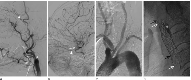
A B C D
Fig. 1. A 69-year-old man with a symptomatic carotid stump.
A. Left common carotid injection angiogram shows the occlusion of internal carotid artery (ICA) with stump (asterisk) and reconstituted ophthal- mic flow into the distal ICA (arrowhead).
B. Retrograde filling (arrow) from the ophthalmic artery (arrowhead) supplies anterior cerebral circulation.
C. Arch aortogram reveals severe elongation of the aortic arch, making an acute angle with the common carotid artery. The left ICA is not seen with the remaining stump (asterisk).
D. Native radiography after a stent graft reveals the self-expandable stent (black arrows) within the stent graft (white arrows) and the excluded stump with contrast stasis (asterisk).
Sung Won Youn, et al
submit.radiology.or.kr J Korean Soc Radiol 2011;65(6):569-572
571
fitted the ECA lumen but not the CCA lumen. Considering that fitting to the CCA lumen without a gap is essential for the exclusion of a stump, an additional larger-caliber balloon or stent may be regarded as the feasible option. We thought that there would be a risk of rupture (9) or recoiling with ad- ditional dilatation of a larger-caliber balloon. Therefore, a self- expandable stent was placed to impose a consistent radial force on the stent graft and to resist collapse in the area of exposure to external compression (10). Final post-stent angioplasty with an 8-mm balloon alone, which can easily enter a 7-Fr. shuttle sheath, was performed at the CCA segment, resulting in both successful apposition to the CCA lumen and exclusion of the stump.
With the exception of a microembolism, the risk of compli- cation was minimized, including vessel rupture during balloon expansion (9) and tissue necrosis from ECA sacrifice. In our patient, special care was taken to position the distal end of the stent graft to preserve the patency of as many ECA branches as possible. Only the superior thyroidal branch was sacrificed, and there was no associated complication. However, cerebral a microembolism via the retrograde collateral pathway of the ophthalmic artery was not prevented. The use of an endovas- cular protection device would have been helpful to prevent a microembolism, but it is likely that a microembolism was in- evitable before placement of the protection device as a result of the repeated struggle to place a shuttle sheath over the acute angle between the arch and the CCA.
In conclusion, sequential self-expandable stent fitting into the CCA lumen upon balloon-expandable stent-graft fitting of the ECA lumen is a useful troubleshooting protocol for the successful exclusion of a symptomatic carotid stump when a self-expandable stent graft is not available and there is incom- patibility between balloon loading stent graft and a 7-Fr. shut- tle sheath.
REFERENCES
1. Barnett HJ, Peerless SJ, Kaufmann JC. “Stump” on internal carotid artery--a source for further cerebral embolic isch- emia. Stroke 1978;9:448-456
2. Kumar SM, Wang JC, Barry MC, Farrell L, Kelly CJ, Fitzger- ald PH, et al. Carotid stump syndrome: outcome from sur- stent (9 × 40 mm; Cordis, Miami, FL, USA) was deployed
over the stent graft. After a Rider PTA catheter (8 × 40 mm;
Leventon S.A., Barcelona, Spain) was expanded at the CCA, the contrast medium no longer filled the stump. Post-stenting DSA showed no stent thrombosis, but sluggish flow was ob- served in the left MCA. Diffusion-weighted MRI revealed new cortical embolisms in the left ACA and MCA territory. A neurologic examination revealed aggravation of the hemipare- ses (right arm, grade 3; right leg, grade 4; left leg, grade 2). The patient was on continuous anticoagulation and triple anti- platelet therapy, and was last seen 6 months after the proce- dure, at which point he remained with no new events and all of his pareses had only slightly improved (all grade 4).
DISCUSSION
A carotid stump is a rare source of microemboli, and all other possible sources of emboli must be excluded before as- suming that the occluded ICA is the cause of symptoms (1-5).
In our patient, DSA excluded any atheromatous lesion in the aortic arch, CCA, and ECA and demonstrated a bulbous stump without trickling flow through ICA. Most of all, the patient was free from new events 6 months after exclusion of the stump.
Management of symptomatic ICA occlusion remains con- troversial. In a case with good retrograde filling of the distal ICA from the ophthalmic artery, anterograde ICA reopening may be a feasible option under balloon protection (6, 7). How- ever, exclusion of stump was performed because this lesion was regarded as chronic occlusion in stage and when a bal- loon protection device was not available.
For the exclusion of the stump, a self-expandable stent graft is the gold standard (3-5). Though a self-expandable stent graft was not available at the time of the procedure, we attempt- ed to place a balloon-expandable stent graft. It was neglected that a balloon-expandable stent can be deformed by compres- sion or cannot sufficiently be expanded, and in such a case, ad- ditional stenting is necessary. Contrary to the manufacturer’s recommendations specifying that the introducer for a Jostent peripheral stent graft should be two sizes larger than the usual introducer size for balloon catheter, a 7-mm balloon catheter loading stent graft could not enter the 7-Fr. shuttle sheath. We switched to a 5-mm balloon catheter loading stent graft, which
Self-Expandable Stenting over a Stent Graft for the Exclusion of a Carotid Stump
submit.radiology.or.kr
J Korean Soc Radiol 2011;65(6):569-572
572
with symptomatic carotid near occlusion. Interv Neuroradi- ol 2010;16:309-316
8. Macdonald S, Lee R, Williams R, Stansby G; Delphi Carotid Stenting Consensus Panel. Towards safer carotid artery stenting: a scoring system for anatomic suitability. Stroke 2009;40:1698-1703
9. Broadbent LP, Moran CJ, Cross DT 3rd, Derdeyn CP. Man- agement of ruptures complicating angioplasty and stent- ing of supraaortic arteries: report of two cases and a re- view of the literature. AJNR Am J Neuroradiol 2003;24:
2057-2061
10. Lownie SP, Pelz DM, Lee DH, Men S, Gulka I, Kalapos P. Ef- ficacy of treatment of severe carotid bifurcation stenosis by using self-expanding stents without deliberate use of angioplasty balloons. AJNR Am J Neuroradiol 2005;26:
1241-1248 gical management. Eur J Vasc Endovasc Surg 2001;21:
214-219
3. Naylor AR, Bell PR, Bolia A. Endovascular treatment of ca- rotid stump syndrome. J Vasc Surg 2003;38:593-595 4. Nano G, Dalainas I, Casana R, Malacrida G, Tealdi DG. En-
dovascular treatment of the carotid stump syndrome. Car- diovasc Intervent Radiol 2006;29:140-142
5. Carrafiello G, Delodovici ML, Piffaretti G, Castelli P. Endo- vascular treatment of carotid stump syndrome. Vasc En- dovascular Surg 2009;43:277-279
6. Suh DC, Kim JK, Choi CG, Kim SJ, Pyun HW, Ahn C, et al.
Prognostic factors for neurologic outcome after endovas- cular revascularization of acute symptomatic occlusion of the internal carotid artery. AJNR Am J Neuroradiol 2007;
28:1167-1171
7. Choi BS, Park JW, Shin JE, Lü PH, Kim JK, Kim SJ, et al. Out- come evaluation of carotid stenting in high-risk patients
목동맥 그루터기의 차단을 위한 인조혈관 스텐트시 부가적인 자가팽창성 스텐트의 설치: 기구부적합시의 문제 해결방법1
윤성원
1· 김호균
1· 도진국
2· 김영환
3목동맥 그루터기는 속목동맥이 막히고 남은 자루모양의 구조로 미세색전의 잠재적인 근본 원인이 될 수 있다. 따라서 재 발하는 미세색전증을 예방하기 위해 이를 혈관내벽으로부터 차단할 필요가 있다. 유증상 목동맥 그루터기를 가진 69세 남자에서 왼쪽 일반목동맥과 대동맥궁 사이의 각도가 예각을 이루어, 7 프렌치 왕복덮개를 왼쪽 목동맥에 간신히 설치하 였으나 7 mm 직경의 인조혈관 스텐트를 얹은 풍선도관이 7 프렌치 왕복덮개 내로 들어가지 못하였다. 5 mm 풍선도관 위에 인조혈관 스텐트를 얹은 다음, 풍선팽창성 인조혈관 스텐트를 설치하였다. 순차적으로 인조혈관 스텐트 위에 자가 팽창성 스텐트를 설치하고, 8 mm 풍선을 확장시켰다. 유증상 목동맥 그루터기를 차단하고자 할 때, 바깥목동맥과 일반 목동맥의 직경 차이로부터 비롯된 기구의 부적합성이 문제가 될 수 있으며, 이때 부가적인 자가팽창성 스텐트 설치로 이 문제를 해결한 경험을 보고하고자 한다.
대구가톨릭대학교 의과대학 1영상의학과학교실, 2신경과학교실, 3계명대학교 의과대학 영상의학과학교실