대한소화기학회지 2002;39:335 - 342
INTRODUCTION3)
Angiogenesis represents the formation of new capillaries from preexisting blood vessels. It is required for tumor
접수: 2001년 9월 1일, 승인: 2002년 3월 19일 연락처: 이완식, 501-757, 광주광역시 동구 학1동 8번지
전남대학교 의과대학 내과학교실 Tel: (062) 220-6292, Fax: (062) 225-8578 E-mail: [email protected]
growth as well as normal embryonic development.1,2 It has been reported that new vessel growth is continuously stimulated during tumor growth, and tumors with a luxuriant vasculature have a higher fraction of dividing cells than tumors with a poorly developed vasculature.3-5Namely, there is a direct correlation between the density of tumor vessels and an adverse prognosis in patients with solid tumors including gastric carcinoma.6 It is also known that tumor angiogenesis is mediated by several angiogenic factors such
Expressions of Angiopoietins-1 and -2 and their Relations with the Stage in Gastric Adenocarcinoma
Wan Sik Lee, M.D., Sung Kyu Choi, M.D., Jong Sun Rew, M.D., Sei Jong Kim, M.D., Young Jin Kim, M.D.*, Chang Soo Park, M.D.†, Gou Young Koh, M.D.‡, and Chan Choi, M.D.†
Departments of Internal Medicine, Surgery*, and Pathology†, Chonnam National University College of Medicine, Gwangju, Korea;
Department of Life Science‡, Pohang University of Science and Technology, Pohang, Korea
위선암에서 안지오포이에틴 1, 2의 발현 및 병기와 림프절 전이와의 관계
전남대학교 의과대학 내과학교실, 일반외과학교실*, 병리학교실†, 포항공과대학 생명과학과‡
이완식·최성규·유종선·김세종·김영진*·박창수†·고규영‡·최 찬†
목적: 종양의 침범 과정에서 혈관 형성은 종양의 성장에 필수적이며, 많은 연구들에의해 혈관 형성 과정이 종양의 성장과 발달, 전이에 중요한 역할을 함이 밝혀졌다. 지금까지 발견된 혈관 형성 촉진인자로는 혈관내피 성장인자(VEGF), 혈소판 유발 생장촉진인자(PDGF) 등이 있다. 안지오포이에틴 1 (Ang-1)과 이에 길항작용을 하는 안지오포이에틴 2 (Ang-2)는 최근 발견된 혈관 형성 촉진인자로 혈관내피세포에 있는 Tie-2 수용체에 작용한다. Ang-1은 혈관내피세포의 Tie-2 수용체를 활 성화시켜 주변부의 평활근세포 및 지지세포들의 집적과 상호작용을 유도하며 Ang-2는 이러한 혈관 주위 지지 세포들과 혈관내피세포 간의 결속력을 약화시켜 신생 혈관 형성을 촉진시킨다. 위선암의 침윤도 및 전이능은 암종의 혈관 형성능 과 밀접한 관계가 있다고 밝혀져 있다. Ang-1과 Ang-2는 이러한 혈관 형성에 관여하는 인자로서 위선암의 악성도와 전이 능과의 관계에 대해 연구하였다. 대상 및 방법: 53예의 조기 위암과 55예의 진행성 위암을 포함한 108예를 대상으로 Ang-1과 Ang-2에 대해 면역조직화학적 염색을 시행하였으며 진행성 위암의 경우 원발 병소와 함께 전이된 림프절에서도 Ang-1과 Ang-2의 발현을 조사하였다. 결과: Ang-1과 Ang-2의 발현은 위선암의 침윤 깊이(T-병기) 및 림프절 전이(N-병기) 정도에 비례하여 높게 나타났다(P<0.001). Ang-1, Ang-2 모두 조기 위암에 비해 진행된 위암에서 더욱 강하고 광범위 한 발현을 보였다(P<0.001). 림프절에 전이한 선암세포들이 원발 병소의 선암세포보다 더 강한 발현 양상을 보였다 (P<0.001). 결론: 이상의 결과로 Ang-1과 Ang-2는 위선암의 침윤 및 전이에 중요한 역할을 할 것으로 생각된다. (Korean J Gastroenterol 2002;39:335-342)
색인단어: 위선암, 혈관 형성, 안지오포이에틴(Angiopoietin), 전이
대한소화기학회지: 제39권 제5호, 2002
as vascular endothelial growth factor, platelet-derived growth factor, and transforming growth factor. Thus, they have crucial roles in development, invasion, and metastasis of malignant cells.
Angiopoietins are a novel family of angiogenesis me- diators that serve as ligands for the vascular endothelial Tie-2 receptor, a member of a family of receptor tyrosine kinases. Angiopoietin-1 (Ang-1) and angiopoietin-2 (Ang-2) specifically bind to Tie-2. It is predominantly expressed in endothelial cells and upregulated in microvessels of malignant tumors. Tie-2 signalling mediates downstream angiogenic events including the stabilization of neovessels.7 Proper regulation of Ang-1 and Ang-2 is absolutely required for normal vascular development. They seem to regulate vascular remodelling and endothelial cell interactions with supporting pericytes and smooth muscles.
Gastric adenocarcinoma causes major health problems and is the second most common fatal carcinoma worldwide.
Moreover, it is the most common neoplasm in the Far East and South America.8 Therapeutic strategy for gastric carcinoma is usually determined by the disease status especially by the presence and the grade of positive regional lymph nodes.9,10
Ang-1 and Ang-2 are involved in the angiogenetic process of the various neoplasms including astrocytomas and thyroid tumors.11,12 There have been a few reports about angio- poietins and their effects on gastric adenocarcinoma; their expression pattern in the tumor cells, and whether they are responsible for the progression of gastric adenocarcinoma.
Since angiogenesis is critical for tumor invasion and metastasis, we hypothesized that Ang-1 and Ang-2 would have effects on the invasion and metastasis of gastric adeno- carcinoma. Thus, we carried out this study to obtain evidence for our hypothesis.
MATERIALS and METHODS
1. Materials 1) Study population
One hundred and eight patients who underwent surgery for gastric adenocarcinoma from September 1997 to April 2000 at Chonnam National University Hospital were selected.
These include 53 cases of early gastric carcinoma (EGC) and 55 cases of advanced gastric carcinoma (AGC). To each
specimen of AGC, corresponding metastatic lymph node specimen was also selected as a metastatic reference of a given case. The selection of the cases was based on the availability of formalin-fixed and paraffin-embedded blocks, the accessibility of the patient data including sex, age, and stage of the disease, and the follow-up information. The patients comprised of 35 men and 18 women in EGC and 39 men and 16 women in AGC. The mean age was 57 in EGC and 59 in AGC.
2) Characteristics of cases
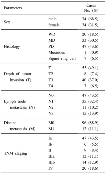
Histologic differentiation of the cases and distribution of cases according to the TNM staging system were as follows (Table 1).
Table 1. Clincopathological Parameters of 108 Patients with Gastric Adenocarcinoma
Parameters Cases
No. (%)
Sex male
female
74 (68.5) 34 (31.5)
Histology
WD MD PD Mucinous Signet ring cell
20 (18.5) 33 (30.5) 47 (43.6) 1 (0.9) 7 (6.5)
Depth of tumor invasion (T)
T1 T2 T3 T4
53 (49.1) 8 (7.4) 40 (37.0) 7 (6.5)
Lymph node metastasis (N)
N0 N1 N2 N3
47 (43.5) 35 (32.4) 11 (10.2) 15 (13.9) Distant
metastasis (M)
M0 M1
96 (88.9) 12 (11.1)
TNM staging
Ia Ib II IIIa IIIb IV
47 (43.5) 6 (5.5) 9 (8.4) 12 (11.1) 14 (12.9) 20 (18.6) WD, well differentiated; MD, moderately differentiated;
PD, poorly differentiated.
336
이완식 외 7인. 위선암에서 안지오포이에틴 1, 2의 발현
2. Methods
1) Immunohistochemistry
Antibodies for Ang-1 and Ang-2 were obtained from Regeneron Pharmaceuticals, Inc. Immunohistochemical staining was performed using the Micro-Probe staining system (Fisher Scientific, Pittsburgh, PA, USA) based on capillary action. Paraffin sections were immunostained with polyclonal synthetic antibodies for the Ang-1 and Ang-2 (Regeneron, Tarrytown, NY, USA) by the avidin-biotin peroxidase complex methods. Sections were deparaffinzed and heated in microwave oven for 7 min to retrieve the antigens and then immersed in 0.6% hydrogen peroxide for 7 min to block the endogenous peroxidase activity. Ang-1 and Ang-2 antibodies were used at concentrations of 1:100, and then incubated with tissues for 12 hours at room temparature. Anti-mouse Ig G (Sigma, St. Louis, MO, USA) labeled with biotin was used as a secondary antibody. After multiple rinses with universal buffer, the streptavidin-alkaline phosphatase detec- tion system (Biomeda, Foster, CA, USA) was applied for 7 min. As a final step, the slides were immersed for 7 min with the enzyme substrate, 3 amino-9-ethylcarbazole (AEC, Sigma). Then, the slides were counterstained with hematoxylin solution for 1 min (Research Genetics). After dehydration, the tissue was sealed with a universal mount (Research Genetics).
2) Interpretation of the immunohistochemical staining The slides were interpreted by two observers without knowledge of the clinical outcomes. The slides that caused different interpretation were reviewed with special interests and eventually reached a full agreements.
Results for the expression of the Ang-1 and Ang-2 were based on both the degree of the staining intensity and percentage of the area in which the postive result was detectable. The staining intensity was scored as 0 for the case of negative staining, 1 for the case of weak staining, 2 for the case of moderate staining, 3 for the case of strong staining. Extent of staining was scored as 0 for the case less than 25% positivity, 1 for the case of 25-50% positivity, 2 for the case of 50-75% positivity, and 3 for the case over 75% positivity.
3) Statistical analysis
All statistical analyses performed in this study were based
on the computerized statistical program, SPSS version 10.0 (SPSS, Chicago, IL, USA). The Spearman's correlation coefficient was used to analyze the staining pattern of the Ang-1 and Ang-2 in gastric adenocarcinoma with reference to the TNM staging system. The relationship between the degree of gastric adenocarcinoma differentiation and expression of the Ang-1 and Ang-2 was analysed by Kruskal-Wallis test. Mann-Whitney U test was used to seek the difference in expression of the Ang-1 and Ang-2 between the EGC and AGC. In AGC, primary sites and metastatic lymph nodes were saparately analysed for the expression of Ang-1 and Ang-2 by the Wilcoxon Signed Ranks test.
RESULTS
1. Immunohistochemical staining of Ang-1 Ang-1 was detected in the 99 of 108 gastric adenocar- cinoma tissues. Positve staining for Ang-1 was observed predominantly in the cytoplasm of the tumor cells (Fig. 1, Fig. 3). Ang-1 was also detected in the blood vessels around the endothelium. Normal glandular portion of the specimens, especially mucosal epithelial cells were frequently stained for the angiopoietins.
In particular, tumor emboli within the dilated vessels were usually stained for Ang-1, and intensity of the staining was stronger than the main tumor masses.
Fig. 1. Expression of angiopoietin 1. (A) A case of early gastric adenocarcinoma that is not stained for angiopoietin 1 antibody (H&E stain, ×200). (B) Advanced gastric adenocarcinoma of poorly differentiated type reveals a strong and extensive immunostaining pattern for angiopoietin-1 (immunohistochemical stain, ×200).
337
A B
The Korean Journal of Gastroenterology: Vol. 39, No. 5, 2002
Fig. 3. Expression of angiopoietin-1 in primary tumor and metastatic lymph node. (A) Advanced gastric adenocarcinoma of well differentiated type reveals strong and extensive immunostaining pattern for angiopoietin-1 (immunohistochemical stain, ×200).
(B) Metastatic lymph nodes reveal strong and extensive immunostain- ing pattern for angiopoietin-1 (immunohistochemical stain, ×200).
2. Immunohistochemical staining of Ang-2 Ang-2 was detected in 100 of 108 gastric adenocar- cinoma tissues. Ang-2 was predominantly observed in the cytoplasm of the tumor cells (Fig. 2, Fig. 4). Ang-2 shows higher activity in the tumor emboli of the peritumoral vascular spaces than Ang-1.
In gastric adenocarcinoma, no significant difference was observed between Ang-1 and Ang-2 with regard to their staining intensity and extent.
Fig. 2. Expression of angiopoietin-2. (A) A case of early gastric adenocarcinoma that is not stained for angiopoietin 2 antibody (immunohistochemical stain, ×200). (B) Advanced gastric adenocarcinoma of poorly differentiated type reveals a medium intensity and medium extent immunostaining pattern for angiopoietin-2 (immunohistochemical stain, ×200).
Fig. 4. Expression of angiopoietin-2 in primary tumor and metastatic lymph node. (A) Advanced gastric adenocarcinoma of moderately differentiated type reveals a strong and medium extent immunostaining pattern for angiopoietin-2 (immunohistochemical stain, ×200).
(B) Metastatic lymph nodes reveal a strong and extensive immunostain- ing pattern for angiopoietin-2 (immunohistochemical stain, ×200).
Table 2. Correlation between the Expression of Angio- poietins-1, -2 and the Pathologic Type of the Gastric Adenocarcinoma (Kruskal-Wallis Test)
Pathologic type No. Mean rank
Ang-1
WD MD PD SIGNET Total
20 33 47 7 107
55.67 56.09 52.30 50.79
Ang-2
WD MD PD SIGNET Total
20 33 47 7 107
59.60 55.73 51.26 48.29
%Ang-1
WD MD PD SIGNET Total
20 33 47 7 107
55.92 56.92 51.32 54.14
%Ang-2
WD MD PD SIGNET Total
20 33 47 7 107
54.88 55.82 53.02 49.50
WD, well differentiated; MD, moderately differentiated; PD, poorly differentiated. Ang-1, intensity of the staining for Ang-1;
Ang-2, intensity of the staining for Ang-2; %Ang-1, percentage of positivity at each tumor cell mass for Ang-1; %Ang-2, percentage of positivity at each tumor cell mass for Ang-2.
Mucinous type was not included because of statistical insignificance.
338
A B
A B
A B
Lee, et al. Expressions of Angiopoietins-1 and -2 in gastric adenocarcinoma
3. Angiopoietin-1,-2 and differentiation of gastric adenocarcinoma
There were no statistical correlations between the expression of the Ang-1 or 2 and the degree of differentiation or cell type of gastric adenocarcinoma (Table 2).
4. Angiopoietin-1, -2 and TNM staging
Both Ang-1 and Ang-2 showed enhanced staining pattern as the T factor advanced. There were significant correlations between the depth of invasion and the expression of Ang-1 and Ang-2. Similar results were demonstrated in the N factor of gastric adenocarcinoma. As the N factor advanced, the primary tumor showed stronger reaction for the Ang-1 and Ang-2. For the M factor, both Ang-1 and Ang-2 showed stronger staining pattern in the primary sites of the tumor undergoing distant metastasis, but there were not significant correlations as the cases of T and N factor (Table 3).
Table 3. Correlation between the Expression of Angio- poietins and the TNM Staging System in Gastric Ade- nocarcinoma (Spearman's Correlation Coefficient)
Ang-1 Ang-2 %Ang-1 %Ang-2
T factor .481* .349* .404* .282*
N factor .449* .406* .417* .389*
M factor .225† .190† .193† .146*
Ang-1, intensity of the staining for Ang-1; Ang-2, intensity of the staining for Ang-2; %Ang-1, percentage of positivity at each tumor cell mass for Ang-1; %Ang-2, percentage of positivity at each tumor cell mass for Ang-2.
* Correlation is significant at the .05 level (2-tailed).
†Correlation is significant at the .01 level (2-tailed).
5. Immunohistochemical staining of Ang-1, -2 in EGC and AGC
Expression rate of Ang-1 and Ang-2 was much higher in the AGC than in the EGC on the basis of their staining intensity and extent (Table 4, Fig. 1, Fig. 2).
6. Expression of Ang-1, -2 at the metastatic lymph nodes of gastric adenocarcinoma compared with their primary sites
Tumor cells in the metastatic lymph nodes showed a stronger and more extensive staining pattern than their primary foci (Table 5, Fig. 3, Fig. 4).
Table 4. Expression of Angiopoietins in Early Gastric Adenocarcinoma and Advanced Gastric Adenocarcinoma (Mann-Whitney U Test)
N Mean rank Significance (2-tailed)
Ang-1
EGC AGC Total
53 55 108
39.18 69.26
.000
Ang-2
EGC AGC Total
53 55 108
42.70 65.87
.000
%Ang-1
EGC AGC Total
53 55 108
41.98 66.56
.000
%Ang-2
EGC AGC Total
53 55 108
45.12 63.54
.001
EGC, early gastric carcinoma; AGC, advanced gastric carcinoma; Ang-1, intensity of the staining for Ang-1;
Ang-2, intensity of the staining for Ang-2; %Ang-1, percentage of positivity at each tumor cell mass for Ang-1;
%Ang-2, percentage of positivity at each tumor cell mass for Ang-2.
Table 5. Comparision of the Expressions Level for Angio- poietins at the Primary and Metastatic Sites of Advanced Gastric Carcinoma (Wilcoxon Signed Ranks Test)
No. Mean rank Significance (2 tailed)
LAng-1>Ang-1* 21 15.67 .003
LAng-2>Ang-2† 29 21.40 .001
%LAng-1>%Ang-1‡ 23 16.67 .001
%LAng-2>%Ang-2§ 31 20.35 .001 Ang-1, intensity of the staining for Ang-1; Ang-2, intensity of the staining for Ang-2; %Ang-1, percentage of positivity at each tumor cell mass for Ang-1; %Ang-2, percentage of positivity at each tumor cell mass for Ang-2.
* Case that shows stronger intensity for Ang-1 at metastatic lymph nodes than at primary sites.
† Case that shows stronger intensity for Ang-2 at their metastatic lymph nodes than at primary sites.
‡ Case that shows more extensive staining pattern for Ang-1 at their metastatic lymph nodes than at primary sites.
§ Case that shows more extensive staining pattern for Ang-2 at their metastatic lymph nodes than at primary sites.
339
대한소화기학회지: 제39권 제5호, 2002
DISCUSSION
Metastasis of tumor is a complex process. The individual tumor cell is separated from the homeotypic environment brought about by the dysfunction of adhesion molecules.
Then, the tumor cell invades into the vascular system, and is arrested in the capillary beds. Subsequently, it adheres to the vessel walls, and extravasates again to the distant organ, finally resulting in angiogenesis and completing the process of metastasis. This complex process involves many growth factors, cytokines and adhesion molecules including cadherin, CD44, nm23 gene, matrix metalloproteinase-2, and plasminogen activator type I.13
In the process of tumor invasion, angiogenesis is essential for tumor growth. Many studies have shown that angiog- enesis plays an important role in the growth, progression, and metastasis of solid tumors.
Conversion of endothelial cell organization into functioning vessels is an essential early developmental process and is central to pathological process such as tumor formation.
Endothelial cell proliferation and organization are under regulation of ligands signaling through endothelial-cell- specific trans-membrane receptor tyrosine kinases such as endothelial growth factor receptor 1 and 214,15 and the Tie-1 and Tie-2 receptors.16
The recent discovery of Ang-1 and its antagonist Ang-2 has provided insight into the molecular and cellular mechanism of blood vessel formation.17,18 Ang-1 and Ang-2 share about 60% amino acid sequence and bind to the enthothelial cell tyrosine kinase receptor, Tie-2 with similar affinity.17,18 Ang-1 mRNA is mainly present in periendothelial cells including vascular smooth muscle cells,17,19 and Ang-2 mRNA is mainly present in endothelial cells.18 Ang-1 remodels primitive vessels and helps to maintain and stabilize mature vessels by promoting interaction between endothelial cells and surrounding cells.
In contrast, Ang-2 is expressed mainly at the site of vascular remodeling, blocking the constitutive stabilizing action of Ang-1. Ang-2 expression reduces matrix contacts and interactions of support cells. These endothelial cells are necessary for neovascularization.20 Ang-1 and the tyrosine kinase receptor Tie-2 are required for normal vascular development of mice. They are essential for myocardium differentiation and recruitment of smooth muscle cells and pericytes.21 The Tie-2 receptor is upregulated in microvessels
of malignant tumors.22 Additionally Ang-1 is widely expressed in many adult and embryonic tissues, whereas Ang-2 is expressed only in adult tissues such as ovaries, uterus, and placenta.23
Tumor angiogenesis is closely related to the severity of gastric adenocarcinoma.24 Namely, the extent of tumor vas- cularization correlates with prognosis and hematogenous metastasis in gastric adenocarcinoma.25 In addition, lymph node metastasis shows significant correlation with the vascularity of the tumor.26 Tumor suppressor gene, p53, plays an important role in controlling tumor angiogenesis by regulating the expression of vascular endothelial growth factor which is an angiogenic inducer.27In the early stage of gastric adenocarcinoma, angiogenesis is an independent factor that impacts on lymph node metastasis.28
Angiopoietin is ralated to the angiogenesis of various malignancy. In astrocytoma, Ang-1 mRNA is localized in tumor cells and Ang-2 mRNA is detected in endothelial cells of hyperplastic and non-hyperplastic vessels by in situ hybridization experiments.11
In our result, angiopoietins were mainly expressed at the cytoplasm of the tumor cells in gastric adenocarcinoma.
Although there were no stastical significances, angiopoietins were more strongly expressed in the well differentiated adenocarcinoma that mimics normal mucosal glands than in the poorly differentiated adenocarcinoma. Angiopoietins were rarely detectable in the compact hypercellular region which had scanty cytoplasm. On the other hand, most of the tumor emboli revealed unexceptionally strong immunoreactivity.
Ang-1 and Ang-2 were also expressed in blood vessels and its supporting cells. Expression of the Ang-1 and Ang-2 was also detected in the normal mucosal gland and surface epithelial cell. In blood vessels, periendothelial smooth muscle cells as well as endothelial cells showed positive reaction for angiopoietin expression. At lymph nodes, angiopoietins were positive at the germinal center.
Ang-1 and Ang-2 were more strongly and extensively expressed in the AGC than in the EGC, and the staining patterns became stronger and more extensive as the stage went higher. In AGC, lymph node metastasis of the primary adenocarcinoma showed stronger and more extensive express- ion for both the Ang-1 and Ang-2 than their primary foci.
These findings suggest that Ang-1 and Ang-2 are associated with the angiogenic process required for invasion and metastasis of gastric adenocarcinoma. Moreover, the fact 340
이완식 외 7인. 위선암에서 안지오포이에틴 1, 2의 발현
that lymph node metastasis is closely related to the angiogenesis of the gastric malignancy suggests that the expression of angiopoietins can be regarded as a marker of the distant metastasis of gastric adenocarcinoma and may predict its poor prognosis.
Until recently, there was only one published data about the Ang-2 and its significance in tumor angiogenesis of the gastric carcinoma.29 In our study, distant metastatic foci were included for detecting expressions of both Ang-1 and Ang-2. Ang-1 was also closely related to the progression and metastasis of gastric adenocarcinoma. These findings might indicate the concurrent and sequential contribution of Ang-1 and Ang-2 for the tumor angiogenesis of gastric carcinoma. We believe that Ang-2 is necessary for neo- vascularization and Ang-1 is required to stabilize it.
As theorized previously, angiopoietins, which thought to be involved in the angiogenesis of the embryo and the some malignancies, have important roles in the progression and metastasis of the gastric adenocarcinoma. This study does not include long term follow-up data about the patients' survival because relatively recent cases were selected. Thus, further clinical follow-up should be needed to determine the role of Ang-1 and Ang-2 for the prognosis of gastric adeno- carcinoma. In addition, although we used only imunohisto- chemical staining method, quantitative data of RNA expression by using northern blot analysis would guarantee more profound evidence. To obtain more evidence for angiopoietins' contribution to the invasion and metastasis of gastric adenocarcinoma, in vivo experiments by transfection of cDNA of Ang-1 and Ang-2 into cell line of low- metastatic potential would be necessary. Finally, Ang-1 and Ang-2 antagonists will be helpful in intervening the angiogenic process of gastric adenocarcinoma.
This study demonstated that angiopoietin 1 and 2 which have major role in the embryogenesis and physiological vascular formation are related to the progression of gastric carcinoma. These findings were based on the observations that angiopoietin 1 and 2 are expressed more strongly and extensively in advanced gastric carcinoma than in early gastric carcinoma. Furthermore, angiopoietin 1 and 2 seemed to participate in the metastatic process of gastric carcinoma because their expression is enhanced at the metastatic lymph node foci. These results show that Ang-1 and -2 have important roles in the invasion and metastasis of gastric adenocarcinoma.
SUMMARY
Background/Aims: In the process of tumor invasion, angiogenesis is prerequisite for tumor growth. Many studies have shown that angiogenesis plays an important role in the growth, progression, and metastasis of solid tumors.
Recently, several angiogenic factors, angiopoietin 1 (Ang-1) and its naturally occuring antagonist angiopoietin 2 (Ang-2), have been identified. They are novel ligands that bind to the Tie-2 receptor on endothelial cells. Ang-1 activates Tie-2 receptor on endothelial cells to promote recruitment and interaction with support cells such as pericytes and smooth muscle cells. In contrast, Ang-2 reduces matrix contacts and interactions of support cells with endothelial cells, which are necessary for the neovascularization process to occur.
Methods: In order to investigate the role of Ang-1 and Ang-2 in invasion and metastasis of the adenocarcinoma of the stomach, 108 cases of gastric adenocarcinomas were selected. These cases include 53 early gastric carcinoma (EGC) and 55 advanced gastric carcinoma (AGC). The specimens were stained with the Ang-1 and Ang-2 antibodies by immunohistochemical staining method. Results: The expression level of the Ang-1 and -2 is statisticaly correlated with the depth of invasion (T factor) and the lymph node metastasis (N factor) (P<0.001). Both Ang-1 and Ang-2 were more strongly and extensively expressed in AGC than in EGC (P<0.001). Carcinoma cells that metastsized to lymph nodes showed a stronger and more extensive staining pattern than their primary counter part of adenocarcinoma (P<0.001). Conclusions: These results indicate that both Ang-1 and Ang-2 are important in invasion and metastasis of gastric adenocarcinoma.
Key words: Gastric adenocarcinoma, metastasis, Neovas- cularization. Angiopoietin
REFERENCES
1. Folkman J. What is the evidence that tumors are angiogenesis dependent? J Natl Cancer Inst 1990;82:4-6.
2. Folkman J, Shing Y. Angiogenesis. J Biol Chem 1992;267:
10931-10934.
3. Viglietto G, Maglione D, Rambaldi M, et al. Upregulation of vascular endothelial growth factor (VEGF) and down regulation of placenta growth factor (PIGF) associated with 341
The Korean Journal of Gastroenterology: Vol. 39, No. 5, 2002
malignancy in human thyroid tumors and cell lines.
Oncogenes 1995;11:1569-1579.
4. Kandel J, Bossy-Wetzel E, Radvanyi F, Klagsbrun M, Folkman J, Hanahan D. Neovascularization is associated with a switch to the export of bFGF in the multistep development of fibrosarcoma. Cell 1991;66:1095-1104.
5. Lyng H, Skretting A, Rofstad EK. Blood flow in six human melanoma xenograft lines with different growth charac- teristics. Cancer Res 1992;52:584-592.
6. Tanigawa N, Amaya H, Matsumura M, et al. Extent of tumor vascularization correlates with prognosis and hematogenous metastasis in gastric carcinomas. Cancer Res 1996;56:
2671-2676.
7. Stewart RJ, Marsden PA. Angiopoietins-1 and -2:
Chromosomal localization and differential expression in human prostate cancer. J Urol 1999;161(suppl):131S.
8. Dupont JB Jr, Lee JR, Burton GR, Cohn I Jr.
Adenocarcinoma of the stomach: review of 1497 cases.
Cancer 1978;41:941-947.
9. Maruyama K, Gunven P, Okabayashi K, Sasako M, Kinoshita T. Lymph node metastasis of gastric cancer. General pattern in 1931 patients. Ann Surg 1989;210:596-602.
10. Jatzko GR, Lisborg PH, Denk H, Klimpfinger M, Stettner HM. A 10-year experience with Japanese-type radical lymph node dissection for gastric cancer outside of Japan. Cancer 1995;76:1302-1312.
11. ZagZag D, Hooper A, Friedlander DR, et al. In situ expression of Angiopoietins in astrocytomas identifies angiopoietin-2 as an early marker of tumor angiogenesis. Exp Neurol 1999;159:391-400.
12. Bunone G, Vigneri P, Mariani L, et al. Expression of angiogenesis stimulators and inhibitors in human thyroid tumors and correlation with clinical pathological features. Am J pathol 1999;155:1967-1976.
13. Chan AO, Luk JM, Hui WM and Lam SK. Molecular biology of gastric carcinoma:from laboratory to bedside. J Gastroenterol Hepatol 1999;14:1150-1160.
14. Fong GH, Rossant J, Gertsenstein M, Breitman ML. Role of the Flt-1 receptor tyrosine kinase in regulating the assembly of vascular endothelium. Nature 1995;376:66-70.
15. Shalaby F, Rossant J, Yamaguchi TP, et al. Failure of blood-island formation and vasculogenesis in Flk-1-deficient mice. Nature 1995;376:62-66.
16. Sato TN, Tozawa Y, Deutsch U, et al. Distinct roles of the receptor tyrosine kinase Tie-1 and Tie-2 in blood vessel formation. Nature 1995;376:70-74.
17. Davis S, Aldrich TH, Jones PF, et al. Isolation of angiopoietin-1, a ligand for TIE2 receptor, by secretion trap expression cloning. Cell 1996;87:1161-1169.
18. Maisonpierre PC, Suri C, Jones PF, et al. Angiopoietin-2, a natural antagonist for Tie-2 that disrupts in vivo angiogenesis.
Science 1997;277:55-60.
19. Suri C, Jones PF, Patan S, et al. Requsite role of angiopoietin-2, a ligand for the TIE2 receptor, during embryonic angiogenesis. Cell 1996;87:1171-1180.
20. Folkman J, D'Amore PA. Blood vessel formation: what is its molecular basis? Cell 1996;87:1153-1155.
21. Dumont DJ, Gradwohl G, Fong GH, et al. Dominant-negative and targeted null mutations in the endothelial receptor tyrosine kinase, Tek reveal a critical role in vasculogenesis of the embryo. Genes Dev 1994;8:1897-1909.
22. Hatva E, Kaipainen A, Mentula P, et al. Expression of endothelial cell-specific receptor tyrosine kinases and growth factors in human brain tumors. Am J Pathol 1995;146:368-378.
23. Tanaka S, Mori M, Sakamoto Y, Makuuchi M, Sugimachi K, Wands JR. Biologic significance of antiopoietin-2 expression in human hepatocellular carcinoma. J Clin Invest 1999;103:
341-345.
24. Tanigawa N, Amaya H, Matsumura M, Lu C, Iki M.
Association between tumor angiogenesis and Borrmann type 4 carcinomas of the stomach. Oncology 1998;55:461-467.
25. Tanigawa N, Amaya H, Matsumura M, et al. Extent of tumor vascularization correlates with prognosis and hematogenous metastasis in gastric carcinomas. Cancer Res 1996;56:
2671-2676.
26. Ichikura T, Uefuji K, Tomimatsu S, Okusa Y, Yahara T, Tamakuma S. Surgical strategy for patients with gastric carcinoma with submucosal invasion: a multivariate analysis.
Cancer 1995;76:935-940.
27. Maeda K, Kang SM, Onoda N, et al. Expression of p53 and vascular endothelial growth factor associated with tumor angiogenesis and prognosis in gastric cancer. Oncology 1998;55:594-599.
28. Xiangming C, Hokita S, Natsugoe S, et al. Angiogenesis as an unfavorable factor related to lymph node metastasis in early gastric cancer. Ann Surg Oncol 1998;5:585-589.
29. Etoh T, Inoue H, Tanaka S, et al. Angiopoietin-2 is related to tumor angiogenesis in gastric carcinoma: possible in vivo regulation via induction of proteases. Cancer Res 2001;61:
2145-2153.
342