Ⅰ. 서 론
종양 내 신생혈관형성(angiogenesis)은 종양의 성장과 progression에서 아주 중요한 역할을 한다. 신생혈관형성 이라는 용어는 1971년 Judah Folkman에 의해 처음으로 사용되었으며 기존의 혈관계로부터 새로운 혈관을 형성함 으로써 종양이 성장할 수 있으며, 종양뿐만 아니라 prolif- erative retinopathies, age-related macular degenera-
tion (AMD), tumorheumatoid arthritis(RA) 등 다양한 질환과 밀접한 관련이 있다고 하였다
1).
종양세포들에 의해 유도되는 성장 인자들이 특이적으로 혈관내피세포(vascular endothelial cell)에 작용하여 신생 혈관형성을 야기하게 되며, 이런 성장 인자들 중 혈관내피 성장인자(vascular endothelial growth factor, VEGF)는 가장 중요한 역할을 하는 단백질이다. 혈관내피세포성장인 자는 처음으로 소의 뇌하수체 여포세포의 배양액에서 혈관 이상구∙김철환
단국대학교 치과대학 구강악안면외과학교실
구강 편평세포암종에서 태반성장인자의 발현
EXPRESSION OF PLACENTA GROWTH FACTOR IN THE ORAL SQUAMOUS CELL CARCINOMA
Sang-Gu Lee, Chul-Hwan Kim
Departments of Oral and Maxillofacial Surgery, College of Dentistry, Dankook University, Cheonan, Korea
Angiogenesis is essential for solid tumor growth and progression. Among the pro-angiogenetic factors, vascular endothelial growth factor(VEGF), also known as vascular permeability factor, is the most impor- tant as a mitogen for vascular endothelium.
The VEGF family of molecules currently consists of six growth factors, VEGF-A, VEGF-B, VEGF-C, VEGF-D, VEGF-E, and placenta growth factor(PlGF). Over-expression of PlGF is associated with angio- genesis under pathological conditions such as ischemia, inflammation, and cancer.
Hence, the goal of this study is to identify the correlation of clinicopathlogical factors and the up-regula- tion of PlGF expression in oral squamous cell carcinoma.
We studied the immunohistochemical staining of PlGF, PlGF gene expression and a real time quantita- tive RT-PCR in 20 specimens of 20 patients with oral squamous cell carcinoma.
The results were as follows.
1. In the immunohistochemical study of poorly differentiated and invasive oral squamous cell carcinoma, the high level staining of PlGF was observed. And the correlation between immunohistopathological PlGF expression and histological differentiation of specimens was significant (Pearson correlation analysis, significance [r] >0.6, P < .05).
2. In the PlGF gene RT-PCR analysis, PlGF expression was more in tumor tissue than in adjacent nor- mal tissue. Paired-samples analysis determined the difference of PlGF mRNA expression level between the cancer tissue and the normal tissue (Student’s t - test, P < .05)
These findings suggest that up-regulation of the PlGF gene may play a role in progression and local metastasis in invasive oral squamous cell carcinoma.
Key words: Placenta growth factor, Squamous cell carcinoma, Vascular endothelial growth factor
내피세포에 특이적으로 작용하여 성장을 촉진시키는 인자 로 발견되었다
2,3).
혈관내피세포성장인자는 46 kDa의 헤파린 결합성 이합 체 단당백(heparin - binding homodimeric glycopro- tein)으로 현재까지 VEGF-A 외에 태반성장인자(placen- ta growth factor, PlGF), VEGF-B, VEGF-C 및 VEGF-D 등이 알려져 왔다
4-7). 이들은 각기 혈관 내피세포 성장인자 수용체-1(VEGF-1 or Flt-1)과 혈관 내피세포 성장인자 수용체-2(VEGF-2 or KDR/Flk-1)와 결합하여 내피세포의 분화 및 증식을 촉진하게 되며 여러 연구들을 통하여 암종의 성장과 증식과정에서 그 발현이 증가됨이 관
찰되었다
8-11).
PlGF는 VEGF와 구조적으로 약 53% 동일성을 지니는 이합체 단당백(dimeric glycoprotein)이다
12, 13). 이들은 VEGFR-1과 결합하지만 VEGFR-2와는 결합하지 않으면 서 다른 VEGF의 활성을 조절하여 신생혈관형성을 자극하 고 혈관의 투과성을 증가시키는 기능을 한다고 여겨지고 있 다. PlGF가 VEGFR-1과 결합함으로써 VEGF를 대체하 게 되며 더 많은 VEGF가 VEGFR-2와 결합이 가능하게 되어 신생혈관형성이 증가하게 되는 것이다
14, 15). PlGF를 제거한 동물실험에서, PlGF의 부재가 혈관의 발육과 정상 적인 배아형성에는 거의 영향을 주지 않았지만, 허혈 (ischemia), 염증(inflammation), 암종(cancer)과 같은 병적 상태에서 이차적인 혈관 형성 및 성장을 감소시킨다는 것을 알 수 있었다
16).
몇몇 갑상선 종양(thyroid tumors)과 신장세포암 (hypervascular renal cell carcinoma)에서 PlGF의 발현 이 보고되었으며, 폐암(non-small cell lung cancer), meningioma에서 그 발현이 증가됨을 보고하였다
17-18). Adini 등
19)은 PlGF의 과발현이 혈관형성, 세포 사멸 (apoptosis)의 저지뿐만 아니라, 종양의 성장에 영향을 준 다고 하였다.
하지만, 현재까지 구강편평세포암종에서 PlGF의 발현 양 상에 대한 연구는 아주 미미하며 특히, PlGF의 발현에 따 른 임상 병리학적 요인들과의 연관성에 대한 연구는 거의 전무하다. 이에 본 연구는 구강편평세포암종에서 PlGF의 발현 양상을 검사하고 환자의 임상적 정보와 조직학적 암종 의 분화도에 따른 PlGF 발현의 차이에 대한 상관관계를 알 아보고자 하였다.
Ⅱ. 연구 재료 및 방법
1. 연구 재료
실험에 사용된 조직편은 단국대학교 치과대학 부속 치과 병원 구강악안면외과에서 구강 편평세포암으로 최종 진단
받은 환자 20명의 수술 후 절제된 조직 20편을 사용하였다.
절제된 조직은 10% neutral buffered formalin으로 8-12 시간 고정 후 통상적인 방법으로 paraffin block으로 만들 어졌다.
2. 연구 방법
1) 면역조직화학적 염색
절취한 조직을 고정한 후 Poly-L-Lysine으로 처리된 슬 라이드에 4 ㎛ 파라핀 절편을 제작하였다. 통상적인 방법으 로 탈 파라핀 후 antigen retrieval을 위하여 0.01 M Citrate buffer (pH 6.0)로 pressure cooker로 15분 처리 한 후 endogenous peroxidase와 nonspecific binding을 막기 위하여 20% 과산화수소용액/methanol에 15분 처리 후 normal goat serum에 20분 처리 하였다.
PlGF에 대한 polyclonal antibody(Abcam Co, USA)를 1:25로 희석한 후 조직에 얹어 4 ℃에서 8시간 이상 incu- bation 하였다. 그 후 PBS(phosphate buffered saline, pH 7.0)로 3회 수세 후, lab Vision Kit 에 있는 일차항체 enhancer 에 20분간 incubation 하였고 PBS로 3회 수세 한 후, Polymer로 40분간 실온에서 incubation 하였다. 역 시 3회 PBS로 수세 후 DAB (Diaminobenzidine)으로 발 색하여 hematoxylin으로 대조염색 후 광학현미경으로 관 찰하였으며, 병리의사에 의해 염색이 weak 또는 negative 일 경우에는 low-level staining으로, 골고루 강한 염색을
나타낼 경우는 high-level staining으로 구분하였다.
2) PlGF RT-PCR 분석
탈 파라핀한 조직 절편에서 종양조직을 취하여 total RNA를 RNA Tissue Kit(inTron, Korea)로 추출한 후 cDNA를 역시 RT - PCR Kit(inTron, Korea)로 합성 후 PCR을 시행하였다. 대조군으로 house keeping gene인 GAPDH의 발현을 조사하였다.
PCR은 2분 동안 95 ℃에서 initial denaturation을 시 행하였고 95 ℃에서 30초 동안, 60 ℃에서 30초 동안, 72
℃에서 30초 동안 35 cycle 처리하였다. PCR product는 capilary electrophoresis 기계(eGene, HAD - GT12)로 분석하였으며, 각각의 primer sequence와 PCR 조건은 다 음과 같았다(Table 1).
3) Real time quantitative RT - PCR 분석
DNA - dye로 SYBR Green, 1:100 stock solution이
PlGF와 GAPDH의 PCR product를 검출하기 위해 사용
되었다. PCR master mix를 사용하여 10 pmol forward
primers와 reverse primers를 가지는 96 - well plate에
서 quantitative PCR이 gradient thermocycler를 사용하
여 5분 동안 95 ℃, 그 다음으로 1분 동안 95 ℃, 45초 동 안 60 ℃, 30초 동안 72 ℃에서 40 cycle 처리, 수행되었 다. 96-chnnel optical unit로 분석하여 GAPDH mRNA copy 수에 대한 PlGF mRNA copy 수의 상대적 비율을 수 치로 계산하였다.
4) 통계학적 분석
면역조직화학적 염색 결과에 따른 PlGF expression lev- el과 암종의 임상적, 조직학적 양상과의 관계를 알아보기 위 해 pearson correlation analysis를 사용하였으며, 유의성 은 연관계수[r] > 0.6, P < .05로 하였다.
또한, PlGF mRNA의 상대적 수준(PlGF/GAPDH)과 암종의 임상적 양상과 종양 인접 정상조직에서 발현의 차이 에 대한 관계를 알아보기 위해 student’s t - test를 사용하 였으며, 유의성은 P < .05로 하였다.
Ⅲ. 연구 결과
1. 면역조직화학적 염색 소견
정상적인 구강편평상피조직에서 PlGF 발현은 거의 관찰 되지 않았으며, 고등도 분화 구강편평상피세포 암종의 경 우, PlGF의 발현이 미약하며 단지 몇몇 침습적인 종양세포 에서만 관찰되었다(Fig. 1, 2).
반면, 중등도 분화 구강편평상피세포 암종의 경우, 침습적 인 종양세포의 cytoplasm에서 mild한 PlGF 발현이 관찰 되었으며, 저등도 분화 구강편평상피세포 암종의 경우, 정 상조직에 비해 상당히 증가된 PlGF 발현을 관찰할 수 있었 다(Fig. 3, 4).
2. 면역조직화학적 염색 결과에 따른 PlGF expres- sion level과 암종의 임상적, 조직학적 양상과의 관계
PlGF low-level staining은 총 20례 중에 14례(70%)였 으며 high-level staining은 6례(30%)였다. 암종의 조직 학적 분화도 양상과 PlGF 발현과의 연관성이 유의하게 나 타났으나 다른 요인들과 PlGF 발현과의 연관성은 유의성 이 없었다(Table 2).
3. PlGF mRNA 발현
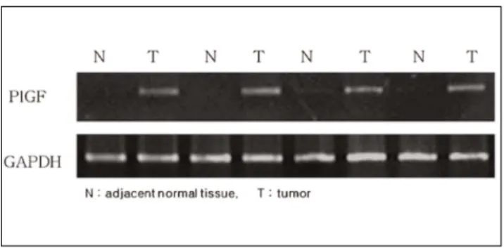
실험에 사용된 총 20개의 조직편 중 종양 인접 정상조직 에서는 PlGF mRNA 발현이 거의 나타나지 않았으나, 모 든 종양 조직에서 뚜렷한 PlGF mRNA 발현을 관찰 할 수 있었다(Fig. 5).
4. PlGF mRNA의 상대적 수준(PlGF/GAPDH)과 암종의 임상적, 병리학적 양상과의 관계
20례의 종양 조직편에서 GAPDH에 대한 PlGF의 상대 적 수치는 0.69에서 1.04로 평균 0.83이었다. 종양 조직에 서 PlGF mRNA 발현이 종양인접 정상조직에서 보다 더 높았으며 이는 통계학적으로 유의성이 있었다(paired t - test, P < .05). 그러나 환자의 성별, 나이, TNM 분류와 같 은 임상적인 요인들과 PlGF mRNA 발현과의 연관성은 유 의하게 나타나지 않았다(Table 3).
Table 1. Sequences of Primers for PlGF RT - PCR
Primer Primer sequence AT
PlGF RT-PCR-forward 5’ -GCGATGAGAATCTGCACTGTGT-3’ 60℃
PlGF RT-PCR-reverse 5’ -ATTCGCAGCGAACGTGCTGA-3’
GAPDH RT-PCR-forward 5’ -CGACCACTTTGTCAAGCTCA-3’ 60℃
GAPDH RT-PCR-reverse 5’ -TGTGAGGAGGGGAGATTCAG-3’
Abbrev. AT: annealing temperature
Fig. 1. Immunohistochemical stain- ings for PlGF of normal oral squa- mous cells (×200)
Fig. 2. Immunohistochemical stain- ings for PlGF of well differentiated oral squamous cell carcinoma (×
200)
Fig. 3. Immunohistochemical stain- ings for of moderate differentiated oral squamous cell carcinoma (×
200)
Ⅳ. 총괄 및 고찰
신생혈관형성은 종양의 성장과 전이를 위해 필수적이며, 형질전환성장인자 - α(TGF - α), 형질전환성장인자 - β (TGF - β), 혈관내피세포성장인자(VEGF), 혈소판유래 내
피세포성장인자(PD - ECGF), 종양괴사인자 - α(TNF - α) 등 다양한 인자들에 의해 영향을 받는다. 그 중 혈관내피 세포성장인자는 혈관내피세포(vascular endothelial cell) 에 작용, 분열을 촉진하여 신생혈관형성을 야기 시키는 중 요한 단백질로 현재까지 VEGF-A, VEGF-B, VEGF-C, Fig. 4. Immunohistochemical stain-
ings for PlGF of poor differentiated and invasive oral squamous cell carcinoma
Fig. 5. PlGF mRNA expression after RT-PCR
Table 2. The Correlation between Immunohistopathological PlGF Expression and Clinical and Pathological Factors
Variable Case(n) PIGF staining pattern r
Low-staining(n=) High-staining(n=) p Sex
Male 11 6 5 -0.373
Female 9 8 1 0.105
Age
60 5 4 1 0.126
60 15 10 5 0.597
Histological differentiation
Well 13 12 1 0 0..6 66 63 3**
Moderate 3 1 2 0 0..0 00 01 1**
Poor 4 1 3
Tumor size
T1 0 0 0
T2 11 9 2 0.44
T3 7 5 2 0.052
T4 2 0 2
Nodal status
N(-) 11 9 2 0.356
N(+) 9 5 4 0.123
Metastasis
M(-) 17 13 4 0.336
M(+) 3 1 2 0.147
TNM stage
I 0 0 0
II 8 7 1 0.473
III 6 5 1 0.035
IV 6 2 4
n = number of patients, r = correlation coefficient, p = P value
*Pearson correlation analysis, significance [r] > 0.6, P < .05
VEGF-D, VEGF-E, 태반성장인자(PlGF) 등 6개의 구성 원들이 밝혀져 있다
20-23).
VEGF - A는 가장 잘 알려진 혈관내피세포성장인자로 혈 관내피세포성장인자 수용기인 VEGFR-1, VEGFR-2와 결합하여 혈관내피세포의 투과성을 증가시켜 새로운 혈관 의 성장을 직접적으로 촉진한다. VEGF-B는 배아시기의 혈관형성에 관여하는 인자이며, VEGF-C와 VEGF-D는 림프관 내피세포에 선택적으로 작용하는 성장인자들이다.
PlGF는 vasculogenesis에 필수적이며 특히, 허혈, 염증, 암종 등에서 이차적인 혈관 형성 및 성장에 관여 한다
24-27).
종양이 성장함에 따라 새로운 혈관 형성이 필요하게 되며 따라서 혈관내피세포성장인자들이 증가되는 반면에 혈관형 성방해인자들은 감소하게 되는 angiogenetic switch가 일 어나게 된다. PlGF는 이런 angiognetic switch에서 가장 중요한 역할을 하며 특이하게도 정상적인 생리 상태에서 보 다 병적인 상태에서 더 많은 기여를 한다고 알려져 있다
28,29)
. 아직 그에 대한 명확한 기전이 밝혀지지 않았지만,
PlGF가 증가하게 되면 VEGFR-1으로 부터 혈관내피세포 성장인자들이 분리되고 PlGF가 그 자리를 대체하게 된다.
그 후 분리된 많은 혈관내피세포성장인자들은 VEGFR-2 와 더 많이 결합할 수 있게 되고 VEGFR-2의 인산화 (phosphorylation)를 일으키게 된다. 혈관내피세포성장인 자 수용기의 인산화에 의해 혈관내피세포 내 신호전달 체계 가 활성화되고 이런 과정에서 세포내 신호전달 체계는 주로 MAPK(mitogen-activated protein kinase) 인산화 효소 신호전달체계가 관여한다고 알려져 있으며 이들은 세포가
외부의 환경변화에 따른 자극들을 인지하여 그 정보를 세포 내부로 전달하는 역할을 담당하는 대표적인 신호전달체계 의 하나로서 세포의 성장, 발생, 분화, 사멸 등을 조절하는 기전 중 하나로, 결국 세포의 분화 및 증식을 촉진하게 된 다. 다시 말해, PlGF는 혈관내피세포내 신호전달 체계에 직접적인 영향을 주지는 않지만, 간접적으로 혈관내피세포 성장인자들과 VEGFR-2의 결합을 조절하는 역할을 한다 고 생각되며 여기서 분명한 것은 PlGF 및 VEGFR-1의 발 현 증가가 혈관내피세포성장인자에 대한 혈관내피세포의 반응을 자극한다는 것이다
30-32).
많은 연구들에서 정상조직과 비교하여 증가한 혈관내피세 포성장인자와 PlGF의 발현이 종양의 성장 및 신생혈관형 성과 연관이 있다고 하였고 실제로 직장암, 폐암, 두경부 암 등에서 혈관내피세포성장인자 및 PlGF의 발현이 증가한다 고 하였다
5,33,34). Wei 등
35)은 직장암에서 PlGF 발현과 환자 의 임상적, 조직학적 양상과의 연관성에 대한 연구에서 PlGF의 증가가 암종의 progression과 연관성이 있다고 하 였으며, 이는 PlGF가 직장암의 예후에 대한 표지자로도 사 용될 가능성이 충분하다고 하였다. 이렇게 다른 부위에 발 생하는 암종과 PlGF와의 연관성에 대한 많은 연구에 비해 구강편평세포암종에서 PlGF 발현과 그에 따른 임상적, 조 직학적 양상과의 연관성에 대한 연구는 찾아보기 어려웠다.
본 연구에서 PlGF 발현을 알아보기 위한 면역조직화학적 검사 결과 정상적인 구강편평상피조직의 경우, 상피 전층에 서 그 발현이 거의 관찰되지 않았고 이러한 양상은 PlGF가 정상적인 생리 상태인 경우, 그 발현이 제한된다는 다른 연 Table 3. Relationship between relative levels of PlGF mRNA(PlGF/G3PDH) and Clinical and Pathological Factors
Variables No. PIGF mRNA
a(mean±SD) P value
Sex
Male 11 0.8745±0.1248 0.084
Female 9 0.7778±0.0877
Stage
I - II 8 0.7588±0.0914 0.259
III - IV 12 0.8792±0.1110
Tumor status
T1 11 0.7873±0.1045 0.492
T2 - 4 9 0.8844±0.1160
Lymph node status
N0 11 0.7709±0.0933 0.347
N1 - 3 9 0.9044±0.1050
Metastasis status
M0 17 0.8106±0.1081 0.977
M1 3 0.9467±0.1210
Tissue
Tumor 20 0.8310±0.1178 0.000*
Adjacent normal tissue 20 0.3960±0.0602 a: PlGF mRNA expression derived from real-time quantitative RT-PCR
*P value derived from paired t test, others derived from independent t test
구들의 결과들과 일치한다고 생각 된다
28,29). 또한 정상조직 세포들과는 달리 구강편평세포암종에서 PlGF의 발현이 증 가하는 것을 관찰 할 수 있었는데, 특이한 점은 조직병리학 적 조직 소견 상 분화가 잘 되어 있고 침습성이 덜한 구강편 평세포암종에서는 정상 세포에 비해 PlGF의 발현 증가가 미미하였으나, 저분화도의 침습적인 구강편평세포암종에서 는 PlGF의 강한 발현이 관찰되었다는 것이다. 이는 PlGF 의 발현 정도가 암종의 분화도 또는 침습성과 상관관계가 있다고 생각해 볼 수 있었으며, 면역조직화학적 염색 결과 에 따른 PlGF expression level과 암종의 임상적, 조직학 적 양상과의 관계를 알아보기 위한 통계학적 분석에서 증명 되었다. 이를 위해 사용된 통계 방법인 pearson correla- tion analysis의 상관 계수 r은 그 값이 0.6 이상일 경우, 연관성이 매우 높다고 해석할 수 있으며, 0.4이상인 경우는 중등도의 연관성을 나타내며, 그 이하인 경우는 아주 낮은 연관성을 나타낸다고 해석할 수 있다. 본 연구에서는 상관 계수(r)를 0.6 이상으로 설정하였기에 조직학적 분화도에 따른 PlGF 발현과의 연관성만이 유의하다고 해석하였다.
TNM Stage에 따른 PlGF 발현과의 연관성에서 비록 p- value가 0.05 보다 작았지만, 상관 계수(r)가 0.473이었으 므로 유의성은 있지만 연관성이 적은 것으로 해석하였다.
면역조직화학적 검사 소견을 증명하기 위한 PlGF 유전자 RT-PCR 분석에서 20편의 종양 조직 샘플 모두에서 PlGF mRNA 발현을 관찰할 수 있었다. 이런 결과는 Kodama 등 36)이 경부편평세포암종의 29 샘플 중 15 샘플(52%)에서 강한 PlGF 유전자 발현이 관찰되었다는 연구 보고와는 차 이가 있는데 아마도 실험에 사용된 암종의 분화도와 침습성 의 차이에 따른 것이라 생각된다.
Lizian 등
37)은 PlGF의 발현과 암종의 임상적, 병리학적 양상과의 관계를 통계적으로 분석하기 위해 real-time RT- PCR을 시행하였다. 본 연구에서도 마찬가지 방법으로 PlGF 유전자의 발현을 정량화하였으며, 그에 따른 PlGF mRNA의 상대적 수준(PlGF/GAPDH)과 암종의 임상적, 병리학적 양상과의 관계를 통계학적으로 분석한 결과, 종양 인접 정상조직과 종양조직에서 PlGF 발현의 차이가 유의 성이 있게 나타났다. 이는 정상조직에서 보다 종양조직에서 PlGF의 발현이 활성화되어 있다는 것이며 이의 증가가 종 양의 신생혈관형성에 관여하리라고 추측할 수 있었다.
결론적으로 PlGF 유전자 발현이 정상조직세포에서 보다 구강편평세포암종에서 증가하였고, PlGF 발현과 구강편평 세포암종의 분화도와 침습성에 따른 상관관계에 유의성이 있음을 확인할 수 있었다. 이러한 결과는 암종의 PlGF가 암종의 progression 및 국소적 metastasis에 관여하리라는 것을 추론케 한다. 향 후, 구강편평세포암종에서 더 많은 PlGF 유전자의 발현 및 임상적 요인들 및 조직학적 양상에
대한 데이터의 축적이 이루어진다면 암종의 예후에 대한 인 지자(indicator)로서 그 사용이 가능하리라 사료된다.
Ⅴ. 결 론
종양의 성장에 있어서 종양 내 신생혈관형성(angiogene- sis)은 아주 중요한 역할을 담당한다. 혈관형성은 종양의 성 장뿐만 아니라 침윤, 원격전이에 있어서 필수 요건이다. 정 상 또는 병적 상태 모두에서 혈관형성을 직접적으로 촉진하 는 가장 강력한 인자는 혈관내피세포성장인자(vascular endothelial growth factor, VEGF)이다. 태반성장인자 (placenta growth factor, PlGF)는 VEGF와 유사성을 가 진 당단백질로 종양조직에서 과발현 되어 종양의 증식과 성 장에 영향을 미친다고 알려져 있다.
이에 본 연구는 구강 편평세포암종에서 PlGF의 발현을 검사하고 그것이 종양 환자의 임상적 양상 및 조직학적 분 화도와 관련이 있는지 알아보고 또한 종양 인접 정상조직과 의 발현 차이를 알아보고자 하였다.
실험에 사용된 조직편은 단국대학교 치과대학 부속 치과 병원 구강악안면외과에서 구강 편평세포암종으로 최종 진 단 받은 환자 20명의 수술 후 절제된 조직 20편을 사용하였 다. 각각의 조직편에서 PlGF의 발현을 확인하기 위해 면역 조직화학적 검사, PlGF RT-PCR 분석 및 real time quantitative RT- PCR을 시행한 후 통계학적으로 검증하 여 다음과 같은 결과를 얻었다.
1. 면역조직화학적 검사; 정상 구강편평상피의 경우, PlGF의 발현이 관찰되지 않았으며, 고등도 분화 구강 편평상피세포 암종의 경우, PlGF의 발현이 미약하며 단지 몇몇 침습적인 종양세포에서만 관찰되었다. 반면 에 저등도 분화 구강편평상피세포 암종의 경우, 증가된 PlGF 발현을 관찰할 수 있었다.
2. RT-PCR 분석 및 PlGF mRNA 발현; 20개의 종양 조직편 모두에서 PlGF의 발현을 관찰할 수 있었으며, 인접 정상조직에서는 PlGF의 발현이 미미하였다.
3. 통계학적으로 검증한 결과, 조직학적 분화도에 따른 PlGF의 발현의 차이가 유의성이 있게 나타났으며 (pearson correlation analysis, [r] > 0.6, P < .05), 또한 종양 인접 정상조직과 종양조직에서 PlGF 발현 의 차이가 유의성이 있게 나타났다(Student’s t - test, P < .05).
이상의 결과에서 보면, 20예의 구강편평세포암종에 대한
면역조직화학적 염색 결과, 저분화도의 침습적인 종양조직
에서 PlGF의 발현이 증가하는 것이 관찰되었으며, PlGF
expression level과 암종의 임상적, 조직학적 양상과의 연
관성을 검증한 결과에서도 조직학적 분화도에 따른 발현양
상의 차이가 유의성이 있게 나타났다.
또한 종양 인접 정상 조직과 비교하여 종양 조직에서 PlGF mRNA의 발현이 증가하는 것을 관찰할 수 있었으 며, PlGF mRNA의 상대적 수준(PlGF/GAPDH)과 암종 의 임상적, 조직학적 양상과의 연관성을 검증한 결과에서도 그 차이가 유의성이 있게 나타났다. 따라서 구강편평세포 암종에서 정상조직에 비해 PlGF의 과발현이 나타나며, PlGF가 암종의 progression에 기여하리라고 사료된다.
References