66
©The Korean Society of Food Science and Technology
전립선 암세포에서 delphinidin에 의한 HIF-1α와
STAT3 억제를 통한 혈관내피 성장 인자 발현 저해 효과
김문현
1,2,†·김미현
3,†·박영자
4· 장영채
1,2·박윤엽
1,2·송현욱
1,2,*
1대구가톨릭대학교 의용생체공학연구소, 2대구가톨릭대학교 의과대학 의학과, 3인제대학교 물리치료학과, 4서라벌대학교 임상병리과
Delphinidin Suppresses Angiogenesis via the Inhibition of HIF-1
α and
STAT3 Expressions in PC3M Cells
Mun-Hyeon Kim1,2,†, Mi-Hyun Kim3,†, Young-Ja Park4, Young-Chae Chang1,2, Yoon-Yub Park1,2, and Hyun-Ouk Song1,2,*
1Research Institute of Biomedical Engineering, CatholicUniversity of Daegu School of Medicine 2Department of Medicine, CatholicUniversity of Daegu School of Medicine
3Department of Physical Therapy, Inje University 4Department of Clinical Pathology, Sorabol College
Abstract Delphinidin is a blue-red pigment and one of the major anthocyanins in plants. It plays an important role in anti-oxidant, anti-inflammatory, anti-mutagenic and anti-cancer properties. In this study, we investigated the inhibitory effects of delphinidin on vascular endothelial growth factor (VEGF) gene expression, an important factor involved in angiogenesis and tumor progression in human prostate cancer. Delphinidin decreased levels of epidermal growth factor (EGF)-induced VEGF mRNA expression in PC-3M cells. The expression of the EGF-induced hypoxia inducible factor-1α (HIF-1α) and signaling transducer and activator of transcription 3 (STAT3) proteins, which are the major transcription factors for VEGF, were inhibited by delphinidin. In addition, delphinidin decreases HRE-promoter reporter gene activity, suggesting that delphinidin can suppress the transcription of HIF-1α under EGF induction, leading to a decrease in the expression of VEGF. Delphinidin specifically suppressed the phosphorylation of Akt, p70S6K, and 4EBP1, but not the phosphorylation of EGFR. Therefore, our results suggest that delphinidin may inhibit human prostate cancer progression and angiogenesis by inhibiting HIF-1α, STAT3 and VEGF gene expression.
Keywords: angiogenesis, delphinidin, HIF-1α, VEGF, STAT3
서
론
안토시아닌(anthocyanin)은 식용 작물에 널리 분포되어 있는 폴 리페놀 계열 중의 하나로 색이 진한 보라색, 적색, 청색을 띠는 색소 성분이다(1). 안토시아닌은 뛰어난 항산화 활성을 갖고 있 을 뿐만 아니라, 항바이러스, 항염증, 항암 등에 효과가 있다고 보고되어 있다(2). 안토시아닌의 일종인 델피니딘(delphinidin)은 블루베리, 체리, 적포도, 적자색 감자, 적자색 양배추 등에 함유 되어 있으며(3), 산화방지제로 사용되고 있다(1). 또한, 델피니딘 은 관절염(4), DNA 손상보호(5), 당뇨병(6), 항염증(7)에 효과가 있으며, 전립샘 암세포의 성장 억제와 세포사멸을 유도하여 항암 에도 영향을 미친다고 알려져 있다(8,9). 최근 연구에 따르면 델 피니딘이 vascular endothelial growth factor receptor-2 (VEGFR2) 을 억제한다(10)는 보고가 있었으나, 암세포의 신생혈관 형성과 관련된 vascular endothelial growth factor (VEGF)와 그에 관한 신호전달 메커니즘에 대한 연구는 아직 부족한 실정이다. 신생혈관생성은 새로운 혈관을 생성하는 생리적인 과정으로 세 포에 산소와 영양분을 공급하는데 있어 필수적이며, 종양 성장, 전이, 그리고 침투에서 중요한 역할을 한다(10,11). 암세포에서 방 출된 대부분의 인자들은 새로운 혈관을 만들어내는 중요한 역할 을 한다고 알려져 있다. 특히 신생혈관생성에서 가장 중요한 인 자인 VEGF는 저산소 및 다양한 성장 인자와 같은 자극에 의해 유도되며(12), 초기 종양성장 단계에도 영향을 미친다(13).VEGF는 주로 hypoxia inducible factor-1 (HIF-1)에 의해 전사 단계에서 조절된다고 보고되었다(14). VEGF의 전사인자인 HIF-1는 hypoxia-response elements (HREs)-promoter에 결합하여 저산 소 환경에서 세포 항상성 유지와 관련된 많은 유전자를 조절한 다. HIF-1은 산소 의존적으로 조절되는 HIF-1α sub-unit과 일정한 양으로 발현되는 HIF-1β sub-unit으로 구성되어 있다(15). 정상 산 소 환경에서 HIF-1α는 oxygen-dependent degradation domain (ODD)의 프롤린(proline) 잔기의 수소화에 의해 von hippel-lindau (VHL)과 빠르게 결합하여 ubiquitination system으로 분해되어
HIF-†Mun-Hyeon Kim and Mi-Hyun Kim contributed equally to this
work.
*Corresponding author: Hyun-Ouk Song, M.D., Ph.D., Department of Medicine, Catholic University of Daegu School of Medicine, Daegu 38430, Korea
Tel: 82-53-650-4599 Fax: 82-53-650-4834 E-mail: [email protected]
Received October 1, 2015; revised December 17, 2015; accepted December 25, 2015
전립선 암세포에서 delphinidin에 의한 HIF-1α와 STAT3 억제를 통한 혈관내피 성장 인자 발현 저해 효과 67
1α의 발현이 감소한다(16,17). 하지만 저산소 환경에서는 HIF-1α 가 분해되지 않고 안정화됨으로써 핵으로 전위되어 HIF-1β와 hetero-dimer를 형성하고, 형성된 HIF-1이 HRE-promoter에 결합함 으로써 VEGF와 같은 표적 유전자들의 전사를 활성화 시킨다. 또 한, HIF-1α 발현은 산소에 비의존적으로 사이토카인(cytokines)과 성장인자에 반응하여 조절된다. 특히 표피생장인자(epidermal growth factor, EGF)는 정상 산소에서 표피생장인자수용체(epidermal growth factor receptor, EGFR)를 통한 신호전달경로를 활성화 함으로써 HIF-1α 발현을 증가시킨다고 보고되었다(18).
암세포에서 주로 신호전달 단백질의 인산화 경로를 통해 HIF-1α 발현이 조절된다고 보고되었다. 그 경로로 p38, c-Jun N-terminal protein kinase (JNK), 그리고 세포밖조절단백질인산화효 소(extracellular regulated protein kinase, ERK)로 이루어진 Mito-gen-activated protein kinases (MAPK) family 신호전달 경로(19)와 phosphatidylinositol 3-kinase (PI3K)/Akt 신호전달 경로(20)가 알려 져 있다. 또한 Akt 하위 표적 유전자인 mammalian target of rapamycin (mTOR)와 p70S6 kinase 1 (p70S6K) 인산화 조절을 통해 HIF-1α 발현을 조절한다고 보고되었다(21,22). 따라서, 본 연구는 델피니딘의 신생혈관생성 억제에 관한 신호 전달 메커니즘을 조사하기 위하여, 인간 전립샘 암세포인 PC3M 세포에서 델피니딘이 EGF로 유도된 신생혈관생성에 주요한 인 자인 HIF-1α와 VEGF 발현에 미치는 영향과 조절메커니즘을 확 인하였다.
재료 및 방법
세포배양과 재료실험에 사용한 PC3M세포(human colorectal carcinoma cell)는 American type culture collection (American type culture collec-tion, Rockville, MD, USA)에서 분양 받아 사용하였으며, 1% 항 생물질(GIBCO-BRL, Invitrogen Co., Carlsbad, CA, USA), 10% fetal bovine serum (FBS) (GIBCO-BRL)가 포함된 RPMI1640 (GIBCO-BRL, Invitrogen Co., Carlsbad, CA, USA) 배지를 사용 하여 37oC, 5% CO
2 조건에서 배양하였다. 델피니딘은 Cayman
Chemical (Cayman Chemical Co., Ann Arbor, MI, USA)에서 구 입하여 다이메틸설폭사이드(dimethyl sulfoxide, DMSO; Sigma Chemical, St. Louis, MO, USA)에 녹여 사용하였다. 그 외 시약 은 Sigma-Aldrich사로 부터 구입하여 사용하였다.
세포 증식 측정
델피니딘 농도에 따른 세포 생존율을 측정하기 위해 세포(1× 10⁴ cells/well)를 96 well plate에 분주하여 24시간 안정화시킨 후, 델피니딘을 농도별로 처리하여 24시간 배양하였다. 배양된 세포 에 WST-1 분석키트(Cayman Chemical, Ann Arbor, MI, USA)를 처리하여 37oC, 5% CO
2 조건에서 4시간 동안 포마진(formazin)
을 용해한다. 반응 후 ELISA 판독기(Molecular Devices, Sunny-vale, CA, USA)로 440 nm에서 흡광도를 측정하였다.
Western blot analysis
세포 내 존재하는 단백질을 분리하기 위하여 total lysis buffer (50 mM Tris, 150 mM NaCl, 5 mM ethylenediaminetetraacetic acid (EDTA), 1 mM dithiothreitol (DTT), 0.5% nonidet P-40, 100 mM phenylmethylsulfonyl fluoride (PMSF), 20 mM aprotinin, 20 mM leupeptin, pH 8.0)를 넣고 4oC에서 30분 간 용해한 후, 원 심분리(12,000 rpm, 10분)하여 상층액을 취하였다. 또한 세포질/핵 내 단백질을 분리하기 위해 세포에 완충용액 A (10 mM Hepes (pH 7.9), 1.5 mM MgCl2, 10 mM KCl, 0.5 mM DTT, 300 mM Saccharose, 0.1% NP-10, 0.5 mM PMSF)를 가하여 4oC에서 5분 용해시킨 후 원심분리(1,000 rpm, 1분)로 pellet (핵 단백질)을 분 리하였다. 분리된 pellet을 완충용액 B (20 mM Hepes (pH 7.9), 20% glycerol, 100 mM KCl, 100 mM NaCl2, 0.2 mM EDTA, 0.5 mM DTT, 0.5 mM PMSF)로 4oC 15분 용해 하여 원심분리 (10,000 rpm, 5 min)로 핵 내 단백질을 분리하였다. 위와 같은 방 법으로 추출한 단백질을 sodium dodecyl sulfate (SDS)-폴리아크 릴아마이드젤(polyacrylamide gel) 전기이동 후 단백질을 나이트로 셀룰로스 막(nitrocellulose membrane, Whatman, GE Health Care Corp., Fairfield, CT, USA)로 전기이동 시켰다. 특이 단백질을 탐 색하기 위하여 5% 탈지우유(skim milk, GIBCO-BRL, Invitrogen Co., Grand Island, NY, USA)가 함유된 TBS-T로 1시간 반응시켜 비 특이적인 단백질을 blocking 후, 특정 단백질에 대한 항체 HIF-1α, HIF-1β, EGFR, p-AKT, p-p70S6K, p-4EBP1, β-actin (Santa Cruz Biotechnology Inc., Dallas, TX, USA)을 2.5% 탈지우유가 함유된 TBS-T에 1:1000으로 희석하여 반응시켰다. 반응된 mem-brane에 특정항체 대한 이차항체를 처리 한 후 enhanced chemil luminoesence (ECL, Amersham Life Science Corp. Arlington Heights, IL, USA)를 사용하여 X-ray film에 감광시켜 특정 단백 질의 발현을 분석하였다.
Reverse transcriptase polymerase chain reaction (RT-PCR) 세포의 RNA를 추출하기 위해 TRlzol (Invitrogen Co., Grand Island, NY, USA)를 사용하였으며, OD260과 OD280에서 흡광도 를 측정하여 동량의 total RNA와 oligo (dT)를 RT premix kit (Bioneer Inc., Daejeon, Republic of Korea)를 사용하여 cDNA를 합성하였다. 합성된 cDNA를 특정 primer (VEGF; forward 5'-ctgagcctctctaccccagg-3', reverse 3'-gagcaggaagaggatgaggg-5') ( β-actin; forward 5'-agggtgtgatgggtatggg-3', reverse 3'-caggatcttcatgag-gtagtc-5') 유전자 중심으로 polymerase chain reaction (PCR) 방법 으로 증폭하여 cDNA를 1.5% 아가로스젤로 전기이동하고 ethidium bromide (EtBr)로 염색한 후 UV하에서 확인하였다.
HRE promoter 활성 분석
pGL3-HRE-luciferase를 사용하여 실험을 수행하였다(23). 6-well plate에 세포(1×10⁴ cells/well) 배양한 뒤, pGL3-HRE-luciferase와 pCMA-β-galactosidase를 DNA transfection reagent (TranslT-LT1 transfection reagent, Mirus, Madison, WI, USA)를 사용하여 12시 간 cotransfection하였다. 배지를 교체하고 명시된 시료를 처리하 여 12시간 배양 후 lucferase와 β-galactosidase의 효소 활성을 분 석키트 (Luciferase assay system, Promega Corp., Madison, WI, USA)를 사용하여 측정하였다. β-Galactosidase는 대조군으로 사용 하였다. 통계 분석 모든 실험결과에 대한 통계처리는 각 실험 군별로 표준차이가 있는가를 검증하기 위해 분산분석을 수행하였으며 다양성 분석 은 던컨시험(Duncan’s test)로 분석하였다. 통계처리 후 p값이 0.05 미만일 경우 (p<0.05) 통계적인 유의성이 있다고 판정하였다.
결과 및 고찰
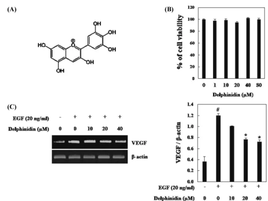
델피니딘의 VEGF의 전사 억제 효과 암 세포에서 저산소 환경은 돌연변이유발을 유도하고 악성 세 포의 침입을 증가 시킬 수 있다(24). 저산소 및 다양한 성장인자 와 같은 자극에 의해 유도되는 VEGF는 혈관내피세포의 성장을 유도함으로써 신생혈관생성에 영향을 미치는 가장 중요한 인자 로 알려져 있다(25-27). 또한, 성장인자 중 하나인 EGF는 산소 농도가 정상 상태일 경우 EGFR 신호전달을 활성화 시킴으로써 신생혈관형성을 증가시킨다(28). 따라서 본 연구에서 성장인자인 EGF를 사용하여 델피니딘이 HIF-1α와 STAT3의 발현과 VEGF의 전사에 미치는 영향을 조사하였다. 델피니딘이 PC3M 사람 전립샘암 세포의 생존율에 영향을 미 치는지 알아보기 위해 WST-1 분석을 수행하였다. 델피니딘 10-80µM의 농도에서 PC3M세포의 생존율을 확인한 결과, 델피니딘 80µM까지 영향을 미치지 않았다(Fig. 1B). 이와 같은 결과로, 델 피니딘을 독성이 없는 농도인 10, 20, 40 µM을 처리하여 RT-PCR 분석을 실시한 결과 EGF의 처리에 의해 VEGF mRNA 수준이 증가하였으며, 델피니딘 10 µM에서 큰 변화가 없었다. 그러나 델 피니딘 20, 40 µM에 의해 EGF로 유도된 VEGF mRNA 수준이 통계적으로 유효하게 감소하였다(Fig. 1C). 따라서 위와 같은 결 과로 델피니딘이 VEGF를 억제시킴으로써 전립샘 암세포의 신생 혈관생성에 억제 효과를 가질 수 있음을 알 수 있었다. 델피니딘의 HIF-1α 발현 억제 효과 저산소 환경에서 세포적응에 중요한 역할을 하는 유전자인 HIF-1α는 성장인자와 사이토카인에 의해 발현이 증가되어 VEGF의 전사를 유도한다고 알려져 있다. 또한 HIF-1α의 활성은 전립샘 암, 유방암 등과 같은 암세포에서 전이를 증가시킨다고 알려져
Fig. 1. Inhibitory effects of delphinidin on EGF-induced VEGF expression in PC3M. (A) Chemical structre of delphinidin. (B) Cell viability was determined by a WST-1 assay. (C) RT-PCR analysis of VEGF mRNA was carried out using total RNA prepared from PC3M cells incubated with 20 ng/mL EGF treatments for 12 h in the presence or absence of the indicated concentrations of delphinidin. Each value represents the mean±SD of three independent experiments and expressed (#p<0.01 vs. controls; *p<0.05 vs. EGF-stimulated cells).
Fig. 2. Inhibitory effects of delphinidin on HIF-1α and STAT3 expression in PC3M. PC3M cells were pretreated with the indicated concentrations of delphinidin for 30min, and then stimulated by EGF treatment for 6 h. Nuclear extracts were subjected to Western blot using antibodies against HIF-1α and HIF-β, STAT3. Each value represents the mean±SD of three independent experiments and expressed (#p<0.01 vs. controls; *p<0.05 vs. EGF-stimulated cells).
전립선 암세포에서 delphinidin에 의한 HIF-1α와 STAT3 억제를 통한 혈관내피 성장 인자 발현 저해 효과 69
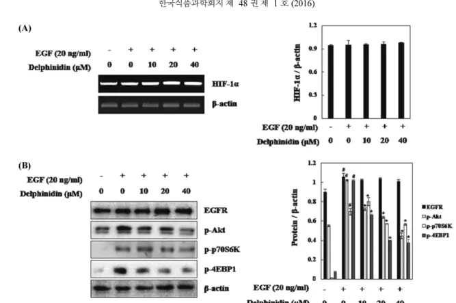
있다(29,30). STAT3 또한 VEGF의 전사를 조절하는 인자로 알려 져 있으며, 최근 연구에 따르면 STAT3 억제에 의해 EGF로 유도 된 HIF-1α가 감소된다고 보고되었다(31). 따라서 HIF-1α와 STAT3 는 암세포 증식과 종양의 신생혈관 형성에 주요한 인자이다. 위 실험을 통해 델피니딘이 VEGF의 발현을 효과적으로 감소시켰으 므로 그 조절 경로를 알아보기 위해 VEGF 전사 인자인 HIF-1 과 STAT-3에 대한 델피니딘의 영향을 확인하였다. 웨스턴블롯분 석을 통해 세포 핵으로 전위 된 HIF-1α, HIF-1-β와 STAT3의 단 백질량을 확인한 결과, PC3M 세포핵 내의 HIF-1α 단백질량이 EGF에 의해 증가하였으며, 델피니딘의 처리에 의해 감소하였다. 또한, STAT3도 동일하게 EGF에 의해 유도되어 델피니딘에 의해 유의적으로 감소한 것을 확인할 수 있었다. 반면 EGF와 델피니 딘은 HIF-1-β에는 영향을 미치지 않았다(Fig. 2). 따라서 델피니 딘은 HIF-1-β에 영향 없이 HIF-1α와 STAT3 세포 핵으로 전위를 감소시킴으로써 VEGF의 전사발현을 조절함을 알 수 있었다. HRE promoter 활성에 대한 델피니딘의 영향 활성화된 HIF-1α는 핵으로 전위되어 HIF-1-β와 복합체를 이루 어 VEGF 프로모터 영역에 결합함으로 VEGF 전사를 유도 시킨 다(32). 따라서 HIF-1 유전자가 결합하는 프로모터 영역인 HRE 프로모터 활성을 측정하기 위해 HRE 프로모터 염기서열이 연결 된 luciferase reporter gene을 PC3M 세포에 transfection하여 HRE-promoter 분석을 수행하였다. 그 결과 EGF로 유도한 PC3M 세포에서 HRE 프로모터 활성이 4배 증가하였으며, 델피니딘에 의해 유의적으로 감소하는 것을 확인하였다(Fig. 3A). 따라서 델 피니딘이 HIF-1α의 발현을 억제시켜 세포 핵 내의 HRE promoter 에 결합하지 못함으로써 VEGF 전사활성을 감소시키는 것을 알 수 있었다. 또한, 델피니딘이 조절한 HIF-1α가 HIF-1α의 핵으로 전위를 조절하는지, 세포 내 HIF-1α 단백질 발현 양을 조절하는 지 확인하기 위하여 세포 전체의 HIF-1α의 발현을 확인하였다. 그 결과, EGF에 의해 유도된 세포 내 단백질 발현이 델피니딘에 의해서 감소되었다(Fig. 3B). 이를 통해 델피니딘이 HIF-1α 단백 질 발현감소를 통해 세포핵으로 전위되는 것을 억제시킴으로써 VEGF의 전사를 조절 한다는 것을 알 수 있었다.
Akt, p70S6K, 4EBP1 경로를 통한 델피니딘의 HIF-1α 단백 질 합성 억제 효능 확인
델피니딘의 VEGF 전사활성 감소 효과가 HIF-1α의 발현 억제 에 의한 것임을 확인하였으므로, 델피니딘의 HIF-1α 조절 경로를 확인하기 위하여 RT-PCR을 통해 HIF-1α mRNA를 확인하였다. 그 결과 HIF-1α mRNA 수준은 EGF에 의해 유도되지 않았을 뿐만 아니라 델피니딘에 의해 조절되지 않았다(Fig. 4A). 이와 같은 결 과는 델피니딘이 1α mRNA 수준에 영향을 미치지 않고, HIF-1α 단백질 합성을 억제하여 VEGF를 조절한다고 예상할 수 있었 다. 많은 연구에 따르면 EGF에 의해 증가된 HIF-1α 단백질 합성 은 PI3K/Akt와 mTOR 신호전달 메커니즘을 통해 유도된다고 보 고되었다(20). mTOR는 p70S6K와 4EBP1과 같은 단백질 합성 조 절인자의 인산화에 관여하는 것으로 알려져 있다. 4EBP1의 인산 화는 eukaryotic initiation factor (elF-4E)의 활성을 감소시켜 단백질 번역을 조절하며(33), p70S6K의 인산화 또한 HIF-1α mRNA의 50-UTR 구역 조절을 통해 HIF-1α 단백질 번역을 유도한다(34). 따라 서 HIF-1α 단백질 합성 경로인 Akt, p70S6K, 4EBP1 인산화와 EGF의 수용체인 EGFR의 발현을 확인해보았다(35). PC3M세포에 EGF을 처리한 결과 Akt, p70S6K, 4EBP1 인산화가 모두 증가하였 으며, EGFR의 발현 또한 증가하였다. 또한 EGF에 의해 인산화된 Akt, p70S6K, 4EBP1 모두 델피니딘에 의해 감소하였다. 하지만 EGF에 의해 유도된 EGFR의 발현은 델피니딘에 의해 영향을 받 지 않았다(Fig. 4B). 이와 같은 결과는 EGF로 유도된 HIF-1α 단 백질 발현에 대한 델피니딘의 억제효능은 Akt, p70S6K, 4EBP1의 인산화 조절에 의한 것이며, EGFR의 발현에는 관여하지 않음을 확인할 수 있었다. 결론적으로, 델피니딘은 Akt, p70S6K, 4EBP1 의 인산화를 조절하여 EGF로 유도된 HIF-1α 단백질 발현을 감소 Fig. 3. Inhibitory effects of delphindin on HRE-promoter activity in PC3M cells. (A) PC3M cells were transiently co-transfected with a reporter gene, pGL3-HRE-Luciferase, and pRL-CMV as a reference. Following incubation for 24 h, the cells were incubated with EGF treatment in the presence or absence of indicated concentration of delphindin. (B) PC3M cells were pretreated with the indicated concentrations of delphindin for 1 h, followed by incubation with 20 ng/mL EGF for 6 h. The expression of HIF-1α were determined by Western blot analysis. Each value represents the mean±SD of three independent experiments and expressed (#p<0.01 vs. controls; *p<0.05 vs. EGF-stimulated cells).
시켰다. 더욱이, 델피니딘이 STAT3와 HIF-1α의 발현 억제를 통해 VEGF 발현을 감소시켜 신생혈관생성 억제효과가 있는 것으로 나 타났다(Fig. 5). 본 연구를 통하여, 델피니딘이 HIF-1α와 VEGF의 억제를 통하여 암세포 증식억제와 혈관형성 억제제로서 사용 가 능성이 있음을 새롭게 확인하였다.
요
약
델피니딘은 양전하를 뛰는 diphenylpropane의 polyphenolic ring 구조를 가진 주요한 안토시아닌 색소 중에 하나이다. 최근 연구 에서 델피니딘은 항산화, 항염증 뿐만 아니라 항암 효능을 가진 다고 보고되었다. 본 연구에서는 전립샘 암에서 종양의 성장과 신생혈관생성에 관련된 중요한 인자인 VEGF 발현에 대한 델피 니딘의 억제 효과를 조사하였다. RT-PCR을 통해 델피니딘을 처 리한 PC3M 전립샘 암세포 세포에서 EGF로 유도한 VEGF mRNA 발현 수준이 감소됨을 확인하였다. 또한 델피니딘은 VEGF 의 전사인자인 HIF-1α와 STAT3가 세포 핵으로 전위되는 것을 효과적으로 억제하였다. 한편 luciferase assay을 통해 HRE-pro-moter 활성을 확인해 본 결과, 델피니딘이 HIF-1α의 전사 활성을 억제시켜 VEGF 발현을 감소시키는 것을 알 수 있었다. 그리고 델피니딘은 EGFR의 발현에는 영향을 미치지 않고, Akt, p70S6K, 4EBP1의 인산화를 특이적으로 억제하는 것으로 나타났다. 결론 적으로 델피니딘이 HIF-1α와 STAT3, VEGF 발현을 억제를 통하 여 암세포 증식억제와 신생혈관형성을 억제하는 역할을 새롭게 확인하였다.
감사의 글
이 연구는 2015년도 대구가톨릭대학교 하비에르 연구비의 지 원으로 이루어졌음.
Fig. 4. Inhibitory effects of delphinidin on EGF-induced signaling leading to the expression of HIF-1α in PC3M cells. (A) RT-PCR analysis of HIF-1α mRNA was carried out using total RNA prepared from PC3M cells incubated with 20 ng/mL EGF treatments for 12 h in the presence or absence of the indicated concentrations of delphinidin. (B) PC3M cells were pretreated with the indicated concentrations of delphindin for 1 h, followed by incubation with 20 ng/mL EGF for 10 min. The expression of EGFR and the phosphorylated levels of Akt, p70S6K and 4EBP1 were determined by Western blot analysis. Each value represents the mean±SD of three independent experiments and expressed (#p<0.01 vs. controls; *p<0.05 vs. EGF-stimulated cells).
Fig. 5. Proposed molecular mechanism for regulation of HIF-1α and VEGF expression by delphinidin in EGF-treated PC3M cells.
전립선 암세포에서 delphinidin에 의한 HIF-1α와 STAT3 억제를 통한 혈관내피 성장 인자 발현 저해 효과 71
References
1. Martin S, Favot L, Matz R, Lugnier C, Andriantsitohaina R. Del-phinidin inhibits endothelial cell proliferation and cell cycle pro-gression through a transient activation of ERK-1/-2. Biochem. Pharmacol. 65: 669-675 (2003)
2. He J, Giusti MM. Anthocyanins: Natural colorants with health-promoting properties. Annu. Rev. Food Sci. Technol. 1: 163-187 (2010)
3. Harborne JB. Flavonoids in the environment: Structure-activity relationships. Prog. Clin. Biol. Res. 280: 17-27 (1988)
4. Haseeb A, Chen D, Haqqi TM. Delphinidin inhibits IL-1beta-induced activation of NF-kappaB by modulating the phosphoryla-tion of IRAK-1(Ser376) in human articular chondrocytes. Rheu-matology (Oxford) 52: 998-1008 (2013)
5. Liu W, Lu X, He G, Gao X, Li M, Wu J, Li Z, Wu J, Wang J, Luo C. Cytosolic protection against ultraviolet induced DNA damage by blueberry anthocyanins and anthocyanidins in hepato-carcinoma HepG2 cells. Biotechnol. Lett. 35: 491-498 (2013) 6. Gharib A, Faezizadeh Z, Godarzee M. Treatment of diabetes in
the mouse model by delphinidin and cyanidin hydrochloride in free and liposomal forms. Planta Med. 79: 1599-1604 (2013) 7. Bae CH, Jeon BS, Choi YS, Song SY, Kim YD. Delphinidin
inhibits LPS-induced MUC8 and MUC5B expression through toll-like receptor 4-Mediated ERK1/2 and p38 MAPK in human airway epithelial cells. Clin. Exp. Otorhinolaryngol. 7: 198-204 (2014)
8. Hafeez BB, Siddiqui IA, Asim M, Malik A, Afaq F, Adhami VM, Saleem M, Din M, Mukhtar H. A dietary anthocyanidin del-phinidin induces apoptosis of human prostate cancer PC3 cells in vitro and in vivo: Involvement of nuclear factor-kappaB signaling. Cancer Res. 68: 8564-8572 (2008)
9. Hafeez BB, Asim M, Siddiqui IA, Adhami VM, Murtaza I, Mukhtar H. Delphinidin, a dietary anthocyanidin in pigmented fruits and vegetables: A new weapon to blunt prostate cancer growth. Cell Cycle 7: 3320-3326 (2008)
10. Lamy S, Blanchette M, Michaud-Levesque J, Lafleur R, Duro-cher Y, Moghrabi A, Barrette S, Gingras D, Beliveau R. Del-phinidin, a dietary anthocyanidin, inhibits vascular endothelial growth factor receptor-2 phosphorylation. Carcinogenesis 27: 989-996 (2006)
11. Shin JM, Jeong YJ, Cho HJ, Park KK, Chung IK, Lee IK, Kwak JY, Chang HW, Kim CH, Moon SK, Kim WJ, Choi YH, Chang YC. Melittin suppresses HIF-1alpha/VEGF expression through inhibition of ERK and mTOR/p70S6K pathway in human cervi-cal carcinoma cells. PLoS One 8: e69380 (2013)
12. Jeong YJ, Cho HJ, Magae J, Lee IK, Park KG, Chang YC. Asco-furanone suppresses EGF-induced HIF-1alpha protein synthesis by inhibition of the Akt/mTOR/p70S6K pathway in MDA-MB-231 breast cancer cells. Toxicol. Appl. Pharmacol. 273: 542-550 (2013)
13. Zhang M, Li W, Yu L, Wu S. The Suppressive Effect of Resver-atrol on HIF-1alpha and VEGF Expression after Warm Ischemia and Reperfusion in Rat Liver. PLoS One 9: e109589 (2014) 14. Semenza GL. Targeting HIF-1 for cancer therapy. Nat. Rev.
Can-cer 3: 721-732 (2003)
15. Huang JH, Lee FS, Pasha TL, Sammel MD, Karakousis G, Xu G, Fraker D, Zhang PJ. Analysis of HIF-1α and its regulator, PHD2, in retroperitoneal sarcomas: Clinico-pathologic implications. Can-cer Biol. Ther. 9: 303-311 (2010)
16. Bruick RK, McKnight SL. A conserved family of prolyl-4-hydroxylases that modify HIF. Science 294: 1337-1340 (2001) 17. Jeong JH, Jeong YJ, Cho HJ, Shin JM, Kang JH, Park KK, Park
YY, Chung IK, Chang HW, Magae J, Kang SS, Chang YC. Ascochlorin inhibits growth factor-induced HIF-1alpha activation
and tumor-angiogenesis through the suppression of EGFR/ERK/ p70S6K signaling pathway in human cervical carcinoma cells. J. Cell. Biochem. 113: 1302-1313 (2012)
18. Cheng JC, Klausen C, Leung PC. Hypoxia-inducible factor 1 alpha mediates epidermal growth factor-induced down-regulation of E-cadherin expression and cell invasion in human ovarian can-cer cells. Cancan-cer Lett. 329: 197-206 (2013)
19. Fan B, Wang YX, Yao T, Zhu YC. p38 Mitogen-activated protein kinase mediates hypoxia-induced vascular endothelial growth fac-tor release in human endothelial cells. Acta. Physiol. Sinica 57: 13-20 (2005)
20. Dery MAC, Michaud MD, Richard DE. Hypoxia-inducible factor 1: Regulation by hypoxic and non-hypoxic activators. Int. J. Bio-chem. Cell Biol. 37: 535-540 (2005)
21. Hudson CC, Liu M, Chiang GG, Otterness DM, Loomis DC, Kaper F, Giaccia AJ, Abraham RT. Regulation of hypoxia-induc-ible factor 1alpha expression and function by the mammalian tar-get of rapamycin. Mol. Cell. Biol. 22: 7004-7014 (2002)
22. Skinner HD, Zheng JZ, Fang J, Agani F, Jiang BH. Vascular endothelial growth factor transcriptional activation is mediated by hypoxia-inducible factor 1alpha, HDM2, and p70S6K1 in response to phosphatidylinositol 3-kinase/AKT signaling. J. Biol. Chem. 279: 45643-45651 (2004)
23. Maxwell PH, Wiesener MS, Chang GW, Clifford SC, Vaux EC, Cockman ME, Wykoff CC, Pugh CW, Maher ER, Ratcliffe PJ. The tumour suppressor protein VHL targets hypoxia-inducible factors for oxygen-dependent proteolysis. Nature 399: 271-275 (1999)
24. Vaupel P, Mayer A. Hypoxia in cancer: Significance and impact on clinical outcome. Cancer Metast. Rev. 26: 225-239 (2007) 25. Harris AL. Hypoxia--a key regulatory factor in tumour growth.
Nat. Rev. Cancer 2: 38-47 (2002)
26. Gupta GP, Massague J. Cancer metastasis: Building a framework. Cell 127: 679-695 (2006)
27. Wang Y, Zhu YD, Gui Q, Wang XD, Zhu YX. Glucagon-induced angiogenesis and tumor growth through the HIF-1-VEGF-depen-dent pathway in hyperglycemic nude mice. Genet. Mol. Res. 13: 7173-7183 (2014)
28. van Cruijsen H, Giaccone G, Hoekman K. Epidermal growth fac-tor recepfac-tor and angiogenesis: Opportunities for combined anti-cancer strategies. Int. J. Cancer 117: 883-888 (2005)
29. Lauzier MC, Michaud MD, Dery MA, Richard DE. HIF-1 activa-tion during tumor progression: Implicaactiva-tions and consequences. Bull. Cancer 93: 349-356 (2006)
30. Lv L, Yuan J, Huang T, Zhang C, Zhu Z, Wang L, Jiang G, Zeng F. Stabilization of snail by HIF-1alpha and TNF-alpha is required for hypoxia-induced invasion in prostate cancer PC3 cells. Mol. Biol. Rep. 41: 4573-4582 (2014)
31. Cho KH, Choi MJ, Jeong KJ, Kim JJ, Hwang MH, Shin SC, Park CG, Lee HY. A ROS/STAT3/HIF-1alpha signaling cascade mediates EGF-induced TWIST1 expression and prostate cancer cell invasion. Prostate 74: 528-536 (2014)
32. Ke Q, Costa M. Hypoxia-inducible factor-1 (HIF-1). Mol. Phar-macol. 70: 1469-1480 (2006)
33. Gingras AC, Gygi SP, Raught B, Polakiewicz RD, Abraham RT, Hoekstra MF, Aebersold R, Sonenberg N. Regulation of 4E-BP1 phosphorylation: A novel two-step mechanism. Genes Dev. 13: 1422-1437 (1999)
34. Jefferies HBJ, Fumagalli S, Dennis PB, Reinhard C, Pearson RB, Thomas G. Rapamycin suppresses 5'TOP mRNA translation through inhibition of p70s6k. EMBO J. 16: 3693-3704 (1997) 35. Mi C, Ma J, Shi H, Li J, Wang F, Lee JJ, Jin X.
4',6-dihydroxy-4-methoxyisoaurone inhibits the HIF-1alpha pathway through inhibition of Akt/mTOR/p70S6K/4E-BP1 phosphorylation. J. Pharmacol. Sci. 125: 193-201 (2014)