J Lung Cancer 2008;7(2):101-102
101
A Case of Pneumatosis Intestinalis Induced by Chemo- therapy in a Patient with Lung Cancer
Pneumatosis intestinalis is an uncommon condition that is characterized by the presence of gas within the bowel wall. We experienced a case of pneumatosis intestinalis after cession of chemotherapy and we herein report on this case. A 58-year old man was admitted to our hospital for the evaluation of incidentally recognized pneumatosis intestinalis. He was diagnosed as having non small cell lung cancer in August 2006 and he received radiation therapy for concomitant brain metastasis and SVC syndrome in September 2006. He achieved a partial response after completing 6 cycles of chemo- therapy with gemcitabine and cisplatin. Newly enlarged lymph nodes were observed on the follow-up CT, and chemotherapy with paclitaxel and carbo- platin was started in July 2007. Due to the lack of a response, the therapeutic regimen was switched to oral erlotinib. After 1 month of treatment, the follow-up CT for response evaluation revealed pneumatosis intestinalis in the ascending colon without any subjective symptoms such as fever or abdominal pain. The laboratory results were within the normal range except for a slight increase of leukocytes. He underwent right hemicolectomy, but he didn’t survive his postoperative acute renal failure and pneumonia. (J Lung Cancer 2008;7(2):101 102)
Key Words: Pneumatosis intestinalis, Chemotherapy, Non small cell lung cancer
Mi-Hye Kwon, M.D.
Sun-Jung Kwon, M.D.
Eugene Choi, M.D.
Moon-Jun Na, M.D. and Ji-Woong Son, M.D.
Department of Internal Medicine, Kon- yang University College of Medicine, Daejeon, Korea
Received: August 5, 2008 Accepted: October 13, 2008
Address for correspondence Ji-Woong Son, M.D.
Department of Internal Medicine, Konyang University Hospital, 685, Gasuwon-dong, Seo-gu, Daejeon 302- 718, Korea
Tel: 82-42-600-8817 Fax: 82-42-600-9090 E-mail: [email protected]
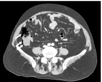
Fig. 1. Abdominal CT shows air in the wall of the ascending colon (arrows).
102 J Lung Cancer 2008;7(2):101-102
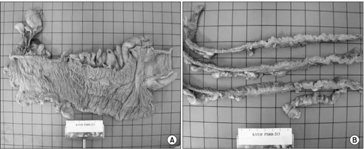
Fig. 2. (A) Gross appearance of the right hemicolectomy specimen. (B) The section shows numerous air-containing cysts.