• 검색 결과가 없습니다.

JSUJournal of Surgical Ultrasound

N/A
N/A
Protected

Academic year: 2021

Share "JSUJournal of Surgical Ultrasound"

Copied!
7
0
0

로드 중.... (전체 텍스트 보기)

전체 글

(1)

The Incidence, Risk Factors, and Clinical Outcomes for

Endovenous Heat-induced Thrombosis after Radiofrequency Ablation

Geunhyeok Yang, Sungsin Cho, Jin Hyun Joh

Department of Surgery, Kyung Hee University Hospital at Gangdong, Kyung Hee University School of Medicine, Seoul, Korea

Received October 10, 2019 Revised November 18, 2019 Accepted November 26, 2019

Purpose: Endovenous radiofrequency ablation (RFA) is a safe, effective treatment for vari- cose veins caused by saphenous reflux. Endovenous heat-induced thrombosis (EHIT) is a unique complication for this procedure. This study evaluated the incidence, risk factors, and clinical consequences of EHIT.

Methods: We retrospectively reviewed the data of patients with varicose veins who under- went radiofrequency ablation (RFA). Duplex ultrasonography (DUS) was performed within 1 week and then 6 months after the procedure. If EHIT was found at the first post- procedural DUS, then monthly surveillance was done. The incidence of EHIT and the risk factors were analyzed. The clinical consequence was finally investigated.

Results: During the study period, a total of 1,247 saphenous veins in 783 patients under- went RFA. Four hundred fifty-seven (58.4%) patients were women. The mean age was 52.9 ± 12.4 years (range: 8–85 years). EHIT was present in 7 (0.6%) saphenous veins in 7 (0.9%) patients. EHIT developed in 6 great saphenous veins (GSV) and 1 small saphenous vein.

EHIT class I, II, and III were 3, 2, and 2 patients, respectively. The diameter of GSV ≥ 6 mm was the significant risk factor for the occurrence of EHIT. Six EHITs spontaneously resolved within 5 weeks after the procedure. One EHIT was resolved in 7 months after the procedure. No incidences of pulmonary embolism occurred.

Conclusion: EHIT was a rare complication after RFA. Moreover, it spontaneously resolved without any clinical sequelae. Thus, performing routine DUS is not recommended to eval- uate EHIT in the asymptomatic patient.

Keywords: Varicose vein, Endovascular, Thrombosis, Ultrasonography, Risk factor Correspondence to:

Jin Hyun Joh

Department of Surgery, Kyung Hee University Hospital at Gangdong, Kyung Hee University School of Medicine, 892 Dongnam-ro, Gangdong-gu, Seoul 05278, Korea Tel: +82-2-440-6261

Fax: +82-2-440-6296 E-mail: [email protected]

INTRODUCTION

Varicose veins has high prevalence. The reported incidence of varicose vein ranges from 1% to 73% for females and 2% to 56% in males.(1) It is part of chronic venous disease, which is reported to have a substantial negative impact on health-related qual-

ity of life.(2) Before the worldwide spread of endove- nous therapy, high ligation and stripping of the sa- phenous vein has been the standard treatment for patients with varicose vein. Nowadays, the endove- nous ablation procedures are the most used techni- ques for the treatment of chronic venous insuf- ficiency.(3)

J Surg Ultrasound 2019;6:64-70

https://doi.org/10.46268/jsu.2019.6.2.64 JSU Journal of Surgical Ultrasound

(2)

Radiofrequency ablation (RFA) is a safe and effec- tive treatment for varicose veins. The RFA showed less periprocedural pain and bruising compared with endovenous laser therapy (EVLT).(4,5) Long-term results have been shown to be equal to those obtained with conventional surgical treatment, while provid- ing faster recovery, less pain, and a better quality of life.(6,7)

Endovenous heat-induced thrombosis (EHIT) is a worrisome complication of this procedure. Thrombotic complications after RFA have been classified 2 main categories: deep venous thrombosis (DVT) and EHIT.

It is a prvoked phenomenon. Indeen it occurs in a very short time soon after a planned and controlled injury of a selected length of the vein opposed to a spontaneous thrombosis which occurs instead in a unpredictable way and in segments not generally continuous.(8) The purpose of this study is to report the incidence, clinical consequence, and risk factors of EHIT after RFA for the varicose vein with larger volume of patients.

METHODS

A retrospective analysis was performed using clin- ical data of the patients who underwent RFA.

Patients were underwent duplex scan at the out pa- tient department and taken history, physical exa- mination.

Ultrasound examination was done with a colorized duplex scan (Vivid E9 Ultrasound system; GE Healthcare, Waukesha, WI, USA). The diameters of the great saphenous vein (GSV) and the small saphe- nous vein (SSV) were measured in supine position.

With B-mode imaging, the inner anechoic diameter of the GSV was measured from the saphenofemoral junction (SFJ) to 5 cm distal to the junction. The SSV diameter was measured in the same manner from the saphenopopliteal junction (SPJ) to 5 cm distal to the junction. The largest diameter was chosen to analyze

the relationship between diameter and stump length.

After evaluation of the diameter, SFJ, SPJ, and truncal vein reflux in response to a Valsalva’s ma- neuver and/or manual distal compression followed by release with upper body elevation or with standing position were identified with duplex scanning. The saphenous reflux was defined as a reflux time ≥ 0.5 second.

The RFA procedure was performed under general or spinal anesthesia at the operating room in all patients. Concurrent high ligation of the saphenous vein was not performed in our center. A phlebectomy around and below knee joint was performed simulta- neously if indicated. RFA using ClosureFast

TM

(Covidien, Mansfield, MA, USA) catheter was done according to the manufacturer’s instructions for use.

The treatment consisted of segmental heating of the great saphenous vein (GSV), small saphenous vein (SSV) or accessory saphenous vein using a catheter with a 7-cm heating element. To reduce EHIT at GSV, the catheter was advanced to 2 cm below the saphenofemoral or saphenopopliteal junction under ultrasound guidance. If there is deep vein confluence with SSV, ablation was performed 2 cm distal from the confluence, and if there is no deep vein con- fluence, ablation was started from the fascial level at SSV. About 10 mL of tumescent solution (normal sal- ine) per one centimeter of truncal vein was injected around the vein before the start of ablation for tissue protection. The temperature of the element was maintained at 120

o

C for 20 seconds per segment us- ing a thermocouple on the heating element, which provided a feedback loop to the generator during withdrawal. Double ablation of the most proximal segment was performed. If the incompetent perfo- rating vein was present at the ablated segment, dou- ble treatment was done. External compression of the treated segment was applied using a surgical towel.

At the completion of procedure, all wounds were

dressed with steri-strips or histoacryl agent and legs

(3)

Table 1. Patient Demographics

Demographics N = 783 (%)

Age (year) mean ± SD (range) 52.9 ± 12.4 (8–84)

Sex (male:female) 326 (41.6%):457 (58.4%)

Indication for treatment, n (%)

Heaviness 427 (54.5)

Night cramping 401 (51.2)

Discomfort 351 (44.8)

Pain 167 (21.3)

Swelling 62 (7.9)

Cosmetic concern 45 (5.7)

Itching 10 (1.3)

Limbs 1,128

Right:Left:Bilateral 201 (25.7):237 (30.3):345 (44.1)

Truncal veins 1,247

1:2:3:4 405 (51.7):311 (39.7):48 (6.1):19 (2.4)

Anesthesia, n (%)

Local 10 (1.3)

MAC 19 (2.4)

Spinal 543 (69.3)

General 211 (26.9)

Tumescent solution (ml), mean ± SD (range) 333.5 ± 142.2 (40–800)

Number of phlebectomy, mean ± SD (range) 7.1 ± 6.28 (0–35)

SD = standard deviation; MAC = monitored anesthesia care.

were placed in fulllength cohesive elastic bandage (Karl Otto Braun GmbH & Co, Wolfstein, Germany) or Rosidal CC (Lohmann & Rauscher). The bandage was exchanged for thigh-length compression stocking after 24 hours. Patients was advised to wear this stocking for a minimum of 2 weeks. Patients were discharged on the postoperative day one with a stocking.

Patients were followed up at 1 week and 1, 6 months after surgery. At all subsequent visits, the patients were examined clinically and with duplex scanning.

At 1 week, duplex scanning was performed to confirm saphenous vein occlusion and to evaluate any compli- cations such as deep vein thrombosis, hematoma, EHIT, or any complications related with surgery. At 1,6 months, a further duplex scan was performed to evaluate any complications if EHIT was detected on the initial duplex scanning.

The incidence of EHIT was evaluation. Risk factors assessed for the development of EHIT included demo-

graphic data, saphenous vein diameter, operation time, amount of tumescent solution, number of phlebectomy. The natural course of EHIT was elucidated.

Statistical analysis was done using independent t-test for comparison of continuous variables and Fisher’s exact test for comparison of nominal variables. All P values were 2-tailed, and P < 0.05 was considered to be statistically significant.

RESULTS

During the study period, a total of 1,247 saphenous veins in 783 patients were performed RFA. Patient demographics are shown in Table 1. The mean age was 52.9 ± 12.4 years with a range of 8 to 84 years.

The female to male ratio was 457:326. The most com-

mon symptom related with varicose vein was heavi-

ness followed by night cramping, discomfort, pain,

swelling, costmetic conern and itching.

(4)

Fig. 1. The natural course of endovenous heat-induced thrombosis The extension of thrombus into the deep vein on one week was slightly retracted on 5 weeks then, resolved on 6 months.

Table 2. Natural Course of Endovenous Heat-Induced Thrombosis

No Age/Sex Ablation Class Course PE symptom

1 39/male GSV 1 Resolved at 5 weeks None

2 38/female GSV 1 Resolved at 5 weeks None

3 66/male GSV 3 Resolved at 5 weeks None

4 45/female GSV 3 Resolved at 5 weeks None

5 67/female GSV 2 Resolved at 5 weeks None

6 64/male GSV 2 Resolved at 6 months None

7 64/female SSV 1 Resolved at 5 weeks None

GSV = great saphenous vein; SSV = small saphenous vein; PE = pulmonary embolism.

Bileteral ablation was done in 345 patients. RFA was done simultaneously if the GSV and SSV in the same leg showed refluxes of ≥ 0.5 second. In one session, one truncal vein was ablated in 405 patients, 2 truncal veins in 311 patients, and 3 truncal veins in 48 patients. Four truncal veins were ablated simul- taneously in 19 patients. The mean injected tumes- cent solution was 333.5 mL. The phlebectomy for the dilated vein was done simultaneous. The mean num- ber of phlebectomy was 7.1.

EHIT detected by postoperative duplex scanning in 7 patients (0.6%). Postoperative duplex scanning re- vealed class 1 EHIT in 3 patients, class 2 EHIT in 2patients, and class 3 EHIT in 2 patients. All EHIT was spontaneously resoluved at 5 weeks except one patient as shown in Fig. 1. In one patient, EHIT was resolved at 6 months (Table 2). Anticoagulation was not used in all patient with EHIT. The evaluation of

pulmonary embolism was not performed because of no symptom related with it.

The clinical characteristics of the patient with and without EHIT was shown in Table 3. The comorbidies, ablation side and treated saphenous vein, or presence of symptom of two groups were similar. The sig- nificant risk factor for the development of EHIT was the diameter of GSV ≥ 6 mm (Table 4).

DISCUSSION

This study showed that the incidence of EHIT after RFA was 0.6%. The extruded thrombus into the deep vein was slowly retracted toward the saphenous vein.

EHIT was typically resolved after 5 weeks after RFA.

In one patient, EHIT lasted until 6 months. The di-

ameter of GSV ≥ 6 mm was the only significant risk

factor for the development of EHIT.

(5)

Table 3. Clinical Characteristics of Patient with/without Endovenous Heat-Induced Thrombosis

Risk factors EHIT (-) EHIT (+) P value*

Patients 776 7

Age (year), mean ± SD 52.8 ± 12.5 51.0 ± 14.4 0.754

BMI (kg/m

2

) mean ± SD 24.8 ± 3.6 22.3 ± 2.3 0.170

Gender, n (%) 1.000

Female 297 (57.2) 4 (57.1)

Male 222 (42.8) 3 (42.9)

Side, n (%) 0.328

Right 198 (25.5) 3 (42.9)

Left 236 (30.4) 1 (14.2)

Bilateral 342 (44.1) 3 (42.9)

Treated vein, n (%) 0.594

GSV 541 (69.7) 5 (71.4)

SSV 101 (13.0) 0

Both 134 (17.3) 2 (28.6)

Comorbidities, n (%)

History of DVT, n (%) 2 (0.3) 0 1.000

Aspirin, n (%) 29 (3.7) 0 1.000

Smoking, n (%) 39 (5.0) 0 1.000

Diabetes, n (%) 39 (5.0) 0 1.000

Hypertension 90 (11.6) 0 0.594

Presence of symptom, n (%) 371 (47.8) 2 (28.6) 0.147

EHIT = endovenous heat-induced thrombosis; SD = standard deviation; BMI = body mass index; GSV = great saphenous vein; SSV = small saphenous vein; DVT=deep vein thrombosis.

*Statistical analysis was done using independent t-test for comparison of continuous variables and Fisher’s exact test for comparison of nominal variables.

Table 4. Analysis of Risk Factors of Endovenous Heat-Induced Thrombosis

Risk factors Number EHIT (-)

Number (%)

EHIT (+)

Number (%) P value*

GSV diameter 527 n = 521 n = 6 0.008

< 6 mm 291 (55.9) 0

≥ 6 mm 230 (44.1) 6 (100)

Operation time 524 n = 519 n = 5 1.000

< 60 min 303 (58.4) 3 (60)

≥ 60 min 216 (41.6) 2 (40)

Tumescent solution 311 n = 307 n = 4 0.230

< 300 mL 70 (22.8) 2 (50)

≥ 300 mL 237 (77.2) 2 (50)

Number of phlebectomy 281 n = 276 n = 5 0.652

< 5 108 (39.1) 1 (20)

≥ 5 168 (60.9) 4 (80)

EHIT = endovenous heat-induced thrombosis, Fisher’s exact test.

*Statistical analysis was done using Fisher’s exact test.

The reported incidence of EHIT was various. This rate was lower than the rates reported by Sufian and colleagues (1.3%) (9) and Nuttawut and colleagures (7%) (10) and lower than the rate reported by Marsh

and colleagues (0.2%).(11) Interestingly, some pre-

vious studies reported a decrease in the incidence of

EHIT that was commensurate with increases in the

number of RFA procedures performed by an in-

(6)

dividual surgeon.(11,12)

In our study, the larger diameter of GSV ≥ 6 mm was the only significant risk factor for the develop- ment of EHIT. This finding was consistent with the reported findings from a study by Sufian and colleagues.(9) The etiology behind why a large vein diameter is associated with higher rates of EHIT rates remains unclear. Kane et al. (13) reported that in a large vein, incomplete closure near the SFJ might oc- cur, which may facilitate stasis of venous flow near the thermally damaged endothelium that allows a thrombus to develop and propagate into the CFV.

The pathogenesis of EHIT was unclear. But it dif- fers markedly from that of standard deep venous thrombosis. It is characterized by adherence to the vein wall and relatively early resolution, and may not cause pulmonary embolism (PE).(14) Some authors prefer the acronym PASTE (postablation superficial thrombus extension) to the term EHIT because noth- ing indicates that heat application is responsible for thrombus propagation.(15)

In this study, all EHIT’s were spontanouely re- gressed. We confirmed that most cases of EHIT dis- appeared spontaneously within 5 weeks except one case whose EHIT was regressed at 6 months. Marsh and colleagues use the single dose of prophylactic lower molecular-weighed heparin (LMWH).(11) Failure of this strategy for prophylaxis has been reported in patients with a history of venous thromboembolism (VTE).(16,17) Preoperative use of anticoagulation might not be recommended due to interference with thrombotic vessel occlusion.(18) The risk of PE from EHIT remains undefined and is uncommon after open varicose vein surgery.(19) We are unaware of any re- ports of patients with PE directly in association with isolated EHIT.

CONCLUSION

The EHIT is a rare complication after RFA. It was

spontaneously resolved without any clinical symptom. Thus, it may be suggested against routine duplex scanning to evaluate EHIT at postoperative period in asymptomatic patients.

CONFLICT OF INTEREST

None.

REFERENCES

1. Lohr J, Kulwicki A. Radiofrequency ablation: evolu- tion of a treatment. Semin Vasc Surg 2010;23:90-100.

2. Carroll C, Hummel S, Leaviss J, Ren S, Stevens JW, Everson-Hock E, et al. Clinical effectiveness and cost-effectiveness of minimally invasive techniques to manage varicose veins: a systematic review and economic evaluation. Health Technol Assess 2013;17:

i-xvi, 1-141.

3. Kim J, Cho S, Joh JH, Ahn HJ, Park HC. Effect of di- ameter of saphenous vein on stump length after ra- diofrequency ablation for varicose vein. Vasc Specialist Int 2015;31:125-9.

4. Shepherd AC, Gohel MS, Brown LC, Metcalfe MJ, Hamish M, Davies AH. Randomized clinical trial of VNUS ClosureFAST radiofrequency ablation versus laser for varicose veins. Br J Surg 2010;97:810-8.

5. Goode SD, Chowdhury A, Crockett M, Beech A, Simpson R, Richards T, et al. Laser and radio- frequency ablation study (LARA study): a randomised study comparing radiofrequency ablation and endove- nous laser ablation (810 nm). Eur J Vasc Endovasc Surg 2010;40:246-53.

6. Lurie F, Creton D, Eklof B, Kabnick LS, Kistner RL, Pichot O, et al. Prospective randomized study of en- dovenous radiofrequency obliteration (closure proce- dure) versus ligation and stripping in a selected pa- tient population (EVOLVeS Study). J Vasc Surg 2003;

38:207-14.

7. Lurie F, Creton D, Eklof B, Kabnick LS, Kistner RL, Pichot O, et al. Prospective randomised study of en- dovenous radiofrequency obliteration (closure) versus ligation and vein stripping (EVOLVeS): two-year fol- low-up. Eur J Vasc Endovasc Surg 2005;29:67-73.

8. Sufian S, Arnez A, Labropoulos N, Lakhanpal S.

Endovenous heat-induced thrombosis after ablation with 1470 nm laser: incidence, progression, and risk factors. Phlebology 2015;30:325-30.

9. Sufian S, Arnez A, Labropoulos N, Lakhanpal S.

Incidence, progression, and risk factors for endove-

nous heat-induced thrombosis after radiofrequency

(7)

ablation. J Vasc Surg Venous Lymphat Disord 2013;1:

159-64.

10. Sermsathanasawadi N, Pitaksantayothin W, Puang- punngam N, Chinsakchai K, Wongwanit C, Mutiran- gura P, et al. Incidence, risk factors, progression, and treatment of endovenous heat-induced thrombo- sis Class 2 or greater after endovenous radio- frequency ablation. Dermatol Surg 2019;45:573-80.

11. Marsh P, Price BA, Holdstock J, Harrison C, Whiteley MS. Deep vein thrombosis (DVT) after venous ther- moablation techniques: rates of endovenous heat-in- duced thrombosis (EHIT) and classical DVT after ra- diofrequency and endovenous laser ablation in a sin- gle centre. Eur J Vasc Endovasc Surg 2010;40:521-7.

12. Sadek M, Kabnick LS, Rockman CB, Berland TL, Zhou D, Chasin C, et al. Increasing ablation distance pe- ripheral to the saphenofemoral junction may result in a diminished rate of endothermal heat-induced thrombosis. J Vasc Surg Venous Lymphat Disord 2013;

1:257-62.

13. Kane K, Fisher T, Bennett M, Shutze W Jr, Hicks T, Grimsley B, et al. The incidence and outcome of en- dothermal heat-induced thrombosis after endovenous

laser ablation. Ann Vasc Surg 2014;28:1744-50.

14. Frasier K, Latessa V. Minimally invasive vein therapy and treatment options for endovenous heat-induced thrombus. J Vasc Nurs 2008;26:53-7.

15. Jones RT, Kabnick LS. Perioperative duplex ultra- sound following endothermal ablation of the saphe- nous vein: is it worthless? J Invasive Cardiol 2014;26:

548-50.

16. van Rij AM, Chai J, Hill GB, Christie RA. Incidence of deep vein thrombosis after varicose vein surgery. Br J Surg 2004;91:1582-5.

17. Enoch S, Woon E, Blair SD. Thromboprophylaxis can be omitted in selected patients undergoing varicose vein surgery and hernia repair. Br J Surg 2003;90:

818-20.

18. Gibson KD, Ferris BL, Polissar N, Neradilek B, Pepper D. Endovenous laser treatment of the small [corrected] saphenous vein: efficacy and complications.

J Vasc Surg 2007;45:795-801; discussion 801-3.

19. Bounameaux H, Huber O. Postoperative deep vein

thrombosis and surgery for varicose veins. BMJ

1996;312:1158.

수치

Table 1. Patient Demographics
Fig. 1. The natural course of  endovenous heat-induced thrombosis The extension of thrombus into the  deep vein on one week was slightly  retracted on 5 weeks then, resolved  on 6 months
Table 3. Clinical Characteristics of Patient with/without Endovenous Heat-Induced Thrombosis

참조

관련 문서

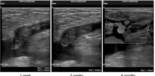
When the glue was injected across a joint, an inflammatory reaction developed in the vein wall as well as the surrounding tissue.. It may be assumed that cyanoacrylate glue

A total of 200 limbs in 148 patients underwent RFA. All saphenous veins with reflux ≥0.5 second were ablated.. Occlusion rate of saphenous vein. Kaplan-Meier analysis showed