심부정맥혈전증이 의심되는 입원 환자에서 임상적 지표 (Wells Score)와 초음파 선별검사와의 비교연구
중앙대학교병원 외과
백두산, 박병욱, 김향경
Comparative Study between the Wells Score and Ultrasonography Screening Test in Patients Hospitalized with Suspected Deep Vein Thrombosis
Doosan Baek, Byeongwook Park, Hyangkyoung Kim
Department of Surgery, Chung-Ang University Hospital, Seoul, Korea
Received October 19, 2017 Revised November 9, 2017 Accepted November 11, 2017
Purpose: Deep vein thrombosis (DVT) is potentially related to fatal complications, such as
pulmonary thromboembolism. This study evaluated the validity of screening methods of DVT, such as the Wells score and D-dimer, to identify the most reliable approach for a diag- nosis of DVT in hospitalized patients.Methods: A retrospective review of the complete medical records from 129 patients who
were referred to our department to rule out DVT was performed. Patients with suspected DVT were categorized according to the Wells score, which was calculated with their past medical history and their presenting symptoms. The risk of DVT suggested by the Wells score and D-dimer testing was then compared with the actual incidence of DVT confirmed by ultrasonography.Results: A total of 34 patients out of 129 patients were diagnosed with DVT (26.4%). The
corresponding DVT incidences in the low, moderate, and high risk groups categorized by the Wells score were 11% (1 of 9), 23.8% (20 of 84), and 36.1% (13 of 36), respectively. No sig- nificant difference was found between the risk of DVT suggested by the Wells score and the actual incidence of DVT confirmed by venous ultrasonography (P = 0.21). The level of the D-dimer was elevated in 81 patients (62.8%). On the other hand, the use of the D-dimer as- say alone was not a reliable screening test, as its elevated level failed to predict the actual in- cidence of suspected DVT (P < 0.001).Conclusion: The overall prevalence of DVT was higher when a patient was judged clinically
to be more likely to have DVT than in the general population. As DVT could not be ruled out completely by the Wells score or D-dimer alone, venous ultrasonography imaging should be performed in patients hospitalized with suspected DVT to confirm the diagnosis.Keywords: Deep vein thrombosis, Wells score, Ultrasonography
Correspondence to:Hyangkyoung Kim
Department of Surgery, Chung-Ang University Hospital, Chung-Ang University College of Medicine, 102 Heukseok-ro, Dongjak-gu, Seoul 06973, Korea
Tel: +82-2-6299-1564 Fax: +82-2-6298-8351 E-mail: [email protected]
ORIGINAL ARTICLE
J Surg Ultrasound 2017;4:62-67
JSU Journal of Surgical Ultrasound
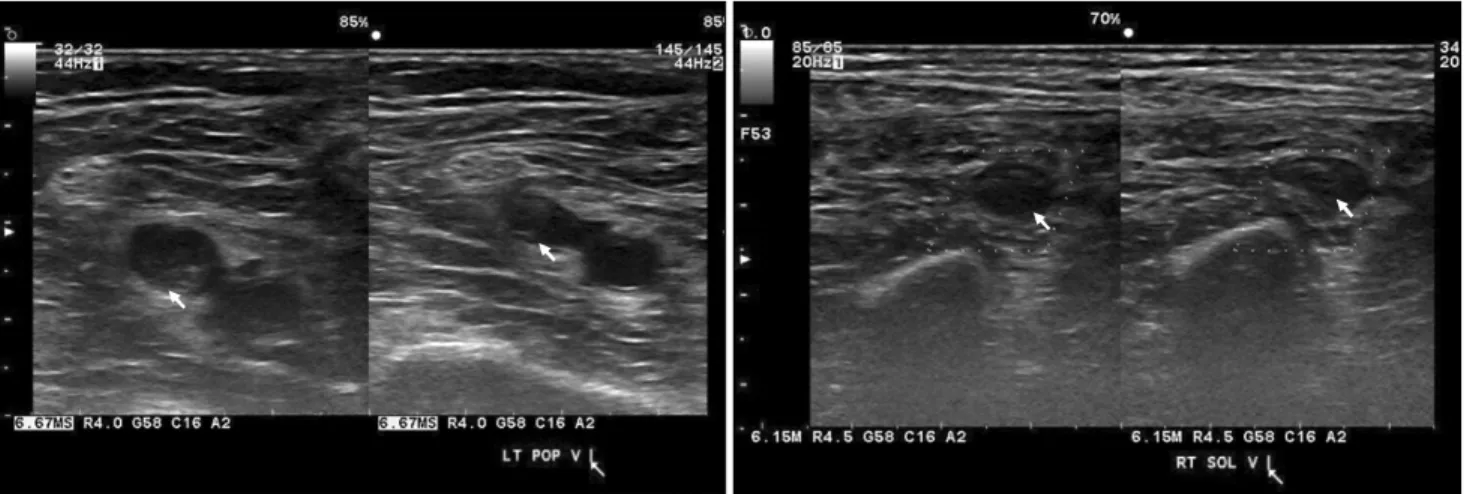
Fig. 1. Popliteal deep vein thrombosis (left), soleus muscle vein deep vein thrombosis (right).
서 론
심부정맥혈전증의 일반적 발생률은 약 1,000명 중 1명 으로 나타나며 수술이나 암등의 위험인자가 생길수록 발 생률은 더욱 증가한다.(1,2) 또한 폐색전증 및 혈전 색전 증 등으로 인한 사망률 및 이환율이 높아질 수 있다.(3) 이러한 심부정맥혈전증을 진단하기 위한 다양한 방법 이 있다. Wells 점수 체계와 같은 임상적 접근은 비용이 들 지 않으나 상대적으로 신뢰도가 떨어지며 정맥조영술은 정확하지만 비용이 높고 침습적이며 조영제와 관련한 부 작용이 있다.(4) 다양한 검사 방법이 제시되면서 검사 비 용을 줄이면서 필요한 검사의 정확성을 높이기 위한 방법 에 대한 연구들이 진행되어 왔다.(5) 여러 병원이나 학회 에서 제시하는 가이드라인에서는 심부정맥혈전증이 의심 되는 경우 Wells 점수로 위험도를 나누고 그 후 D-dimer 나 초음파 검사를 시행하여 확인하는 알고리즘을 추천하 고 있다.(6) 하지만 입원중인 환자는 수술력 및 기저질환 이 있거나 침상안정군이 많기 때문에 낮은 위험도 군이 상 대적으로 적어 Wells 점수로 위험도를 나누어 평가하는 것에 대한 효율성은 논란이 있다.(7-9)
이번 연구는 단일 센터에서 입원중인 환자 중 임상적으 로 심부정맥혈전증의 위험성이 있다고 판단된 환자에서 Wells 점수와 D-dimer 수치를 측정하고 초음파로 확인한 심부정맥혈전 빈도를 비교하여 Wells 점수와 D-dimer의 선별검사로서 통계적 의미를 보고 가장 효율적으로 심부 정맥혈전증을 감별할 수 있는 방법을 확인하고자 하였다.
방 법
본 연구는 본원 임상시험심사위원회의 승인을 받은 후 후향적, 단일 센터로 진행되었다(1709-006-16101). 2016 년 1월 1일부터 2016년 9월 30일까지 타과에서 심부정맥 혈전증이 의심되어 의뢰된 총 144명 환자를 대상으로 하 였으며 이중 의무기록이 정확하지 않은 3명과 외래에서 의뢰된 5명의 환자, 상지 심부정맥혈전증이 의심되는 7명 를 제외한 129명의 환자군을 연구에 포함시켰다.
환자의 총장골정맥부터 시작하여 원위부로 초음파 탐 촉자를 이용하여 2-3 cm 간격으로 압박과 이완을 반복하 면서 정맥의 혈전 여부를 확인하는 압박초음파로 심부정 맥혈전을 확인하였고 진단은 자발적인 정맥혈류가 소실 되고 호흡에 따른 변화가 없거나 valsalva나 프로브를 통 한 원위부 압박으로 인한 반응이 없을 시로 하였다(Fig.
1). 초음파는 숙달된 외과 전문의가 시행하였다.
의무 기록은 의뢰 되었을 시 증상, 수술력 과거력 및 D-dimer 수치증가 여부를 조사하였고 심부정맥혈전증 은 초음파로 진단하였다.(10) 환자들은 효소-연결면역형 광측정검사(enzyme-linked immunofluorescence as- say, ELFA)를 이용하여 혈청 D-dimer를 측정하였고, D-dimer의 농도가 0.5 mg/L 이상인 경우를 양성으로 정 의하였다. 또한, Wells 점수를 계산하여 심부정맥혈전증 위험도에 따라 환자군을 낮음(Wells 점수 = 0), 중간 (Wells 점수 = 1 or 2), 높음(Wells 점수 ≥3)으로 나누었 다.(6) Wells 점수 위험도와 초음파를 통한 실제 심부정맥 혈전증 진단여부를 비교하였으며 통계적 차이를 확인하
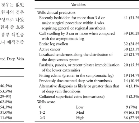
Table 1. Characteristics of 129 Patinets with Suspected Deep Vein Thrombosis
Variables
Sex
Male 60 (46.5%)
Female 69 (53.5%)
Age, years, mean (range) 71.15 (29–95)
D-dimer elevation 81 (62.8%)
Hypertension 70 (54.3%)
Diabetes mellitus 40 (31.0%)
Cardiovascular disease history 15 (11.6%)
Table 2. Wells Score Classification According to Clinical Predictors and Risk Level
Variables Wells clinical predictors
Recently bedridden for more than 3 d or major surgical procedure within 4 wks requiring general or regional anesthesia
41 (31.2%)
Calf swelling by 3 cm or more when compared with the asymptomatic leg
39 (30.2%)
Entire leg swollen 32 (24.8%)
Active cancer 30 (23.3%)
Localized tenderness along the distribution of the deep venous system
23 (21.7%)
Paralysis, paresis, or recent plaster immobilization of the lower extremities
20 (15.5%) Pitting edema (greater in the symptomatic leg) 19 (14.7%) Previously documented deep vein thrombosis 14 (10.9%) Alternative diagnosis as likely or greater than that
of deep vein thrombosis
4 (3.1%)
Collateral superficial veins (nonvaricose) 3 (2.3%) Wells score
0 Low 9 (7%)
1–2 Mod 84 (65.1%)
≥3 High 36 (27.9%)
였다.
연속형변수는 독립표본 T검정으로 분석하였고 Wells 점수에 따른 위험도와 심부정맥혈전증 발생여부의 비교 는 χ2 검정을 시행하였고 초음파와 D-dimer 검사 방법 비교는 McNemar 검정을 시행하였다. P값은 0.05보다 낮 을 시 통계적인 의미가 있다고 정의하였다. 데이터 분석은 IBM IPSS Advanced statistics version 20 (IBM, Armonk, New York, USA)을 이용하였다.
결 과
기본 환자 특성 및 Wells 임상 증상에 따른 빈도수와 위 험도를 Table 1, 2에 정리하였다. 연구에 포함 된 총 129명 의 환자에서 남자는 60명(46.5%), 여자는 69명(53.5%)이 었으며 나이는 평균 71.15세(29-95)였다. 임상경과 중 다 리 부종이 발생한 환자가 68명(52.7%), 주요 수술 혹은 침 상 안정 등의 위험인자가 있는 경우가 61명(47.3%)이었 다. 심부정맥혈전증은 34명(26.4%)에서 확인되었다. 부 위에 따라서는 좌측 10명, 우측 16명, 양측 8명이었고 총 장골정맥 1명, 외장골정맥 6명, 대퇴정맥 4명, 슬와정맥 2명, 비골정맥 2명, 경골정맥 6명, 넙치근정맥 13명이었다.
서혜부 상방 심부정맥혈전증 역시 초음파로 진단하였 다. 이번 연구 환자군에서 전산단층촬영을 통하여 심부정 맥혈전증이 의심되어 초음파를 시행한 경우는 있었으나 초음파를 시행한 후 전산단층촬영을 진행한 경우는 없었 다. 전산단층촬영으로 폐혈전색전증이 생긴 환자의 경우 는 총 5명이었고 초음파로 확인할 시 전부 양성으로 나왔 다. 초음파로 심부정맥혈전증이 보이지 않은 환자 중 호흡 곤란이 생겨 주혈관 이환이 의심되어 추가로 흉부 색전증 전산단층촬영을 시행한 경우는 2건이 있었으나 폐색전증
이나 심부정맥혈전증은 관찰되지 않았다.
Wells 점수에 따라 분류할 시 고위험도(Wells 점수≥3) 는 36명(27.9%), 중등도위험도(Wells 점수 = 1 or 2)는 84 명(65.1%), 낮은 위험도(Wells 점수 = 0)는 9명(7%)이었 다(Table 2). 고위험도 환자군에서 가장 흔한 Wells 점수 임상적 예측인자로는 최근 3일 이상 누워 있었거나, 최근 4주이내 전신 마취나 부위 마취를 필요로 한 주요 수술을 받은 경우가 25명으로 가장 많았고 심부정맥 주행을 따라 국소적 압통이 있는 경우가 15명으로 두번째로 많았다. 중 등도 위험도 환자군에서 흔한 Wells 점수 임상적 예측인 자로는 정상적 다리와 비교했을 때 장딴지가 3 cm 이상 커 진 경우가 27명으로 가장 많았고 사타구니 아래 다리 전체 가 붓는 경우가 20명으로 다음으로 많았다. 낮은 위험도 환자의 수는 9명으로 6명이 비특이적 D-dimer의 증가였 고 3명은 심부정맥혈전증의 위험인자 없이 다리 부종으로 의뢰된 환자였다. Wells 점수 중등도 위험군에서 장딴지 심부정맥혈전증이 확인된 환자는 12명이었고 고위험군에 선 9명이었다.
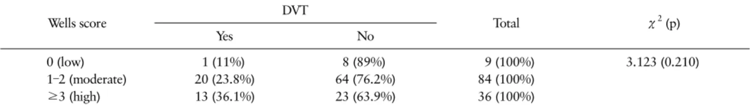
Wells 점수 위험도와 실제 심부정맥혈전증의 진단률을 비교하였을 때 통계적으로 유의한 차이가 없었다(P = 0.210) (Table 3). 고위험도에서 심부정맥혈전증이 진단
Table 4. McNemar Test between Low Extremity Ultrasonography and D-dimer Test
US finding D-dimer elevation
Total McNemar test (p)
Yes No
Positive 25 (30.9%) 9 (18.8%) 34 P < 0.001
Negative 56 (69.1%) 39 (81.2%) 95
Table 3. Comparison between Wells Score and DVT Confirmed by Ultrasonography
Wells score DVT
Total χ
2(p)
Yes No
0 (low) 1 (11%) 8 (89%) 9 (100%) 3.123 (0.210)
1–2 (moderate) 20 (23.8%) 64 (76.2%) 84 (100%)
≥3 (high) 13 (36.1%) 23 (63.9%) 36 (100%)
된 환자는 총 13명(36.1%), 중등도 위험도에서 진단된 환 자는 총 20명(23.8%)을 차지하였다. 그러나 저위험도군 환자 중 1명(11%)에서 초음파를 통해 넙다리혈관 심부정 맥혈전증이 진단되었는데 다리 부종으로 의뢰된 환자였 다(Table 3).
D-dimer가 상승된 환자는 총 81명(62.8%)이었고 25 명(30.9%)에서 심부정맥혈전증이 확인되었다. D-dimer 상승소견을 보인 심부정맥혈전증 환자에서 서혜부인대상 방 근위부 심부정맥혈전증은 4명, 서혜부인대 하방에서 슬와까지의 심부정맥혈전증은 4명, 단독 비골정맥혈전증 은 17명에서 진단되었다. D-dimer 상승소견없이 심부정 맥혈전증으로 진단된 환자는 총 9명이었으며 서혜부인대 상방 근위부 심부정맥혈전증은 3명, 서혜부인대 하방에 서 슬와까지의 심부정맥혈전증은 2명 단독 비골정맥혈전 증은 4명에서 진단되었다. McNemar 검정을 통해 확인하 였을 시 초음파로 진단된 심부정맥혈전증과 D-dimer 상 승 여부는 통계적으로 유의한 차이가 있었다(P = 0.001) (Table 4).
고 찰
이번 연구에서는 심부정맥혈전증이 의심되는 입원 환 자군에서 초음파로 심부정맥혈전증의 발생률을 확인하였 다. 임상의에 의해 d-dimer증가나 다리의 부종 혹은 임상 적 예측인자들로 선별된 환자군 129명 중 확진을 받은 환 자는 34명(26.4%)으로 일반적 발생빈도가 1,000명 중 1 명인 것에 비해 높은 발생률을 보임을 확인할 수 있었
다.(11)
심부정맥혈전증을 위한 방법으로 Wells 점수, D-dim- er, 초음파 그리고 정맥조영술등이 있다. 심부정맥혈전증 Wells 점수는 특별한 장비 없이 증상, 과거력 및 수술력등 만을 이용하여 심부정맥혈전증을 선별검사하기에 유용한 도구이다.(12) 하지만 입원환자의 경우 2가지 이상의 증 상 및 질병을 가지는 경우가 많고 비특이적 신체적 특징이 더 흔하기 때문에 낮은 위험도의 비중이 적고 대부분 중간 및 높은 위험도에 속해있는 사람이 많다. 이런 이유로 입 원중인 환자에서 Wells 점수를 선별검사로 적용하기에는 적절하지 않다고 보고되었다.(7,9) 이번 연구에서도 마찬 가지로 총 129명 환자 중 9명만 낮은 위험도를 가진 환자 였으며 Wells 점수로 분류하였을 시 위험도가 높은 군에 서 심부정맥혈전증의 발생 빈도는 더 높은 경향은 보였으 나 위험도와 실제 심부정맥혈전증의 발생 사이에 통계적 의미는 없었다.
낮은 위험도의 환자군 중 1명에서 심부정맥혈전증이 진 단되었다. 이 환자는 신장질환으로 인한 전신 부종이 있고 양하지 둘레의 차이가 있지 않았으나 초음파상 오른쪽 대 퇴정맥에 심부정맥혈전증이 관찰되었다. 낮은 위험도 환 자군 수가 적어 통계적 의미를 확인하기 어려우나 낮은 위 험도에서도 심부정맥혈전증이 생길 수 있다는 것을 의미 하며 추후 많은 환자군으로 보강된 연구가 필요할 것이다.
D-dimer는 비침습적 선별검사이며 종류에 따라 다르 지만 민감도는 높고 특이도는 낮은 특징을 가진다.(13) Wells 점수와 같이 사용할 시 효율성이 더 높아지는 것으 로 알려져 있어서(14) D-dimer와 Wells 점수를 겸용한 선
별검사는 심부정맥혈전증을 조기 발견하기위해 임상의들 에게 추천되는 방법이다.(15) 하지만 D-dimer는 민감도 가 높지만 특이도가 낮은 특성이 있고 심부정맥 혈전증 없 이도 연령의 증가, 외상, 수술의 기왕력, 급성 감염, 결핵, 뇌혈관 질환, 악성종양, 만성 신부전, 급성 관동맥 증후 군, 심부전, 호흡기 질환등에서도 올라갈 수 있어서 d-dimer 상승만으로는 심부정맥혈전증의 여부를 감별하 기 어렵다.(16) 이번 연구에 포함된 환자군에서도 D-dimer 상승과 심부정맥혈전증 여부 사이에는 통계적으로 유의 한 상관관계가 없음을 확인하였다.
이번 연구에서 확인할 수 있었듯이 Wells 점수와 D-dimer만으로는 심부정맥혈전증을 완전히 배제하기 어렵기 때문에 추후 초음파나 정맥조영술등의 확진 검사 가 필요하다. 정맥조영술은 민감도가 가장 높은 방법이나 조영제가 필요하여 부작용의 우려가 있고 방사선에 노출 되며 비용이 비싸다는 단점이 있다.(4) 반면, 초음파는 민 감도와 특이도가 높고 비교적 이동성이 좋고 정맥조영술 과 같은 방사선의 위험 및 조영제 위험이 없으며 어디에서 든 쉽고 간단히 시행할 수 있어 여러 번 쉽게 반복할 수 있 어 임상적으로 의심되는 환자에서 초음파를 시행하는 것 이 유리할 것이다.
이번 연구의 제한점으로 첫째, 후향적 설계로 적은 수 의 환자군이 포함되었다는 것이다. 적은 수의 환자군으로 낮은 위험도의 환자군이 상대적으로 적어 심부정맥혈전 증의 발생빈도에 대한 정확한 결과를 도출하기 어려웠다.
둘째로, 임상의가 심부정맥혈전증을 의심하여 초음파를 시행한 환자군을 대상으로 하였기 때문에 다른 환자군과 비교가 되지 않아 선택 편견이 개입되어 있을 수 있다. 추 후 다중센터, 전향적, 많은 환자군을 가지고 추후 연구를 한다면 더 정확한 결과를 얻을 수 있을 것이라 생각한다.
결 론
이번 연구에서는 심부정맥혈전증이 의심되는 환자군에 서 심부정맥혈전증 발생률이 26.4%정도 차지하는 것을 확인하였다. 그리고 Wells 점수와 D-dimer 만으로는 심 부정맥혈전증을 배제하기 어렵다는 것도 확인하였다. 따 라서 임상적으로 의심되는 경우에 확진을 위한 초음파 검 사가 필수적으로 시행되어야 할 것이다.
REFERENCES
1. Anderson FA Jr, Wheeler HB, Goldberg RJ, Hosmer DW, Patwardhan NA, Jovanovic B, et al. A pop- ulation-based perspective of the hospital incidence and case-fatality rates of deep vein thrombosis and pulmonary embolism. The Worcester DVT Study. Arch Intern Med 1991;151:933-8.
2. Silverstein MD, Heit JA, Mohr DN, Petterson TM, O'Fallon WM, Melton LJ 3rd. Trends in the incidence of deep vein thrombosis and pulmonary embolism: a 25-year population-based study. Arch Intern Med 1998;158:585-93.
3. Jaff MR, McMurtry MS, Archer SL, Cushman M, Goldenberg N, Goldhaber SZ, et al. Management of massive and submassive pulmonary embolism, iliofe- moral deep vein thrombosis, and chronic thromboem- bolic pulmonary hypertension: a scientific statement from the American Heart Association. Circulation 2011;123:1788-830.
4. Goodacre S, Stevenson M, Wailoo A, Sampson F, Sutton AJ, Thomas S. How should we diagnose sus- pected deep-vein thrombosis? QJM 2006;99:377-88.
5. Fancher TL, White RH, Kravitz RL. Combined use of rapid D-dimer testing and estimation of clinical probability in the diagnosis of deep vein thrombosis:
systematic review. BMJ 2004;329:821.
6. Streiff MB, Agnelli G, Connors JM, Crowther M, Eichinger S, Lopes R, et al. Guidance for the treat- ment of deep vein thrombosis and pulmonary embolism. J Thromb Thrombolysis 2016;41:32-67.
7. Price EL, Minichiello T. The Wells deep vein thrombo- sis score for inpatients: not the right tool for the job.
JAMA Intern Med 2015;175:1118-9.
8. Tafur A. A low Wells score and a negative D-dimer was not safe in patients with cancer for ruling out DVT. Evid Based Med 2014;19:188.
9. Silveira PC, Ip IK, Goldhaber SZ, Piazza G, Benson CB, Khorasani R. Performance of Wells score for deep vein thrombosis in the inpatient setting. JAMA Intern Med 2015;175:1112-7.
10. Lee JH, Park KH. Ultrasonography for the diagnosis of lower extremity deep vein thrombosis. J Surg Ultrasound 2015;2:65-8.
11. Kesieme E, Kesieme C, Jebbin N, Irekpita E, Dongo A.
Deep vein thrombosis: a clinical review. J Blood Med 2011;2:59-69.
12. Wells PS, Hirsh J, Anderson DR, Lensing AW, Foster G, Kearon C, et al. Accuracy of clinical assessment of deep-vein thrombosis. Lancet 1995;345:1326-30.
13. Di Nisio M, Squizzato A, Rutjes AW, Büller HR, Zwinderman AH, Bossuyt PM. Diagnostic accuracy of D-dimer test for exclusion of venous thromboembo- lism: a systematic review. J Thromb Haemost 2007;5:
296-304.
14. Tamariz LJ, Eng J, Segal JB, Krishnan JA, Bolger DT, Streiff MB, et al. Usefulness of clinical prediction rules for the diagnosis of venous thromboembolism: a systematic review. Am J Med 2004;117:676-84.
15. Bates SM, Jaeschke R, Stevens SM, Goodacre S, Wells PS, Stevenson MD, et al. Diagnosis of DVT: Antith- rombotic therapy and prevention of thrombosis, 9th
ed: American college of chest physicians evidence- based clinical practice guidelines. Chest 2012;141(2 Suppl):e351S-418S.
16. Hong MY, Lee C, Yoo SY, Shin DH, Cheong SS, Kwon JH, et al. Cut-off value and factors associated with a false positive D-dimer result for venous throm- boembolism in Koreans. Korean J Med 2013;84:372-8.