PGHN
Original Article
Wilson’s Disease in Bangladeshi Children: Analysis of 100 Cases
Md. Rukunuzzaman
Department of Paediatric Gastroenterology and Nutrition, Bangabandhu Sheikh Mujib Medical University, Dhaka, Bangladesh
Purpose: To evaluate clinical and laboratory profile of Wilson’s disease (WD) in children.
Methods: This cross sectional study was conducted at Bangabandhu Sheikh Mujib Medical University Hospital.
Bangladesh, over a period of 3 years. One hundred consecutive children of WD between 3 to 18 years of age were evaluated.
Results: Mean age was 8.5±1.5 years. Male female ratio was 2:1. Ninety-one percent of patients were Muslim and 9% Hindu. A total of 53% cases of hepatic WD presented between 5 to 10 years of age and most of the neurologic WD manifested in 10-15 years age group. Sixty-nine children presented only with hepatic manifestations, 6 only with neurological manifestations, 14 with both hepatic and neurological manifestation, 10 children was asymptomatic and 1 patient presented with psychiatric features. WD presented as chronic liver disease (CLD) in 42%, CLD with portal hypertension in 34%, acute hepatitis in 20% and fulminant hepatic failure in 4% cases. Stigmata of CLD were found in 18% patients. Keiser-Fleischser ring was found in 76% total patients. Elevated serum transaminase was found in 85% cases, prolonged prothrombin time in 59% cases and hypoalbuminaemia in 53% cases. A total of 73%
patients had low serum ceruloplasmin, basal urinary copper of >100 μg/day was found in 81% cases and urinary copper following penicillamine challenge of >1,200 μg/day was found in 92% cases.
Conclusion: Majority of studied WD children presented with hepatic manifestation of which 76% presented with CLD.
Any child presented with jaundice after the age of 3 years should be investigated for WD.
Key Words: Bangladesh, Child, Wilson’s disease
Received:December 13, 2014, Revised: March 24, 2015, Accepted:May 18, 2015
Corresponding author: Md. Rukunuzzaman, Department of Pediatric Gastroenterology and Nutrition, Bangabandhu Shiekh Mujib Medical University, Shahabag, Dhaka-1205, Bangladesh. Tel: +880-2-55165760-95, Fax: +880-2-9661063, E-mail: [email protected]
Copyright ⓒ 2015 by The Korean Society of Pediatric Gastroenterology, Hepatology and Nutrition
This is an openaccess article distributed under the terms of the Creative Commons Attribution NonCommercial License (http://creativecommons.org/licenses/by-nc/4.0/) which permits unrestricted noncommercial use, distribution, and reproduction in any medium, provided the original work is properly cited.
INTRODUCTION
Wilson’s disease (WD) is an autosomal recessive condition affecting copper metabolism. Impaired ex-
cretion of copper through bile and decrease incorpor- ation into ceruloplasmin causing excessive copper accumulation in different organs like liver, brain and cornea. In WD main defect remains in copper trans-
Table 1. Scoring System Developed at the 8th International Meeting on Wilson’s Disease
Characteristic Score
KF rings (slit lamp examination)
Present 2
Absent 0
Neuropsychiatric symptoms suggestive of WD
Present 2
Absent 0
Coombs negative haemolytic anaemia (+high serum copper)
Present 1
Absent 0
Urinary copper (in the absence of acute hepatitis)
Normal 0
1-2×ULN 1
>2×ULN 2
Normal, but >5×ULN one day after challenge with 2 2×0.5 g D-penicillamine
Liver copper quantitative
Normal –1
Upto 5×ULN 1
>5×ULN 2
Rhodanine positive hepatocytes (if quantitative Cu measurement is not available)
Absent 0
Present 1
Serum ceruloplasmin (normal >20 mg/dL)
Normal 0
10-20 1
<10 2
Mutation analysis
Disease causing mutations on both chromosomes 4 Disease causing mutations on one chromosome 1 No disease causing mutation detected 0 Assessment of the WD-diagnosis score
4 or more: diagnosis of WD highly likely
2-3: diagnosis of WD probable, do more investigations 0-1: diagnosis of WD unlikely
KF: Kayser Fleischer, WD: Wilson’s disease, ULN: upper limit of normal.
porter protein adenosine triphosphatase (ATP) 7B, a P-type ATPase resulting from gene mutation in chro- mosome 13. The worldwide prevalence of WD is 1 in 30,000 and the heterozygote carrier rates are about 1 in 90 persons [1]. Prevalence of WD in Bangladesh is not known.
The clinical presentation of WD varies widely.
Before 10 years of age, 83% patients of WD present with hepatic manifestations and 17% with neuro- psychiatric manifestations; between 10 and 18 years, 52% present with hepatic and 48% with neuro- psychiatric symptoms. After the age of 18 years, 75%
patients present with neuropsychiatric features and 25% patients develope liver disease [2]. The hepatic presentation also varies. Children may be asympto- matic and transaminasaemia may be an incidental finding [3]. Patient may presents with compensated or decompensated chronic liver disease (CLD). Some- times splenomegaly due to portal hypertension may be the only finding [4]. Acute liver failure may be one of the presentations of WD and usually follow a bad outcome [5]. Patients provisionally diagnosed as au- toimmune hepatitis and not responding to standard therapy should be investigated for WD because raised serum immunoglobulins level and autoanti- bodies may be found in both the conditions [6].
Coomb’s negative haemolytic anaemia is a frequent feature of WD. Hemolysis in WD may be acute or chronic, as single episode or recurrent [3]. Neuropsy- chiatric features usually develope in adult but may occur in children [7]. Early neurological features are behavior abnormalities, deterioration of school per- formance or difficulties in activities requiring hand- eye coordination [7,8]. Other neurological manifes- tations include deterioration of handwriting, drool- ing, dysarthria, dystonia, and spasticity. Seizures are infrequent in WD [9]. Common psychiatric abnor- malities are depression, anxiety or even frank psy- chosis [10]. WD with neurological manifestation may or may not have symptomatic liver disease [11].
Occassionally patient of WD may present with renal stones, early osteoporosis, cardiomyopathy, pan- creatitis, hypo parathyroidism, etc [12,13].
Prompt treatment is needed to prevent pro-
gression of neurological and hepatic damage. For this purpose, immediate diagnosis is needed. There is no single test for diagnosis of WD. Due to absence of definitive diagnostic test, identification of cases of WD is based on the combination of clinical finding, laboratory parameter and mutation analysis. Unfor- tunately, genetic diagnosis is not available in Ban- gladesh. WD disease can be diagnosed by using the scoring system developed at the 8th International Meeting on Wilson’s Disease in Leipzig, 2001 (Table 1) [14]. If proper treatment can be started before irre-
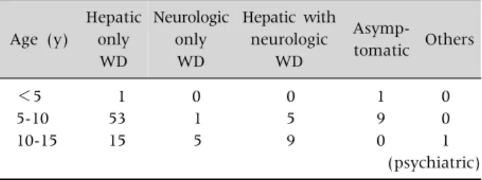
Table 2. Different Type of Presentation of Wilson’s Disease (WD) in Different Age Group (n=100)
Age (y)
Hepatic only
WD
Neurologic only WD
Hepatic with neurologic
WD
Asymp- tomatic Others
<5 1 0 0 1 0
5-10 53 1 5 9 0
10-15 15 5 9 0 1
(psychiatric) Values are presented as number.
versible liver damage or neurological impairment, near normal life expectancy is possible [4].
MATERIALS AND METHODS
It was a cross sectional descriptive study. The study was conducted at the Department of Paediatric Gastroenterology and Nutrition, Bangabandhu Sheikh Mujib Medical University, Dhaka, Bangladesh, dur- ing the period of January, 2011 through December, 2013. One hundred consecutive cases of WD were studied. Patients of either sex or age range from three to eighteen years were studied. Patients pre- sented with liver disease, seeking treatment at the Department of Paediatric Gastroenterology and Nutrition in the University Hospital, were inves- tigated for infective, metabolic, autoimmune hep- atitis and celiac disease. Detailed medical and family history was taken. Thorough physical examination was done including presence of jaundice, hepato splenomegaly, ascites and signs of liver failure.
Neurologic and psychiatric assessments were done.
Slit lamp examination of eyes by an ophthalmologist was done in all cases to identify Kayser Fleischer (KF) rings and/or sunflower cataract. Complete blood count was done to see haemoglobin level and features of haemolysis. Liver function tests were done to see the extent and progression of liver disease. Serum ceruloplasmin, 24 hours urinary cop- per as baseline and after penicillamine challenge were done. Coomb’s test was done in all the cases of WD. All the above mentioned tests were performed using standard laboratory methods. Liver biopsy was not done because of unavailability of testing of hep- atic copper in Bangladesh. Sibs of diagnosed wilso- nian patients were also investigated similarly. WD was diagnosed on the basis of diagnostic scoring sys- tem proposed in Leipzig, 2001 (Table 1) [14]. Diag- nosis of WD was done on the basis of presence of a score of 4 or more. Hepatic copper estimation and mutation analysis cannot be done because of un- availability of the tests in Bangladesh. CLD was diag- nosed on the basis of long history of jaundice, pres- ence of stigmata of CLD and biochemically higher as-
partate aminotransferase (AST) than alanine ami- notransferase (ALT) with low serum albumin. Acute hepatitis was diagnosed as acutely occurring jaun- dice with prodromal symptoms in previously appa- rent healthy individual. Acute hepatic failure was di- agnosed by prolongation of prothrombin time more than 20 seconds not responded to parenteral vitamin K1, ratio of alkaline phosphatase to total serum bilir- ubin of <4 and ratio of aspartate aminotransferase to ALT of >2.2. Statistical analysis was done using SPSS Statistics ver. 18.0 (IBM Co., Armonk, NY, USA). Frequency analysis was done and results were expressed as mean, median, range and percentage.
RESULTS
During the study period 100 patients of WD were analysed. Scores of the patients of WD according to the scoring system at 8th International Meeting on WD were 4 to 5 in 64% patients, 6 to 7 in 22% and >7 in 14% patients. Study population was 4 to 15 years with mean age 8.5±1.5 years. Male female ratio was about 2:1. Ninty one percent of patients were Muslim and 9% were Hindu. Consanguinity of mar- riage was found in 30% of cases. Seven parents were first degree cousin. Family history of CLD was found in 15% of patients. Regarding presentation of WD 69% patients presented only with hepatic manifes- tation, 6% only with neurological features and 14%
manifested both hepatic and neurological disease.
The age of hepatic WD was between 5 to 10 years and that of neurological WD was after 10 years.
Asymptomatic child was identified during family
Table 3.Hepatic Presentation of Wilson’s Disease (Including Hepatic Only and Hepatic with Neurological Presentation) (n=
83)
Types of hepatic disease n (%)
Chronic liver disease 35 (42.17)
Chronic liver disease with portal hypertension 28 (33.73)
Acute hepatitis 17 (20.48)
Fulminant hepatic failure 3 (3.61)
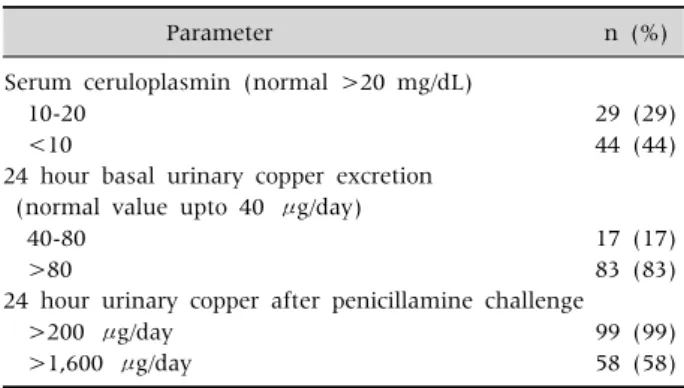
Table 5. Biochemical Parameters of Copper Metabolism (n=
100)
Parameter n (%)
Serum ceruloplasmin (normal >20 mg/dL)
10-20 29 (29)
<10 44 (44)
24 hour basal urinary copper excretion (normal value upto 40 μg/day)
40-80 17 (17)
>80 83 (83)
24 hour urinary copper after penicillamine challenge
>200 μg/day 99 (99)
>1,600 μg/day 58 (58)
Table 4. Liver Function Tests (n=100)
Parameter Mean±SD
Median (maximum-
minimum) Serum ALT 108.05±102.74 70.0 (20-450) (normal 15-40 U/L)
Serum AST 124.60±72.84 120.0 (15-290) (normal 15-37 U/L)
Serum bilirubin 3.86±3.73 1.90 (0.8-14) (normal 0.3-1.1 mg/dL)
PT (control 11.8 sec) 21.93±8.67 20.50 (13-50) Serum albumin 3.09±0.88 3.50 (1.50-4.50) (normal 3.5-6.5 g/dL)
SD: standard deviation, ALT: alanine aminotransferase, AST:
aspartate aminotransferase, PT: prothrombin time.
screening (Table 2).
Hepatic presentations of WD in this study include CLD with or without portal hypertention, acute hep- atitis and fulminant hepatic failure. Among the 83 hepatic cases (including hepatic only and hepatic with neurological features) CLD with or without por- tal hypertension is the main presentation (Table 3).
Jaundice was present in 45% patients. Sixty one per- cent of patients had hepatomegaly and 35% had splenomegaly. Ascites was present in 38% patients.
Stigmata of CLD was in only 18% patients. Common stigmata was thenar and hypothenar wasting (n=
8). Spider angioma was found in two patients.
Clubbing was found in only one patient. Gynaeco- mastia was also found in one patient. KF ring was found in 76% of the total patients. KF ring was pres- ent in 84% of the hepatic only wilsonian patients and in 90% of neurologic wilsonian patients. Asympto- matic and psychiatric patient had no KF rings.
Sunflower cataract was found in 2 cases. Both pa- tients with sunflower cataract presented with hep- atic manifestation. One patient later developed pan- creatitis.
Biochemical tests revealed higher serum AST level than ALT level. Prothrombin time ranges from nor- mal to 50 seconds (Table 4).
Serum cereloplasmin was normal in 27% patient and rest of them had low serum ceruloplasmin.
Baseline 24-hour urinary copper excretion was more than normal in 100% patients. Twenty-four-hour urinary copper after penicillamine challenge >200 μg/day was in 99% cases and >1,600 μg /day in 58%
cases (Table 5).
In 28 cases esophageal varices was found by upper gastrointestinal tract (GIT) endoscopy. WD patient
with hepatic presentations were given zinc sulphate along with penicillamine. All patients with neuro- logical manifestation as well as asymptomatic cases were maintained on zinc.
DISCUSSION
This study included 100 patients who were ana- lysed clinically and biochemically. Mean age was 8.5±1.5 years. Wilson disease is rare before 5 years of age though there is report of occurance of disease at 3 years of age [15]. The youngest patient in present study was a boy of 4 years who was asymptomatic and identified during family screening. Kleine et al.
[16] evaluated 28 Brazilian children, whose mean age at diagnosis was 10 years. Male are more than the female in the present study. Similar observation
was also made by some other researchers [17-20].
Merle et al. [21] found female preponderance. Some observer found no sex difference about the occur- rence of WD [7]. Male preponderance may be due to the facts that parents give more importance for male child to seek medical care. Ninety-one percent of pa- tients were Muslim and 9% Hindu. In Bangladesh, Hindus are 10% of total population. Disease fre- quency is almost equal in both religious groups.
Consanguinity of marriage was found in 30%
patients. Consanguinity of marriage is a feature of WD as because it is an autosomal recessive condition.
Saito found WD to be more prevalent among the child of consanguineous parents in Japan [22].
Among the hepatic patients, 69% had only hepatic involvement and 14% had both hepatic and neuro- logic manifestation similar to observation of El- Karaksy et al. [17]. But a lower prevalence of 65%
and 22% respectively was reported by Medici et al.
[23]. High prevalence may be explained by the age of study group, where hepatic manifestations predom- inate in childhood. Moreover, this study was con- ducted at Department of Paediatric Gastroenterol- ogy and Nutrition where most of the patients come with hepatic complaints.
CLD is the main presentation of WD. Following copper deposition, hepatocytes become destroyed and result in fibrosis and cirrhosis. Portal hyper- tension is the result of distortion of hepatic architec- ture. Though WD may present with acute hepatitis, they ultimately develope chronicity [2]. WD causes hepatic failure in about 5% patients [4]. History re- vealed that 34% patient had gastrointestinal hae- morrhage due to ruptured esophageal varices. GIT haemorrhage may also be due to coagulopathy re- sulted from impaired coagulation factor synthesis, qualitative or quantitative deficiency of platelets or due to peptic ulcer resulted from gastric erosion.
The prevalence of gynaecomastia in cirrhotic pa- tients in adults has been reported in one study to be 44% [24]. Spider angioma was found in 33% cases and clubbing in 24% cases of cirrhosis [25,26]. In the present study thenar and hypothenar wasting was found in 8%, spider angioma in 2% and clubbing and
gynaecomastia in 1% cases each. Spider angioma and gynaecomastia is due to impaired estradial me- tabolism in liver [25]. Clubbing is present in primary biliary cirrhosis and CLD with hepatopulmonary syndrome [27]. Clubbing develops due to release of platelet derived growth factor from aggregated pla- telets in nail bed [26]. Presence of KF rings are reli- able sign in WD but it may also be found in chronic active hepatitis, primary biliary cirrhosis, chronic cholestasis and cryptogenic cirrhosis. KF rings con- sist of copper granules deposited in the descemet’s membrane in limbic region of cornea. Sunflower cat- aracts are usually associated with KF rings and they are found in anterior lens capsule with spoke radia- tion towards the lens periphery [28]. ALT was raised in 92% patients. Serum AST level is higher than serum ALT in this study. It is a feature of WD. Serum AST to ALT ratio >2.2 is a feature of fulminant hepatic fail- ure due to WD [1]. ALT and AST are cytosolic enzyme and level may be normal or low in liver failure and in advanced liver disease. Ioris found normal ALT in 5.5% of patients [29]. El-Karaksy et al. [17] found median serum bilirubin of 2.9 mg/dL and median se- rum albumin of 2.4 gm/dL in his study on hepatic WD. In present study, median total serum bilirubin was 1.9 mg/dL, median prothrombin time 20.50 sec- onds and median serum albumin of 3.5 gm/dL. Liver function was not altered in a good number of patients. This may be due to the fact that these pa- tients were picked up earlier in the disease course.
Seventy three percent patients had lower than nor- mal serum ceruloplasmin. Ceruloplasmin is an acute phase reactant produced in liver. Low serum cer- uloplasmin was found in WD, protein losing enter- opathy, nephrotic syndrome, malnutrition, Menke’s dis- ease, etc [30]. Greater than normal may be found in pregnancy, oral contraceptives usage, acute and chronic inflammation, juvenile idiopathic arthritis, Alzheimer’s disease, schizophrenia and obsessive compulsive disorder [30-32]. Dhawan et al. [33] re- ported normal serum ceruloplasmin in 20% patient.
In our study basal 24 hour urinary copper of more than 40 μgm and post penicillamine 24 hours urinary copper more than 200 μgm (more than 5 times upper
limit normal) was found in almost all the patients.
In penicillamine challenge test, 58% had 24 hours urinary copper of >1,600 μgm/day. El-Karaksy et al. [17] found basal urinary copper more than >100 μgm/day in 53% patients. After penicillamine chal- lenge it was found that 24 hours urinary copper ex- cretion of <1,000 μgm/day in 15%, 1,000 to 1,600 μgm/
day in 36.4% and >1,600 μgm/ day in 48.5% patients.
Limitations of this study are, hepatic copper estima- tion and mutation analysis could not be included in diagnostic scoring system.
WD is a treatable metabolic cause of liver disease.
Majority of studied WD children presented with hep- atic manifestation of which 76% presented with CLD. Any child with jaundice after 3 years of age should be evaluated for WD.
REFERENCES
1. European Association for Study of Liver. EASL Clinical Practice Guidelines: Wilson's disease. J Hepatol 2012;
56:671-85.
2. Sokol RJ, O’connor JA. Copper metabolism and copper storage disorders. In: Suchy FJ, Sokol RJ, Balistreri WF, eds. Liver disease in children. 3rd ed. NewYork:
Cambridge University Press, 2007:626-52.
3. Roberts EA, Schilsky ML; American Association for Study of Liver Diseases (AASLD). Diagnosis and treat- ment of Wilson disease: an update. Hepatology 2008;47:
2089-111.
4. Rosencrantz R, Schilsky M. Wilson disease: patho- genesis and clinical considerations in diagnosis and treatment. Semin Liver Dis 2011;31:245-59.
5. Korman JD, Volenberg I, Balko J, Webster J, Schiodt FV, Squires RH Jr, et al; Pediatric and Adult Acute Liver Failure Study Groups. Screening for Wilson dis- ease in acute liver failure: a comparison of currently available diagnostic tests. Hepatology 2008;48:1167- 74.
6. Czaja AJ, Freese DK; American Association for the Study of Liver Disease. Diagnosis and treatment of au- toimmune hepatitis. Hepatology 2002;36:479-97.
7. Lang C, Müller D, Claus D, Druschky KF. Neuropsycho- logical findings in treated Wilson's disease. Acta Neurol Scand 1990;81:75-81.
8. Wolf TL, Kotun J, Meador-Woodruff JH. Plasma cop- per, iron, ceruloplasmin and ferroxidase activity in schizophrenia. Schizophr Res 2006;86:167-71.
9. Pandey RS, Swamy HS, Sreenivas HS, John CJ.
Depression in Wilson's disease. Indian J Psychiatry 1981;23:82-5.
10. Sahoo MK, Avasthi A, Sahoo M, Modi M, Biswas P.
Psychiatric manifestations of Wilson's disease and treatment with electroconvulsive therapy. Indian J Psychiatry 2010;52:66-8.
11. Kumar MK, Kumar V, Singh PK. Wilson's disease with neurological presentation, without hepatic involve- ment in two siblings. J Clin Diagn Res 2013;7:1476-8.
12. Subrahmanyam DK, Vadivelan M, Giridharan S, Balamurugan N. Wilson's disease-a rare cause of renal tubular acidosis with metabolic bone disease. Indian J Nephrol 2014;24:171-4.
13. Fatima J, Karoli R, Jain V. Hypoparathyroidism in a case of Wilson's disease: Rare association of a rare disorder. Indian J Endocrinol Metab 2013;17:361-2.
14. Ferenci P, Caca K, Loudianos G, Mieli-Vergani G, Tanner S, Sternlieb I, et al. Diagnosis and phenotypic classification of Wilson disease. Liver Int 2003;23:139- 42.
15. Wilson DC, Phillips MJ, Cox DW, Roberts EA. Severe hepatic Wilson's disease in preschool-aged children. J Pediatr 2000;137:719-22.
16. Kleine RT, Mendes R, Pugliese R, Miura I, Danesi V, Porta G. Wilson's disease: an analysis of 28 Brazilian children. Clinics (Sao Paulo) 2012;67:231-5.
17. El-Karaksy H, Fahmy M, El-Raziky MS, El-Hawary M, El-Sayed R, El-Koofy N, et al. A clinical study of Wilson's disease: The experience of a single Egyptian Paediatric Hepatology Unit. Arab J Gastroenterol 2011;12:125- 30.
18. Jha SK, Behari M, Ahuja GK. Wilson's disease: clinical and radiological features. J Assoc Physicians India 1998;46:602-5.
19. Taly AB, Meenakshi-Sundaram S, Sinha S, Swamy HS, Arunodaya GR. Wilson disease: description of 282 pa- tients evaluated over 3 decades. Medicine (Baltimore) 2007;86:112-21.
20. Tryambak S, Sumanta L, Radheshyam P, Sutapa G.
Clinical profile, prognostic indicators and outcome of Wilson's disease in children: a hospital based study.
Trop Gastroenterol 2009;30:163-6.
21. Merle U, Schaefer M, Ferenci P, Stremmel W. Clinical presentation, diagnosis and long-term outcome of Wilson's disease: a cohort study. Gut 2007;56:115-20.
22. Saito T. An assessment of efficiency in potential screen- ing for Wilson's disease. J Epidemiol Community Health 1981;35:274-80.
23. Medici V, Trevisan CP, D'Incà R, Barollo M, Zancan L, Fagiuoli S, et al. Diagnosis and management of Wilson's
disease: results of a single center experience. J Clin Gastroenterol 2006;40:936-41.
24. Cavanaugh J, Niewoehner CB, Nuttall FQ. Gyneco- mastia and cirrhosis of the liver. Arch Intern Med 1990;150:563-5.
25. Li CP, Lee FY, Hwang SJ, Chang FY, Lin HC, Lu RH, et al. Spider angiomas in patients with liver cirrhosis:
role of alcoholism and impaired liver function. Scand J Gastroenterol 1999;34:520-3.
26. Epstein O, Dick R, Sherlock S. Prospective study of peri- ostitis and finger clubbing in primary biliary cirrhosis and other forms of chronic liver disease. Gut 1981;22:
203-6.
27. Karnath B. Stigmata of chronic liver disease. Hospital Physician 2013;39:14-6.
28. Deguti MM, Tietge UJ, Barbosa ER, Cancado EL. The eye in Wilson's disease: sunflower cataract associated with Kayser-Fleischer ring. J Hepatol 2002;37:700.
29. Iorio R, D'Ambrosi M, Marcellini M, Barbera C, Maggiore G, Zancan L, et al; Hepatology Committee of
Italian Society of Paediatric Gastroenterology Hepatol- ogy and Nutrition. Serum transaminases in children with Wilson's disease. J Pediatr Gastroenterol Nutr 2004;39:331-6.
30. Hellman NE, Gitlin JD. Ceruloplasmin metabolism and function. Annu Rev Nutr 2002;22:439-58.
31. Ziakas A, Gavrilidis S, Souliou E, Giannoglou G, Stiliadis I, Karvounis H, et al. Ceruloplasmin is a better predictor of the long-term prognosis compared with fi- brinogen, CRP, and IL-6 in patients with severe un- stable angina. Angiology 2009;60:50-9.
32. Virit O, Selek S, Bulut M, Savas HA, Celik H, Erel O, et al. High ceruloplasmin levels are associated with ob- sessive compulsive disorder: a case control study.
Behav Brain Funct 2008;4:52.
33. Dhawan A, Taylor RM, Cheeseman P, De Silva P, Katsiyiannakis L, Mieli-Vergani G. Wilson's disease in children: 37-year experience and revised King's score for liver transplantation. Liver Transpl 2005;11:441-8.