pISSN: 0378-6471⋅eISSN: 2092-9374
http://dx.doi.org/10.3341/jkos.2014.55.8.1208
Original Article
섬유주세포에서 베바시주맙과 라니비주맙이 일산화질소합성효소의 발현에 미치는 영향
Effect of Bevacizumab and Ranibizumab on the Expression of eNOS in Trabecular Meshwork Cells
김세은⋅김재우
Se Eun Kim, MD, Jae Woo Kim, MD, PhD
대구가톨릭대학교 의과대학 안과학교실
Department of Ophthalmology, Catholic University of Daegu School of Medicine, Daegu, Korea
Purpose: To compare the effects of bevacizumab and ranibizumab on the expression of eNOS in cultured human trabecular meshwork cells (HTMC).
Methods: Primarily cultured HTMC were exposed to 0.5 mg/mL bevacizumab and ranibizumab using 1% serum-containing me- dia for 30 minutes. Expression of eNOS mRNA was assessed with RT-PCR. Additionally, after exposure to 20 ng/mL of vascular endothelial growth factor (VEGF) and 0.5 mg/mL bevacizumab and ranibizumab, the production of nitric oxide was assessed with Griess assay.
Results: VEGF increased the production of nitric oxide in HTMC. Bevacizumab and ranibizumab decreased the expression of eNOS mRNA and production of nitric oxide (p < 0.05) in HTMC. The decrease in eNOS mRNA expression and production of ni- tric oxide was not significant between bevacizumab and ranibizumab (p > 0.05).
Conclusions: In HTMC, both bevacizumab and ranibizumab decreased the expression of eNOS mRNA with no significant differ- ence observed between the two drugs.
J Korean Ophthalmol Soc 2014;55(8):1208-1212
Key Words: Bevacizumab, eNOS, Nitric oxide, Ranibizumab, Trabecular meshwork
■Received: 2014. 1. 25. ■ Revised: 2014. 2. 14.
■Accepted: 2014. 6. 24.
■Address reprint requests to Jae Woo Kim, MD, PhD Department of Ophthalmology, Daegu Catholic University Medical Center, #33 Duryugongwon-ro 17-gil, Nam-gu, Daegu 705-718, Korea
Tel: 82-53-650-4728, Fax: 82-53-627-0133 E-mail: [email protected]
* This work was supported by the grant of Research Institute of Medical Science, Catholic University of Daegu (2013).
ⓒ2014 The Korean Ophthalmological Society
This is an Open Access article distributed under the terms of the Creative Commons Attribution Non-Commercial License (http://creativecommons.org/licenses/by-nc/3.0/) which permits unrestricted non-commercial use, distribution, and reproduction in any medium, provided the original work is properly cited.
혈관내피성장인자(Vascular endothelial growth factor, VEGF)는 신생혈관생성의 중요한 매개체로서 주로 혈관내 피세포에서 발현되는 특이 수용체에 결합하여 내피세포의
증식과 이동, 세포외기질의 분해와 같은 신생혈관생성작용 을 나타낸다.1 또한 VEGF는 혈관보호작용도 나타내는데 이는 지속적인 일산화질소(Nitric oxide, NO)의 생성과 연 관이 있으며 혈관내피세포와 평활근세포에서 VEGF는 NO 합성효소를 활성화시켜 다양한 작용을 나타내게 된다.2,3 임 상적으로 신생혈관억제 등을 목적으로 항혈관내피성장인 자로 만든 약제를 사용하고 있는데 이러한 약제의 부작용 의 하나로 안압상승이 초래될 수 있다.4
섬유주를 통한 방수유출을 조절함에 있어서 NO가 중요한 역할을 하는데 섬유주세포에서도 NO합성효소(endothelial NO synthase, eNOS)가 발현될 뿐만 아니라5-7 NO는 섬유주를 이완시켜 방수유출을 촉진하는 것으로 알려졌으며8,9 녹내
장이 있는 경우에는 NO합성효소의 활성이 감소되어 있는 것으로 알려졌다.10
따라서 안구 내에 항혈관내피세포를 주입할 경우 eNOS의 발현과 NO의 생성을 감소시켜 섬유주를 수축시킴으로써 섬 유주를 통한 방수유출을 감소시켜 안압상승을 유발할 가능성 이 있으나 이에 대한 연구는 아직 자세히 되어 있지 않다.
본 연구에서는 사람의 섬유주세포를 일차배양하여 임상 적으로 사용되는 항혈관내피성장인자 제제인 베바시주맙 과 라니비주맙에 노출시킨 후 이들 약제가 섬유주세포에서 eNOS의 발현에 미치는 영향을 알아보고 두 약제 간의 차 이를 비교해보고자 하였다.
대상과 방법
세포배양
안구은행에서 얻은 사후 6시간 이내에 적출한 안구의 앞 방각에서 섬유주를 벗겨내어 폴리라이신(Sigma, USA)으로 처 리한 배양접시에 옮긴 후 항생제(Gibco, Invitrogen, Carlsbad, CA, USA)와 15% 우태아혈청(Hyclone, USA)이 포함된 Dulbecco’s modified Eagle’s medium 배지(DMEM, Gibco, Invitrogen, Carlsbad, CA, USA)를 사용하여 5% CO2 배양기 에서 초대배양하였다. 섬유주세포가 이식된 조직편 주위로 자라나온 것을 확인한 후 섬유주조직의 이식편을 제거하고 배양을 계속하였으며 세포가 배양접시에 충만해지면 10%
우태아혈청(Gibco, Invitrogen, Carlsbad, CA, USA)을 포함한 배지로 1:3의 비율로 트립신 처리하여 계대배양하였다.
약물처리
24 well 배양접시에 2x10⁴ cells/mL의 농도로 각 well에 세포를 분주한 후 24시간 동안 배양기에 넣어 세포를 부착 시킨 후 배지를 제거하고 나서 1% 혈청이 포함된 DMEM배 지를 이용하여 실험을 시행하였다. VEGF (Sigma, St Louis, MO, USA)가 NO의 생성에 미치는 영향을 확인하기 위하여 phosphatidylinositol 3-kinase 저해제인 500 nM wortmanin (Sigma, St Louis, MO, USA)과 1 μM protein kinase B (Akt) 저해제(1,3-dihydro-1-(1-((4-(6-phenyl-1H-imida- zo[4,5-g]quinoxalin-7-yl)phenyl)methyl)-4-piper- idinyl)-2H-benzimidazol-2-one trifluoroacetate, Sigma, St Louis, MO, USA)에 2시간 동안 먼저 노출시킨 후 염류용 액으로 씻어낸 다음 배지를 교환한 후 20 ng/mL의 VEGF 에 30분간 노출시켰다. 또한 VEGF의 작용을 억제하는 베 바시주맙(Avastin, Genetech, USA)과 라니비주맙(Lucentis, Novatis, USA)에도 0.5 mg/mL의 농도로 각각 노출시켜 다 음의 실험을 시행하였다.
eNOS mRNA 발현을 측정하기 위한 RT-PCR
베바시주맙과 라니비주맙에 노출시킨 섬유주세포에서 Trizol (Invitrogen, Carlsbad, CA, USA)을 이용하여 RNA를 분리한 후 분리된 RNA에서 eNOS mRNA의 발현을 RT-PCR 을 이용해 확인하였다. 섬유주세포에서 분리한 RNA와 Oligo dT primer, Nuclease-free water를 혼합하여 만든 RNA de- naturation mix를 70℃에 5분간 denaturation 시키고 얼음에 5분간 둔 다음 Prime RT premix (Genet bio, Seoul, Korea) 와 혼합하여 42oC에서 1시간, 70oC 10분간 반응시켜 cDNA 로 합성하였다. 합성한 cDNA에 2XGoTaq Green Master Mix (Promega, Fitchburg, WI, USA)와 10 pmol의 forward primer (ctg gct ttc cct tcc agt tc, 225 bp), reverse primer (cct tcc aga tta agg cgg ac, 225 bp)를 각각 혼합하여 DNAEngine cycler (Bio-Rad, Hercules, CA, USA)를 사용 하여 94℃에서 5분, 94℃ 30초, 54oC 30초, 72oC 30초로 총 30 cycles를 시행한 후 57oC에서 5분간 반응시켰다. 증폭된 PCR product를 1% agarose gel에 전기 영동하여 DNA band 를 multi Gauge v2.02 (Fujifilm, Tokyo, Japan)을 이용하여 분석하였다. 이때 β-action을 internal standard로 사용하였다.
Griess assay
섬유주세포에서 NO의 생성은 Griess assay를 이용하여 측정하였다.11 VEGF, 베바시주맙, 라니비주맙에 노출시킨 다음 배지에 동량의 Griess reagent (Sigma, St Louis, MO, USA)를 섞은 후 96-well plate에 옮겨 spectrophotometer (FLUOstar Optima, BMG Labtech, Offenburg, Germany)로 540 nm에서 흡광도를 측정하였다. 이때 표준치를 구하기 위해 sodium nitrite (Sigma, St Louis, MO, USA)를 단계적 으로 희석하여 사용하였다.
통계적 처리
모든 실험은 3계대에서 5계대 사이의 세포를 이용하였고 대조군은 약물처리를 하지 않은 군으로 3회 이상 시행하였 으며, eNOS mRNA의 비교는 측정한 값을 평균±표준편차 로, NO의 생성은 평균±표준오차로 나타내어 unpaired t-test 를 사용하여 유의성을 비교하였으며 유의수준은 0.05%로 정하였다.
결 과
세포배양
초대배양 7일째부터 섬유주조직의 이식편 주위로 섬유주 세포가 자라 나오기 시작하였으며 섬유주세포의 확인은 세 포들이 밀집해서 단층을 형성하며 세포들 사이에 분지를
Nitrite (m)µ 20
15
10
5
0
Control VEGF VEGF+W VEGF+A
*
† †
Figure 1. Effects of vascular endothelial growth factor
(VEGF) on the production of nitric oxide in trabecular mesh- work cells. VEGF increased Nitric oxide (NO) production (*p< 0.05). Both Akt inhibitor (A) and wortmanin (W) inhibit VEGF-induced NO production (†p < 0.05).
20
15
10
5
0
Nitrite (m)µ
Control VEGF Bevacizumab Ranibizumab p = 0.376
Figure 2. Effects of bevacizumab and ranibizumab on the pro-
duction of nitric oxide in trabecular meshwork cells. Both bev- acizumab and ranibizumab decreased Nitric oxide (NO) pro- duction (*p < 0.05). NO production between bevacizumab and ranibizumab was not different significantly (p = 0.376).VEGF = vascular endothelial growth factor.
Control Bevacizumab Ranibizumab
Control Bevacizumab Ranibizumab
β-actin
Figure 3. Expression of eNOS mRNA after exposed to bev-
acizumab and ranibizumab. β-actin used as internal standard.120 100 80 60 40 20 0
Relative expression of eNOS mRNA (%)
Control Bevacizumab Ranibizumab
*
*
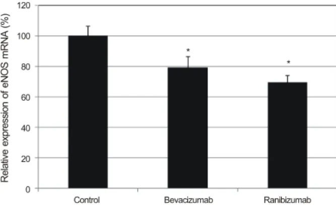
Figure 4. Effects on the expression of eNOS mRNA after ex-
posed to bevacizumab and ranibizumab in trabecular meshwork cells. Both bevacizumab and ranibizumab decreased eNOS ex- pression (*p < 0.05), and the degree of eNOS expression was not different significantly between bevacizumab and ranibizu- mab (p = 0.067).내어 서로 연접하며 약간 길다란 모양의 세포체를 가지는 편평한 모양의 특징적 형태학적인 양상과 섬유주 조직의 이식편 주위에서 위성양상으로 자라나는 섬유주세포의 특 징적인 성장양상으로 확인하였다.12,13
VEGF가 섬유주세포에서 NO의 생성에 미치는 영향 VEGF는 약물처리를 하지 않은 대조군에 비하여 배지에 서의 nitrite 생성을 유의하게 증가시켰다(p<0.05) (Fig. 1).
500 nM wortmanin과 1 μM Akt 저해제를 전처치한 후 VEGF에 노출시켰을 때 배지에서의 nitrite 생성이 각각 유 의하게 감소하였다(p<0.05).
anti-VEGF가 섬유주세포에서 NO의 생성에 미치는 영향 약물처리를 하지 않은 대조군에 비하여 베바시주맙과 라 니비주맙은 배지에서의 nitrite 생성을 유의하게 감소시켰다
(p=0.025, 0.020) (Fig. 2). 그러나 NO 생성 감소의 정도를 비교해보면 베바시주맙과 라니비주맙은 유의한 차이를 보 이지 않았다(p=0.376).
anti-VEGF가 eNOS mRNA의 발현에 미치는 영향 약물처리를 하지 않은 대조군에 비하여 베바시주맙과 라니 비주맙 모두 eNOS mRNA의 발현을 감소시켰다(Fig. 3, 4).
그러나 베바시주맙에 비해 라니비주맙은 eNOS mRNA의 발현을 조금 더 감소시키기는 하였으나 통계적으로 유의할
* *
정도의 차이는 보이지 않았다(p=0.067).
고 찰
본 연구의 결과는 사람의 섬유주세포에서 베바시주맙과 라니비주맙 모두 eNOS의 활성을 감소시키고 NO의 생성을 억제할 가능성이 있음을 보여주고 있다.
VEGF는 당뇨망막증과 망막정맥폐쇄에 의한 신생혈관을 촉진하는 작용과 혈관투과성을 촉진하는 작용을 하며 그 작용기전에 NO가 관여하는 것으로 알려졌다.2,3 VEGF는 혈관내피세포에서 eNOS의 활성을 증가시키고 혈관평활근 세포에서 단백분해효소(matrix metalloproteinase)의 활성을 증가시키며14 여러 성장인자의 작용에도 영향을 미친다. 혈 관내피세포에서 VEGF가 작용하는 신호전달경로는 tyrosine kinase와 phosphatidylinositol 3-kinase가 관여한다.1,15 저자 들은 섬유주세포에서도 이러한 경로가 작용한다는 보고를 한 바 있으며,16 본 실험에서도 wortmanin과 Akt 저해제가 VEGF에 의한 NO의 생성을 억제한다는 것을 확인하였다.
안구 내의 여러 가지 신생혈관질환에 대해 다양한 종류 의 항VEGF 항체를 개발하여 치료에 이용하고 있으며 그중 베비시주맙과 라니비주맙이 많이 사용되고 있는데 두 약제 의 구조적 차이와 효과를 비교한 다양한 연구들이 이미 보 고되어 있다.17,18
베바시주맙은 재조합 단일클론 IgG 항체이며 라니비주 맙은 단일클론 항체의 Fab fagmenrt (IgG1)로 되어 있다.4 이러한 약제들이 안구 조직에 미치는 영향은 이미 보고되 어 있는데17 임상적으로 사용되는 농도에서는 베바비주맙 은 독성을 유발하지 않는다는 보고도 있으나 고농도에 노 출될 경우 섬유주세포에서 베바시주맙에 의해 독성이 유발 되며, 라니비주맙에 비해 베바시주맙이 독성을 유발한다는 보고도 있다.18,19,20
이러한 약제들의 부작용의 하나로 안압상승이 초래될 수
있는데,4,21,22 안압상승의 기전은 두 약제의 구조적 차이나
염증반응 등이 제시되기는 하였으나 아직 명확하게 밝혀지 지 않았다.23 그중의 하나로 생각해 볼 수 있는 것은 an- ti-VEGF는 NO의 생성을 억제하는 작용이 있으므로 섬유주 에 노출될 경우 방수유출을 억제함으로써 안압상승을 유발 하는 원인이 될 가능성도 있다.24,25 따라서 본 연구에서는 두 약제가 eNOS의 발현과 NO 생성 억제에 미치는 영향을 알아보고, 또한 두 약제 중 어느 약제가 더 많은 영향을 미 치는지 알아보고자 하였다.
본 실험의 결과에서 베비시주맙과 라니비주맙 모두 eNOS 의 발현과 NO 생성을 억제하였으므로 섬유주를 수축시켜 안압상승을 초래할 가능성도 있을 것이다. 또한 이러한 기
전에 근거하여 두 약제가 안압상승에 미치는 영향을 간접 적으로 비교하기 위하여 eNOS의 발현과 NO 생성을 비교 한 결과 라니비주맙이 베바시주맙에 비해 eNOS의 발현을 좀 더 억제하기는 하였으나 유의할 정도의 차이를 나타내 지는 않았으며, NO의 생성억제 정도에 있어서도 두 약제 간에 차이를 보이지 않았다. 따라서 임상적으로는 NO의 생 성억제에 의해 안압상승에 미치는 영향은 두 약제 간에 차 이가 없을 가능성이 있을 것으로 생각한다. 본 연구의 단점 으로는 단백질의 발현을 비교해 보지는 못했으나 이전의 연구에서 eNOS의 발현과 그에 의한 단백질의 증가가 일치 하는 것으로 보고되어 있으므로 단백질의 발현도 증가할 것으로 추정된다.2
결론적으로 섬유주세포에서 베바시주맙과 라니비주맙은 eNOS의 활성을 감소시키고 NO의 생성을 억제하여 안압을 상승시킬 가능성이 있다. 또한 이러한 안압상승의 기전에 근거하면 두 약제 간에 안압상승의 정도는 차이를 나타내 지 않았다. 따라서 베바시주맙과 라니비주맙 모두 섬유주 를 통한 방수유출을 감소시킬 가능성이 있다. 그러나 모든 경우에서 안압상승이 유발되는 것은 아니며, 또 다른 기전 에 의해서도 안압상승을 유발할 수 있으므로 실험실 내에 서 행한 본 연구의 결과가 임상적인 의미를 가지기 위해서 는 향후 보다 자세한 연구가 필요할 것으로 생각한다.
REFERENCES
1) Ferrara N, Davis-Smyth T. The biology of vascular endothelial growth factor. Endocrine Rev 1997;18:4-25.
2) Bouloumié A, Schini-Kerth VB, Busse R. Vascular endothelial growth factor up-regulates nitric oxide synthase expression in en- dothelial cells. Cardiovasc Res 1999;41:773-80.
3) Dulak J, Józkowicz A, Dembinska-Kiec A, et al. Nitric oxide in- duces the synthesis of vascular endothelial growth factor by rat vascular smooth muscle cells. Arteioscler Thromb Vasc Biol 2000;
20:659-66.
4) Good TJ, Kimura AE, Mandava N, Kahook MY. Sustained ele- vation of intraocular pressure after intravitreal injections of an- ti-VEGF agents. Br J Ophthalmol 2011;95:1111-4.
5) Nathanson JA, McKee M. Identification of an extensive system of nitric oxide-producing cells in the ciliary muscle and outflow path- way of the human eye. Invest Ophthalmol Vis Sci 1995;36:1765-73.
6) Geyer O, Podos SM, Mittag T. Nitric oxide synthase activity in tis- sues of the bovine eye. Graefes Arch Clin Exp Ophthalmol 1997;
235:786-93.
7) Meyer P, Champion C, Schlotzer-Schrehardt U, et al. Localization of nitric oxide synthase isoforms in porcine ocular tissues. Curr Eye Res 1999;18:375-80.
8) Wiederholt M, Sturm A, Lepple-Wienhues A. Relaxation of tra- becular meshwork and ciliary muscle by release of nitric oxide.
Invest Ophthalmol Vis Sci 1994;35:2515-20.
9) Behar-Cohen FF, Goureau O, D’Hermies F, Courtois Y. Decreased
= 국문초록 =
섬유주세포에서 베바시주맙과 라니비주맙이 일산화질소합성효소의 발현에 미치는 영향
목적: 섬유주세포에서 항혈관내피생성인자인 베바시주맙과 라니비주맙이 eNOS의 발현에 미치는 영향을 비교해 보고자 하였다.
대상과 방법: 사람의 섬유주세포를 일차배양한 후 1% 혈청이 포함된 배지를 이용하여 베바시주맙과 라니비주맙에 0.5 mg/mL의 농도 로 각각 30분간 노출시킨 후 RT-PCR을 이용하여 eNOS mRNA의 발현을 조사하였다. 또한 20 ng/mL 혈관내피생성인자와 0.5 mg/mL 베바시주맙과 라니비주맙에 각각 노출시킨 후 Griess assay를 이용하여 일산화질소의 생성량도 함께 측정하였다.
결과: 섬유주세포에서 베바시주맙과 라니비주맙은 eNOS mRNA의 발현과 일산화질소의 생성을 감소시켰으며(p<0.05), eNOS의 발현 정도와 일산화질소 생성 정도에서 베바시주맙과 라니비주맙은 유의한 차이를 보이지 않았다.
결론: 섬유주세포에서 베바시주맙과 라니비주맙은 eNOS의 발현을 감소시켰으며, 그 정도에 있어서는 두 약제에서 차이를 나타내지 않았다.
<대한안과학회지 2014;55(8):1208-1212>
intraocular pressure induced by nitric oxide donors is correlated to nitrite production in the rabbit eye. Invest Ophthalmol Vis Sci 1996;37:1711-5.
10) Nathanson JA, McKee M. Alterations of ocular nitric oxide syn- thase in human glaucoma. Invest Ophthalmol Vis Sci 1995;36:
1774-84.
11) Green LC, Wagner DA, Glogowski J, et al. Analysis of nitrate, ni- trite and [15N]nitrate in biological fluids. Anal Biochem 1982;
126:131-8.
12) Polansky JR, Weinreb RN, Baxter JD, Alvarado J. Human tra- becular cells. I. Establishment in tissue culture and growth characteristics. Invest Ophthalmol Vis Sci 1979;18:1043-9.
13) Alvarado JA, Wood I, Polansky JR. Human trabecular cells. II.
Growth pattern and ultrastructural characteristics. Invest Ophthalmol Vis Sci 1982;23:464-78.
14) Wang H, Keiser JA. Vascular endothelial growth factor upregulates the expression of matrix metalloproteinases in vascular smooth muscle cells: role of flt-1. Circ Res 1998;83:832-40.
15) Xia P, Aiello LP, Ishii H, et al. Characterization of vascular endo- thelial growth factor’s effect on the activation of protein kinase C, its isoforms, and endothelial cell growth. J Clin Invest 1996;98:
2018-26.
16) Yoon SH, Kim JW. A study of the pathway of nitric oxide pro- duction by nitroglycerin in trabecular meshwork cells. J Korean Ophthalmol Soc 2013;54:1429-34.
17) Spitzer MS, Yoeruek E, Sierra A, et al. Comparative antiproliferative and cytotoxic profile of bevacizumab (Avastin), pegaptanib (Macugen)
and ranibizumab (Lucentis) on different ocular cells. Graefes Arch Clin Exp Ophthalmol 2007;245:1837-42.
18) Kahook MY, Ammar DA. In vitro effects of antivascular endothe- lial growth factors on cultured human trabecular meshwork cells. J Glaucoma 2010;19:437-41.
19) Kim SH, Kim JW. Effect of bevacizumab on survival and pro- duction of nitric oxide in trabecular meshwork cells. J Korean Ophthalmol Sci 2009;50:1404-8.
20) Kernt M, Welge-Lüssen U, Yu A, et al. [Bevacizumab is not toxic to human anterior- and posterior-segment cultured cells].
Ophthalmologe 2007;104:965-71.
21) Kahook MY, Kimura AE, Wong LJ, et al. Sustained elevation of in- traocular pressure associated with intravitreal bevacizumab injections.
Ophthalmic Surg Lasers Imaging 2009;40:293-5.
22) Bakri SJ, McCannel CA, Edwards AO, Moshfeghi DM. Persistent ocular hypertension following intravitreal ranibizumab. Graefes Arch Clin Exp Ophthalmol 2008;246:955-8.
23) Ladas JG, Yu F, Loo R, et al. Relationship between aqueous humor protein level and outflow facility in patients with uveitis. Invest Ophthalmol Vis Sci 2001;42:2584-8.
24) Schuman JS, Erickson K, Nathanson JA. Nitrovasodilator effects on intraocular pressure and outflow facility in monkeys. Exp Eye Res 1994;58:99-105.
25) Wang RF, Podos SM. Effect of the topical application of nitro- glycerin on intraocular pressure in normal and glaucomatous monkeys. Exp Eye Res 1995;60:337-9.