서 론
파킨슨병( P D )은 5 0대 이후에 발병하여 점차 진행되는 퇴 행성 신경질환으로서, 진전(resting tremor), 경직( r i g i d i-
ty), 운동완서(bradykinesia) 등이 대표적인 증상이나 이 외에도 보폭이 좁고 끄는 듯한 걸음걸이, 높낮이가 없는 음 성, 무표정 등의 증상을 나타낸다.1이 질병은 중뇌 내의 흑 질(substantia nigra pars compacta, SNc)에 위치한 도파민성 신경세포(dopaminergic neuron)의 점진적인 퇴화로 인하여 발병한다. 이러한 퇴화는 S N c에서 신경섬유 를 투사하고 있는 선조체( s t r i a t u m )에서 신경전달체인 도 파민의 결핍을 초래한다. 이런 병원적인 퇴화는 선조체 부위 에서 도파민의 감소의 직접적인 원인이 되며 도파민의 감소 가 80% 이상에 달할 경우 P D의 병세를 나타내며 일반인에 서도 8 0세에 이르러서는 약 50% 정도의 감소를 보이지만
Tyrosine Hydroxylase 조절부위의 인위적 변이가 단백질의 안정성과 효소적 특성에 미치는 영향
계명대학교 의과대학 신경과학교실, 서울대학교 의과대학 약리학교실*, 가천의과대학 신경과학연구소†
유영수 이상도 임정근 김용식* 남은주* 주완석* 이이언† 이영재†
Effects of Deletions in the Regulatory Domain on the Stability and Enzymatic Characteristics of Tyrosine Hydroxylase
Young-Soo Yoo, M.D., Sang-Doe Yi, M.D., Jung-Kun Lim, M.D., Yong-Sik Kim, M.D.*, Eun-Joo Nam, M.S.*, Wan-Suk Joo, Ph.D.*, Uhn Lee, M.D.
†, Young-Jae Lee, Ph.D.
†Department of Neurology, Keimyung University, Department of Pharmacology, Seoul National University*, Neuroscience Research Institute, Gachon Medical School†
Background : Various vectors have been developed and tried for the delivery of tyrosine hydroxylase (TH) in order to supplement dopamine, which is severely deficient in Parkinson’s disease, however, none of the protocols tried have yielded fruitful results that can be applied directly to humans. One of the problems revealed from previous trials was a short duration of expression of the delivered gene, that is, tyrosine hydroxylase. Methods : To extend the stability and to improve the enzymatic characteristics of the protein, part of the regulatory domain was deleted via PCR technique.
The cDNA for regulatory domain-deleted THs (dTH) were sub-cloned into a retroviral vector and the resulting recom- binant retrovirus was used to infect NIH-3T3. After selection, expression levels of TH were determined by Western blot analysis and the enzymatic characteristics were examined. Results : The deletion increased steady state expression level of TH protein by 7-fold for d19TH (TH with amino acids #2-19 are deleted) and 3-fold for d31TH (TH with amino acids #2-31 are deleted. The elevated expression level of d19TH is likely due to the enhanced stability of the protein as determined by a treatment of cycloheximide. The activity of d19TH was also increased approximately by 3-fold but no increase of the L-dopa production was observed. However, the production of L-dopa was dramatically increased when GTP cyclohydrolase I (GTPCH I) was co-transfected suggesting that the activity of d19TH is dependent on the presence of cofactor. d19TH seem to be free of feedback inhibition at low concentration of dopamine (10 nM~1 µM) but more sensitive to the inhibition at high concentration of dopamine (10 mM). Conclusions : The deletion of 18 amino acids on the regulatory domain increases the stability of the protein, reduces the activity, and frees it from the feedback inhibi- tion by the end product.
J Korean Neurol Assoc 20(1):60~66, 2002
Key Words : Parkinson’s disease, Tyrosine hydroxylase, Regulatory domain, Stability, L-dopa production, Feedback inhibition.
Manuscript received August 29, 2001.
Accepted in final form November 22, 2001.
* Address for correspondence Young-Jae Lee, Ph.D.
Neuroscience Research Institute, Gachon Medical School Gilsang-myun, Gangwha-gun, Inchon, 417-840, Korea Tel : +82-32-930-5045 Fax : +82-32-930-5007 E-mail : [email protected]
이 경우 병세는 나타나지 않는다.2
파킨슨병의 치료는 약물 치료 외에 수술, 조직 이식 등이 시도되고 있다. 이식 조직으로는 태아의 중뇌조직( f e t a l mesencephalic tissue),3 , 4경동맥소체(carotid body),5 m i c r o c a r r i e r에 쌓인 부신피질(adrenal medulla) 조직6 외에도 도파민을 생산하도록 유전적 변형이 된 세포 등이 이용된다. 그러나 이들은 실용성, 윤리적인 문제 혹은 도파 민 생성의 한시성 등의 문제점들을 갖고 있어서 이를 해결 하기 위하여서는 많은 연구를 필요로 하고있다. Tyrosine hydroxylase (TH)를 이용한 유전자 변형세포의 이식법 역시 TH 발현이 한시성인 문제가 있다.
도파민의 합성은 주로 중뇌에서 일어나며 c a t e c h o l a m i n e 생합성의 첫 단계는 tyrosine hydroxylase (tyrosine 3- monoxygenase, EC 1.14.16.2)에 의한 t y r o s i n e의 수 산화이다. 그 산물(L-dopa, L-3,4-dihydroxyphenylala- n i n e )은 다시 aromatic amino acid decarboxylase ( A A D C )에 의해 도파민으로 전환되어 n o r e p i n e p h r i n e과 epinephrine 등의 합성에 선구물질로 작용한다.7 이 생합성 과정에서 T H는 가장 첫 번째 효소이며 철분과 조효소 (tetrahydrobiopterin, BH4)을 필요로 하고 t y r o s i n e , L-dopa, catecholamine, 인산화 등에 의해 대단히 엄격하 게 조절된다.8
T H는 N - t e r m i n a l의 조절부위와 C-terminal 쪽의 효소 부위로 이루어져 있으며 sodium dodecyl sulfate-poly- acrylamide gel electrophoresis (SDS-PAGE) 상에서 약 60 kD으로 나타난다. 이의 조절부위에는 네 개의 s e r- ine/threonine (amino acid # 8, 19, 31, 40)이 있고 이들은 각기 다른 k i n a s e에 의해 인산화된다.8 , 9 S e r 8은 cell cycle dependent kinase (cdc2/cyclinA)에 의해 인산화되고 이것은 TH 활성과는 무관한 것으로 생각되고있 다.1 0 한편 S e r 1 9는 calmodulin-dependent protein kinase II (CaM-PKII)11 혹은 MAP-kinase activated protein kinase (MAPKAP kinase)에 의해 인산화되지 만1 2 이의 역할 또한 정확히 알려져 있지 않다. Ser31은 protein kinase C (PKC)와 mitogen-activated pro- tein kinase (MAP kinase 1)와 MAP kinase 21 3에 의 해 인산화될 수 있으며 H a y c o c k1 4과 Mitchell 등1 5은 PC12 cell에서 P K C의 활성제인 p h o b o l e s t e r에 의해 S e r 3 1이 인산화되고 이로 인하여 그 활성이 2배정도 증가 된다고 발표하였다.1 6 이와 같이 p h o b o l e s t e r에 의한 P K C 의 활성화, 이에 의한 T H의 인산화, PKC에 의한 M A P k i n a s e의 활성화 등은 T H의 활성이 다른 신호전달체계와
“c r o s s - t a l k”에도 관여할지 모른다는 가능성을 시사한다.
S e r 4 0은 다양한 kinase (PKA, PKC, CaM-PKII)에 의해 인산화될 수 있으며 그 다양한 k i n a s e의 수만큼 이의 조절 도 복잡하게 이루어진다. 일반적으로 protein kinase A ( P K A )에 의한 S e r 4 0의 인산화는 조효소( B H4)에 대한 미 카엘리스 상수( Km)을 줄이고 도파민 등 c a t e c h o l a m i n e에 대한 억제상수( Ki)를 높여서 전체적인 효소의 활성을 높이는 것으로 알려져 있다.1 7 , 1 8 그러나 T H를 P K C나 C a M - P K I I로
인산화하였을 때는 위의 결과와 다르게 나타나고 있다.1 9 인산화된 T H는 쉽게 p r o t e a s e의 표적이 되어 분해된 다.2 0 이는 필요 이상의 도파민 생성을 억제하여 도파민이 자가산화 됨으로 일어나는 신경세포에 대한 손상을 줄일 수 있도록 하기 위함이다.2 1 조효소 B H4는 TH 특이성 p h o s- p h a t a s e를 활성화시켜 T H를 탈 인산화하고 p r o t e a s e의 공격을 피할 수 있도록 하여 단백질의 안정성을 증가시킬 수 있다.2 0 , 2 1 즉 T H의 s e r i n e에 첨가된 인산기는 p r o- t e a s e의 표적이 될 수 있다는 점이다. 본 연구에서는 T H 단백질의 비 인산화가 본 단백질의 안정성을 향상시킬수 있 는지를 확인하기 위하여 조절부위의 인산화 자리를 삭제하 고 steady state에서 이들의 발현정도, 활성 및 L - d o p a 의 생성을 비교하였다. 또한 c y c l o h e x i m i d e를 처리한 세 포에서 일정시간 후 잔여 단백질의 양을 확인함으로 인산화 자리의 삭제가 단백질의 안정성에 미치는 영향을 조사하고 end product 인 도파민에 의한 feedback inhibition 여부를 조사하였다.
대상과 방법
1. TH의 조절부위에 인위적 변이의 도입과 r e t r o v i r a l v e c t o r의 제작
조절부위에 인위적 변이를 가진 TH (dTH)는 이후의 s u b c l o n i n g을 용이하게 하기 위해 PCR 증폭 시 H i n d III 제한효소 자리를 삽입한다. 각 s e r i n e / t h r e o n i n e까지 잘라지는 변이는 아래의 각 p r i m e r와 wTHxh (5’
GCGCACCTCGAGGCGCAC 3’)를 사용하여 P C R로 만 든다. Primer의 염기서열 중 Hind III와 i n i t i a t i o n c o d o n까지는 주형( t e m p l a t e )과 수소결합을 이루지 못하 므로 P C R의 처음 2~3 cycle은 낮은 온도( 5 5°C )에서 a n n e a l i n g하여 P C R하고 나머지 25 cycle은 온도를 높 여( 6 3°C) 특이성을 높인다.
- Serine 19 까지 잘라지는 변이
dTH19D; 5’ A C A A G C T T G C C A T G G A G C T G- GACGCCAAGCAGGCA 3’
- Serine 31 까지 잘라지는 변이
dTH31D; 5’ A C A A G C T T G C C A T G C C G C G G T T CATTGGGCGCAGG 3’
위의 각 P C R에 의해 만들어진 PCR 산물은 p C R 2 . 1 ( I n v i t r o g e n )에 s u b c l o n e하고 염기서열을 확인한 후 Hind III로 잘라서 같은 효소로 처리된 pLNCX (retrovi- ral vector)에 s u b c l o n e한다. 본 실험에서 w T H는 w i l d type TH를 나타내며 P C R에 의해 serine 19까지 잘라진 돌연변이 T H는 d 1 9 T H로 serine 31까지 잘라진 돌연변 이 T H는 d 3 1 T H로 명명하였다.
2. Transfection과 재조합 r e t r o v i r u s의 생산
5×1 05의 PA317 (virus producing cell line)은 6 cm 배양접시에 HAT 배양액으로 배양하다가 t r a n s f e c t i o n 시 O P T I - M E M으로 갈아준다. 12 μg의 D N A는 500 μl의 C a C l2/HEPES (125 mM CaCl2, 0.74 mM Na2H P O4,
140 mM NaCl, 25 mM HEPES) 용액에 부유하고 상온에 서 3 0분 정도 두어 침전이 생기게 한다. 이는 세포 위에 한 방울씩 떨어뜨려 전체를 덮은 후 다시 4시간 동안 3 7°C에서 배양하고 P B S로 두 번 씻은 후 일반 배양액(HAT 배양액) 으로 갈아준다. 다음날 배양액을 일반 배양액으로 갈아주고 다시 하루 밤 지난 후 viral conditioned media는 0.45 μ m로 여과하여 불순물을 제거하고 - 8 0°C에서 보관한다.
3. 세포주의 유전자 변형(Virus 감염)
1×1 05의 감염될 세포( N I H - 3 T 3 )는 하루 전에 10 cm 배양접시에 배양하고 다음날 viral conditioned media 1 ml와 polybrene(4 μg / m l )을 더한다. 37°C 배양기에 서 밤새 감염시킨 후 viral media는 버리고 P B S로 씻는 다. 감염된 세포는 하루 더 배양한 후 TH 재조합 v i r u s로 감염된 경우 400 μg/ml G418을 GTPCH I 재조합 v i r u s로 감염된 경우는 200 μg/ml hygromycin B를 포 함한 선택 배양액(selection media) 으로 교환한다. 한편 각종 T H와 GTPCH I을 이중발현하는 세포주 w T H G C N , d19THGCN, d31THGCN의 경우 이들 두 가지 항생제를 모두 포함한 배양액을 사용한다. 선택 동안 배양액은 1주 일에 2번씩 갈아준다.
4. 단백질 발현의 측정(Western blot analysis) 세포는 t r y p s i n으로 처리한 후 수거하여 h e m a c y t o m e- t e r로 세포의 수를 세고 1×1 06 세포당 100 μl의 S D S lysis buffer (62.5 mM Tris, pH 6.8, 10% glyc- erol, 5% β-mercaptoethanol, 2.3% SDS)로 용해하고 1 0초간 s o n i c a t i o n한다. SDS-PAGE는 10%, stacking g e l은 pH 6.8, running gel은 pH 8.8로 한다.
P o l y m e r i z e된 g e l에 250 μg의 total protein에 해당하는 시료를 l o a d한 후 glycine running buffer (200 mM glycine, 30 mM Tris-B, pH 8.3, 1% SDS) 에서 120V, 18 mA로 3 ~ 4시간 영동한다. 영동이 끝난 g e l은 T o w b i n’s buffer (192 mM glycine, 25 mM Tris, pH 8.3, 0.1% SDS, 10% methanol)에서 1000 mA 로 1시간 동안 polyvinylidene difluoride (PVDF) m e m b r a n e으로 옮긴다. 단백질이 옮겨진 m e m b r a n e은 5% skim milk/TBST (10 mM Tris, pH 8.0, 150 mM NaCl, 0.1% Triton) 에서 한 시간 이상 b l o c k하고 1 : 1 0 0 0으로 희석시킨 1차 항체(anti-TH, 자가제조)로 다 시 한 시간 동안 처리한다. Membrane은 T B S T에서 4번 ( 5분, 5분, 15분, 15분) 이상 씻은 후 horseradish per- oxidase (HRP)가 결합된 2차 항체(Goat anti-rabbit, 1:2000, Amersham)로 다시 한 시간 처리하고 위와 같 은 방법으로 4번 이상 씻는다. 다 씻은 m e m b r a n e은 E C L kit (Amersham)로 발색하고 X-ray film에 노출한다.
5. TH의 활성 측정
시료는 5×1 06의 세포를 300 μl의 lysis buffer (10 mM potassium phosphate, pH 6.2, 0.2% Triton-
X 1 0 0 )로 l y s e하고 1 0초간 s o n i c a t e한다. 12,0000 xg 로 4°C 에서 1 5분간 원심분리한 후 상층액만 떠내어 측정 에 사용한다. 측정의 균일성을 도모하기 위해 각 시료는 bovine serum albumine (BSA) standard를 기준으로 각 시료 당 단백질 양을 Bio-Rad protein assay kit (BIO RAD)를 사용하여 측정하여 표준화시킨다. 활성의 측 정을 위해서는 먼저 반응당 5 μC i의 L - [ 3 , 5 -3H] tyrosine 을 질소가스 아래서 3 0분간 말린다. 건조된 r a d i o a c t i v e t y r o s i n e은 반응 당 50 μl의 반응액에 부유한다. 반응액은 5 mM DTT, 1 mM D,L-6-methyl-5,6,7,8-tetrahy- dropterin (6-MPH4), 165 μM tyrosine, 50 mM 2- [N-morpholino] ethanesulfonic acid, 3,000 units/ml catalase로 제조한다. 반응은 50 μl의 단백질 시료와 50 μl의 반응액을 섞어 3 7°C에서 2 0분간 반응시키 고 1N HCl로 만든 7.5% charcoal suspension 1 ml을 섞어서 반응을 중단시킨다. Reaction/charcoal mixture 는 v o r t e x한 후 원심분리하여 h y d r o x y l a t i o n에 의해 유 리된3H와 t y r o s i n e을 분리한다. 이때 t y r o s i n e은 c h a r- c o a l과 단단히 결합하여 침전물에 남게되고 유리된3H는 상 층액에 떠 있게 된다. 각 t u b e에서 일정 양(500 μl 정도)의 상층액을 떠내어 scintillation counting fluid와 섞어 방 사능을 측정한다. Feedback inhibition 여부는 활성측정 전에 도파민을 첨가하고 상온에서 1 0분간 incubation 한 후 활성을 측정한다.
6. L-dopa의 측정
배양접시에 자란 세포는 25 mM HEPES, 100 mM E D T A가 첨가된 E a r l’s balanced salt solution ( E B S S )으로 3 7°C에서 3 0분간 extract 한다. 용액과 c e l l은 따로 harvest 하고 0.4 M perchloric acid/0.1 mM EDTA되게 한 후 - 8 0°C에서 보관한다. Alumina e x t r a c t i o n시에는 위의 용액 300 μl에 20 mg의 a l u m i- n a와 315 μl의 Tris 용액(3 M Tris-base, 10 mM EDTA, 100 mM sodium sulfite, pH 8.0)을 첨가하 고 2분간 vortex 하고 다시 3 0초간 원심분리한다. 상층액 은 버리고 p e l l e t은 증류수로 4번 이상 씻은 후 300 μl의 0.1 M perchloric acid를 첨가한다. 다시 2분간 v o r t e x 한 후 1 5분간 원심분리하고 100 μl의 상층액은 C18 col- u m n을 이용하여 reverse-phase HPLC로 분리하고 ESA Coulochem II electrochemical detector를 이용 하여 L - d o p a의 양을 측정한다.
7. Stability 측정
1×1 06 세포를 p 6 0의 배양접시에 배양한 후 7 0 % e t h a n o l에 녹인 cycloheximide (5 μg / m l )를 첨가하고 4시간 혹은 1 0시간 후에 세포는 t r y p s i n을 처리하여 수거 한다. Pellet은 P B S로 씻고 50 μl의 SDS lysis solution 을 첨가한 후 s o n i c a t e하여 l y s e한다. Cycloheximide 처리 후 각 시간별로 남아있는 TH 단백질의 양은 w e s t- ern blot analysis로 측정한다.
결 과
조절부위의 인산화 자리의 제거가 단백질의 s t e a d y state 발현에 미치는 영향은 Western blot analysis로
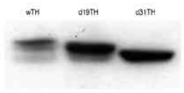
확인하였다. 단백질의 발현은, 정확한 이유는 알 수 없으 나, 상당히 큰 편차( 3 ~ 6 0배)를 보이고 있었으며 7차례의 실험에서 얻은 평균치는 d19TH 단백질의 발현이 w T H에 비해 약 7배 정도 증가하였다(Figure 1). 또한 조절부위 의 인위적 변이는 그 변이의 크기에 따라 T H의 발현이 다 르게 나타났으며 d 3 1 T H의 경우 w T H에 비해 약 3배의 TH 단백질을 발현하고 있었다(Figure 1). 한편 s t e a d y s t a t e에 있어서 단백질의 발현 정도는 t r a n s l a t i o n과 d e g r a d a t i o n의 평형으로 유지되며 어느 한쪽의 변화는 steady state의 발현정도에 영향을 미칠것으로 기대된다.
이를 확인하기 위하여 w T H와 d 1 9 T H를 발현하는 세포에 cycloheximide (CHX)를 처리하고 4시간 혹은 1 0시간 후에 세포를 수거하여 남아있는 TH 단백질의 양을 확인한 결과 w T H는 CHX 처리 4시간 후 겨우 감지할 수 있을 정 도만 남아있었고 10 시간이 지난 후 에는 거의 감지되지 않았다(Figure 2). 그러나 d 1 9 T H의 경우 CHX 처리 4 시간 후에는 대부분이 그대로 남아 있었으며 1 0시간이 지 났어도 거의 반 정도가 남아 있어서 d19TH 단백질이 w T H에 비해 안정성이 확연히 뛰어난 것으로 확인되었다.
한편 T H의 활성의 증가는 d 1 9 T H의 경우 w T H에 비해 약 3배 정도 높은 활성을 보였다(Figure 3). 그러나 wTH, d19TH 혹은 d 3 1 T H만을 발현하는 세포주는 아주 작은 양의 L-dopa (45~80 nM/mg protein)를 생산하 였으며 서로간에 통계적인 유의성을 나타내지 못하였다 (Figure 4). 이는 조효소의 부족으로 인하여 최소한의 활 성만 나타났기 때문이라 볼 수 있다. 그러나 wTHGC 혹은 d19THGC 세포주에서 GTPCH I에 의해 충분한 조효소가 공급되었을 때 L - d o p a의 생산은 7 ~ 9배( 3 2 0 ~ 4 5 0 nM/mg protein) 증가하였다. 한편 T H단백질의 활성은 Figure 1. The effect of deletions on the expression of TH pro-
teins. In order to see the effect of a removal of phosphorylation sites on the regulatory domain on the steady state expression level of TH protein, 18 amino acids (amino acid #2 through amino acid #19, d19TH) or 30 amino acids (amino acid #2 through amino acid #31, d31TH) are removed from the regula- tory domain using PCR technique and the resulting cDNAs were expressed in NIH-3T3 cells using retroviral transfection system. Total amount of TH proteins in each cell lines are determined using western blot analysis and densitometric analysis of the band intensities shows that d19TH expresses approximately 7-fold and d31TH approximately 3-fold more proteins than wTH. Shown above is the typical of 7 separate experiments. TH: tyrosine hydroxylase, wTH: wild type tyro- sine hydroxylase, d19TH: tyrosine hydroxylase with amino acids #2-19 are deleted, d31TH: tyrosine hydroxylase with amino acids #2-31 are deleted, PCR: polymerase chain reaction
Figure 2. The effect of deletion on the stability of TH protein.
To examine stability of TH proteins, cells are treated with cycloheximide and were harvested 4 or 10 hours after the treatment. After lysis, approximately 250 µg of total protein, measured using Bradford reagent, from each time point is loaded and the leftover TH protein is determined via western blot analysis. The deletion on the regulatory domain enhances the stability of the protein that approximately half of the d19TH protein is left 10 hrs after the treatment while wTH protein is almost completely degraded 4 hrs after the treat- ment. TH: tyrosine hydroxylase, wTH: wild type tyrosine hydroxylase, d19TH: tyrosine hydroxylase with amino acids
#2-19 are deleted
Figure 3. The activities of TH with deletion on the regulatory domain with or without GTPCH I. To measure the activities of both TH proteins with or without GTPCH I, cell lysates equiv- alent to 250 µg of total protein are used from each cell lines as enzyme source. The assay reveales that either deletion of 18 amino acids or presence of GTPCH I increases the activity by approximately 3-fold. However, effect of the deletion and the presence of GTPCH I are not additive, suggesting that deletion alone can increase the activity up to a maximum level which cells can tolerate. TH: tyrosine hydroxylase, GTPCH I: gua- nine triphosphate cyclohydrolase I
활성측정 전에 첨가한 도파민의 양에 따라 활성이 낮아졌다 (Figure 5). 이 경우 w T H는 도파민에 의한 f e e d b a c k i n h i b i t i o n에 의해 비교적 예민하게 반응하여 낮은 농도의 도파민에서 활성이 급격히 낮아져서 10 nM의 도파민이 첨 가된 경우 첨가되지 않은 경우에 비해 7 8 %의 활성을 나타 내었고 1 μM의 도파민이 첨가된 경우 7 3 %로 낮아졌다.
그러나 d 1 9 T H는 10 nM의 도파민이 첨가된 경우 9 6 %의 활성을 유지하고 있었으며 비교적 높은 농도의 도파민( 1 0 0 μM) 이 첨가된 후 활성이 5 6 %로 낮아졌다.
고 찰
파킨슨병은 약물 치료의 한시성과 부작용으로 인하여 세 포를 이용한 치료(cell therapy)혹은 도파민의 생성을 유 도할 수 있는 유전자 치료(gene therapy)의 필요성이 대 두되었다. 세포치료법에는 태아의 뇌조직을 환자의 뇌에 이 식하는 방법(fetal tissue graft)이 가장 먼저 시도되었으 며 그 효과 또한 상당히 장기간 지속되었으나 본 시술을 위 해서는 8 ~ 1 2주된 태아 1 0여 개체가 필요하여3 , 4 윤리적인 면에서나 실제적인 면에서 대단히 불합리한 방법이라 할 수 있겠다. 이외에 부신피질 세포,6 carotid body를 이용한 방법5 등이 시도되었으나 성공적이지는 못 하였다. 한편 유 전자를 이용한 치료법에는 도파민성 세포의 생존에 관여하 는 B D N F2 2 , 2 3 혹은 G D N F2 4 , 2 5 등의 신경영양인자( n e u- rotrophic factor), 도파민의 생성을 촉진할 수 있는
T H2 6와 A A D C2 7등이 시도되었다. 이 중에서 가장 널리 실 험된 것은 T H였으나 이의 효과 또한 만족할 만큼 장기간 지속되지는 못하였다. 이런 치료용 유전자의 발현을 장기화 하여 실용화를 이루기 위해서는 이들 유전자를 옮기는데 쓰 일 매개체(vector 혹은 l i p o s o m e의 구성요소), 형질변환 에 쓰일 세포의 특성 등이 깊이 있게 연구되어야 되겠지만 치료에 쓰일 유전자의 발현과 발현된 단백질의 활성 및 안 정성에 대한 연구가 우선되어야 할 것이다.
T H의 활성은 매우 다양한 방법으로 조절되며 이들 중 활 성에 가장 큰 영향을 미치는 두 가지가 조절부위의 인산화 와 생성된 도파민에 의한 feedback inhibition이다.8 T H의 조절부위에 위치한 s e r i n e은 이의 인산화에 따라 T H의 활성을 2배정도 향상시킬 수 있으나 인산화된 단백 질은 p r o t e a s e에 의해 쉽게 분해된다.8 그러므로 조절부위 의 인산화 자리를 포함한 d e l e t i o n은 T H의 인산화를 불가 능하게 만듦으로 낮은 활성을 보이는 반면 분해가 늦게 되 므로 비교적 높은 단백질 발현을 보일 것으로 기대된다. 본 실험에서 두 개의 인산화 자리가 삭제된 d 1 9 T H에서 발현 되는 단백질의 양은 w T H에 비해 7배 이상이나 높게 나타 났으며 약 3배 정도의 높은 활성을 보였다. 이것은 T H의 발현과 활성 측정시 각 세포주에서 얻은 전체 단백질의 양 을 측정하여 각 실험 시 동량을 사용하였으므로 d 1 9 T H를 발현하는 세포주로 만든 시료에는 w T H를 발현하는 세포주 로 만든 시료에서 보다 약 7배 정도의 많은 TH 단백질이 있다고 할 수 있다. 그러므로 전체의 활성이 약 3배 정도 증가하였으나 단위 단백질 당 활성을 비교하였을 때는 약 Figure 5. Feedback inhibition of TH activity by exogenously added dopamine. Modulation of TH activity by dopamine is examined using a wide rage of dopamine concentration added 10 minutes prior to the initiation of TH activity assay. At lower concentration of dopamine added, d19TH is more resis- tant to feedback inhibition but at higher concentration of dopamine, wTH becomes more resistant to the inhibition. The squares represent the activities of d19TH with a various amount of dopamine and the rhombuses represent those of wTH. The activity at each point is expressed as percentage of the activity without dopamine as 100%. TH: tyrosine hydroxy- lase, wTH: wild type tyrosine hydroxylase, d19TH: tyrosine hydroxylase with amino acids #2-19 are deleted
Figure 4. The effect of deletions and GTPCH I on the L-dopa production. Cells grown in a similar condition are harvested, lysed in 0.4 M perchloric acid, and filtered through 0.4 µM fil - ter. Catecholamines are extracted using alumina and the puri- fied samples are analysed in HPLC using C18 column fol- lowed by a detection with ESA Coulochem II electrochemical detector. Cells expressing wTH, d19TH or d31TH produce a basal level of L-dopa but a supply of cofactor BH4 by GTPCH I dramatically increase the intracellular L-dopa levels. The inability of the cells producing L-dopa without GTPCH I could be due to the shortage of cofactor. TH: tyrosine hydrox- ylase, wTH: wild type tyrosine hydroxylase, d19TH: tyrosine hydroxylase with amino acids #2-19 are deleted, d31TH: tyro- sine hydroxylase with amino acids #2-31 are deleted, GTPCH I: guanine triphosphate cyclohydrolase I, BH4: tetrahydro- biopterin
43% 정도( 3배÷7배)로 감소했다고 할 수 있다. 조절 부위 의 인산화는 T H의 활성에 중요한 요인이므로 인산화 자리 의 삭제는 활성감소의 한 요인이 될 수 있으며 본 실험의 결과는 기존의 연구와 일치한다고 할 수 있다.1 7 , 1 8 한편 wTH 단백질의 활성은 GTPCH I으로 인한 조효소 B H 4의 공급에 따라 약 3배 정도 증가(wTH 와 w T H G C의 경우) 한 반면 d 1 9 T H의 경우 GTPCH I에 의해 충분한 조효소 가 공급되어도 활성은 더 이상 향상되지 않았다( d 1 9 T H와 d 1 9 T H G C의 경우). 이는 d 1 9 T H의 활성이 조효소에 의 해 향상되지 않았다기 보다 활성측정 시 첨가하는 B H4의 analogue MPH4에 의해 세포가 나타낼 수 있는 최대한으 로 활성화되었으므로 더 이상의 조효소가 이의 활성을 더 증진시키지 못한 것으로 추정할 수 있다.
생성된 도파민에 의한 feedback inhibition은 필요 이 상의 도파민이 생겨나는 것을 막아서 이로 인하여 세포가 산화되는 것을 방지한다. 그러므로 활성이 높은 T H에 의한 과다한 도파민의 생산은 파킨슨병의 유전자 치료에는 오히 려 불리한 요소로 작용할 수 있으며, 낮은 농도이나 지속적 인 T H의 활성이 더 유리한 조건으로 사료된다. 조절부위에 인위적 변이를 가진 T H가 도파민에 의한 feedback inhi- b i t i o n여부는 cell lysate에는 각기 다른 농도의 도파민을 첨가하고 T H의 활성을 측정하였다. 그 결과 d 1 9 T H의 활 성은 낮은 농도의 도파민에 의해서는 feedback inhibi- t i o n을 적게 받아서 비교적 높은 활성을 유지하고 있지만 이의 농도가 증가하였을 때는 w T H보다 feedback inhi- b i t i o n에 더 예민해 졌다. 이는 d 1 9 T H에서 삭제된 부분이 과다하게 생성된 도파민에 의한 feedback 과 관여하는 부 분이라고 추론할 수 있으나 이에 대해서는 보다 더 정밀한 실험이 행해져야 할 것이다.
본 연구에서는 조절부위의 인위적 변이가 TH 단백질과 본 효소의 효소적 특성에 미치는 영향에 대하여 연구하였으 며 그 결과 조절부위의 변이는 TH 단백질의 발현 혹은 이의 안정성에 큰 영향을 미치고 있음을 발견하였다. 이러한 조건 은 현재 시도되고 있는 유전자치료의 결점을 한결 보완하여 보다 실용적인 방법의 개발에 공헌할 것으로 기대된다.
R E F E R E N C E S
11. Youdim MBH, Riederer P. Understanding Parkinson’s disease. Scientific American 1997;January:752-59.
12. Chiueh CC, Miyake H, Peng MT. Role of dopamine autoxidation, hydroxyl radical generation, and calcium overload in underlying mechanism involved in MPTP- induced Parkinsonism. Adv Neurol 1993;60:251-258.
13. Brundin P, Nilsson OG, Lindvall O, Astedt B, Bjorklund A. Behavioral effects of human fetal dopamine neurons grafted in a rat model of Parkinson’s disease. Exp Brain Res 1986;65:235-240.
14. Bjorklund A. Better cells for brain repair. N a t u r e 1 9 9 3 ; 3 6 2 : 4 1 4 - 4 1 5 .
15. Espejo EF, Montoro RF, Armengol JA, and Lopez-Barnneo J. Cellular and functional recovery of Parkinsonian rats after
intrastriatal transplantation of carotid body cell aggregates.
N e u r o n 1 9 9 8 ; 2 0 : 1 9 7 - 2 0 6 .
16. Borlongan CV, Saporta S, Sanberg PR. Intrastriatal trans- plantation of rat adrenal chromaffin cells seeded on micro- carrier beads promote long-term functional recovery in hemiparkinsonian rats. Exp Neurol 1998;151:203-214.
17. Kopin IJ, The Pharmacology of Parkinson’s disease: An update. Ann Rev Pharmacol Toxicol 1993;32:467-495.
18. Kumer SC, Vrana KE. Intricate regulation of tyrosine hydroxylase activity and gene expression. J Neurochem 1996;67:443-462.
19. Grima B, Lamouroux A, Blanot F, Biguet NF, Mallet J.
Complete coding sequence of rat tyrosine hydroxylase mRNA. Proc Natl Acad Sci 1985;82:617-621.
10. Ramsey AJ, Fitzpatrick PF. Effects of phosphorylation of serine 40 of tyrosine hydroxylase on binding of cate- cholamines: Evidence for a novel regulatory mechanism.
Biochemistry 1998;37:8980-8986.
11. Campbell DG, Hardie DG, Vulliet PR. Identification of the four phosphorylation sites in the N-terminal region of tyro- sine hydroxylase. J Biol Chem 2 6 1 ; 1 9 8 6 : 1 0 4 8 9 - 1 0 4 9 2 . 12. Sutherland C, Alterio J, Campbell DG, LeBourdelles B,
Mallet J, Haavik J, Cohen P. Phosphorylation and activa- tion of human tyrosine hydroxylase in vitro by mitogen- activated protein (MAP) kinase and MAP-kinase-activated kinases 1 and 2., Eur J Biochem 1993;217:715-722.
13. Haycock JW, Ahn NG, Cobb MH, Krebs EG. ERK1 and ERK2, two microtubule-associated protein 2 kinases, mediate the phosphorylation of tyrosine hydroxylase at serine-31 in situ. Proc Natl Acad Sci 1992;89:2365-2369.
14. Haycock JW. Phosphorylation of tyrosine hydroxylase in situ at serine 8, 19, 31, and 40. J Biol Chem 1 9 9 0 ; 2 6 5 : 1 1 6 8 2 - 1 1 6 9 1 .
15. Mitchell JP, Hardie DG, Vulliet PR. Site-directed phos- phorylation of tyrosine hydroxylase after KCl depolariza- tion and nerve growth factor treatment of PC12 cells. J Biol Chem 1990;265:22358-22364.
16. Halloran SM, Vulliet PR. Microtubule-associated protein kinase-2 phosphorylates and activates tyrosine hydroxy- lase following depolarization of bovine adrenal chromaffin cells. J Biol Chem 1994;269:30960-30965.
17. Zigmond RE, Schwarzschild MA. Rittenhouse AR. Acute regulation of tyrosine hydroxylase by nerve activity and by neurotransmitter via phosphorylation. Ann Rev Neurosci 1989;12:415-461.
18. Daubner SC, Lauriano C, Haycock JW, Fitzpatrick PF.
Site-directed mutagenesis of Serine 40 of rat tyrosine hydroxylase. J Biol Chem 1992;267:12639-12644.
19. Funakoshi H, Okuno S, Fujisawa H. Differential effects on acitvity caused by phosphorylatin of tyrosine hycroxylase at serine-40 by three multifunctional protein kinases. J Biol Chem 1991;58:2044-2052.
20. Vrana KE, Roskoski R. Tyrosine hydroxylase inactivation following cAMP-dependent phosphorylation activation. J
Neurochem 1983;40:1692-1700.
21. Ribeiro P, Kaufman S. The effect of tetrahydrobiopterin on the in situ phosphorylation of tyrosine hydroxylase in rat striatal synaptosomes. Neurochem Research 1 9 9 4 ; 1 9 : 5 4 1 - 5 4 8 .
22. Levivier M, Przedborski S, Bencsics C, Kang UJ. Intrastri- atal implantation of fibroblast genetically engineered to pro- duce brain-derived neurotrophic factor prevents degenera- tion of dopaminergic neurons in a rat model of Parkinson’s disease. J Neurosci 1 9 9 5 ; 1 5 : 7 8 1 0 - 7 8 2 0 .
23. Lucidi-Phillipi CA, Gage FH, Shylts CW, Jones KR, Reichardt LF, Kang UJ. Brain-derived neurotrophic fac- tor-transduced fibroblasts: Production of BDNF and effects of grafting to the adult rat brain. J Comp Neurol 1995;354:361-376.
24. Choi-Lundberg DL, Lin Q, Chang, YN, Chiang YL, Hay CM, Mohajeri H, Davidson BL, Bohn MC. Dopaminergic
nbeurons protected from degeneration by GDNF gene therapy. Science 1997;275:838-841.
25. Mandel RJ, Spratt SK, Snyder RO, and Leff SE. Midbrain injection of recombinant adeno-associated virus encoding rat glial cell line-derived neurotrophic factor protects nigral neurons in a progressive 6-hydroxydopamine- induced degeneration model of Parkinson’s disease in rats.
Proc Natl Acad Sci 1997;94:14083-14088.
26. Lundberg C, Horellou P, Malet J, Bjorklund A. Generation of DOPA-producing astrocytes by retroviral transduction of the human tyrosine hydroxylase gene: In vitro charac- terization and in vivo effects in the rat Parkinson model.
Exp Neurol 1996;139:39-53.
27. Moffat M, Harmon S, Haycock J, O’Malley K. L-dopa and dopamine-producing gene cassettes for gene therapy approaches to Parkinson’s disease. Exp Neurol 1 9 9 7 ; 1 4 4 : 6 9 - 7 3 .