CASE REPORT
Asymptomatic adults with isolated, unilateral right pulmonary
vein atresia: multidetector CT findings
1
Y KIM,
MD
,
PhD,
2I R YOO,
MD,
2M I AHN,
MD,
PhDand
2D H HAN,
MD,
PhD1Department of Radiology, School of Medicine, Ewha Women’s University, and2Department of Radiology, Seoul St.
Mary’s Hospital, College of Medicine, The Catholic University of Korea, Seoul, Korea
ABSTRACT. We report two cases of a very rare congenital anomaly, i.e. isolated unilateral pulmonary vein atresia. The patients were asymptomatic and the diagnosis was made using multidetector CT (MDCT), which also showed cyst formation in the right lung. Asymptomatic adult cases or association with cystic lung lesions have never been reported in this condition before.
Received 12 March 2010 Revised 24 June 2010 Accepted 26 July 2010 DOI: 10.1259/bjr/51344661
’2011 The British Institute of Radiology
Unilateral pulmonary vein atresia without associated congenital heart disease is a rare condition [1]. Patients with congenital unilateral pulmonary vein atresia are usually symptomatic and present with recurrent epi-sodes of pneumonia or haemoptysis in infancy or childhood [1]. Although a subclinical course of this anomaly has been recently reported in a 12-year-old boy, so far no asymptomatic adult cases have been described to our knowledge [1]. Also, the presence of pulmonary cysts has never been reported in this condition. We therefore report the CT findings of two asymptomatic adult patients with unilateral pulmonary vein atresia, with associated pulmonary cysts.
Case report
Case 1
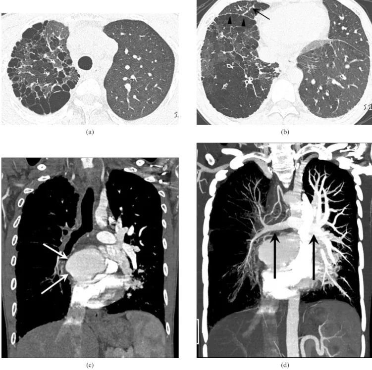
A previously healthy 23-year-old male visited the emergency room because of vomiting lasting 1 day. The chest radiograph obtained at the time showed diffuse interstitial infiltration in the right lung field. Multidetector CT (MDCT) showed a heterogeneous lung density, with areas of normal density, ground-glass opacity and small air-cysts (Figure 1a). The cysts were multiple, but predominantly distributed in the sub-pleural regions. Beaded thickening of the interlobular septum and nodularity along the fissures and costal pleura were noted in the right lower lung (Figure 1b). The volume of the right lung was not diminished. CT also showed a complete lack of the right pulmonary vein and enlargement of the left. The margin of the left atrium
where the right pulmonary vein was expected to be was entirely smooth, with no vascular structures connected to it (Figure 1c). The right pulmonary artery was slightly small, with poor contrast enhancement compared with the left pulmonary artery. The mediastinal branches of the right bronchial artery were markedly dilated, suggesting the presence of systemic arterial flow into the hypoplastic pulmonary arteries (Figure 1d). Anoma-lous pulmonary venous return was excluded on the basis of the absence of pulmonary venous structures con-nected to systemic venous circulation on CT.
Fibre-optic endoscopy revealed findings compatible with reflux oesophagitis and chronic superficial gastritis. The patient recalled being told that his chest radiography showed a ‘‘small right pulmonary artery’’. He did not complain of any respiratory, cardiac or other systemic symptoms, and refused further work-up. The patient was in good health 5 years after the initial emergency room visit.
Case 2
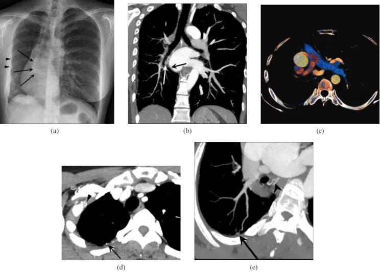
A 39-year-old female with rheumatoid arthritis recently underwent screening chest radiography in preparation for a clinical trial (Figure 2a). This showed a small right hemithorax with significant ipsilateral mediastinal shift, reduced vessel size in the hilar region and mild interstitial opacities in the periphery. Apparent thickening of the pleura was noted along the lateral margin of the right lung. Her shoulder radiographs, which were obtained 2 years previously and reviewed retrospectively, showed the same findings. MDCT was requested because pulmonary artery hypoplasia was suspected. CT revealed a lack of pulmonary vein, small right pulmonary artery, increased diameter of the bronchial artery (Figure 2b) and increased contrast enhancement of the right pulmonary artery compared with the left (Figure 2c). The mediastinal window image
Address correspondence to: Dae Hee Han, The Catholic University of Korea, Department of Radiology, Seoul St. Mary’s Hospital, 505 Banpo-dong, Seocho-gu, Seoul 137-701, Korea. E-mail: lepolder@ gmail.com
demonstrated a hypertrophied intercostal artery and collateral from the right subclavian artery (Figure 2d). Maximum intensity projection showed that the distal end of the intercostal branch runs into a peripheral branch of the pulmonary artery (Figure 2e). Anomalous pulmonary venous connection to systemic venous circulation was not demonstrated on CT.
The patient reported that she had been told about her congenital heart anomaly a long time ago, and did not
have any respiratory or cardiac symptoms. The patient refused further work-up.
Discussion
To our knowledge, fewer than 40 cases of unilateral pulmonary vein atresia without associated structural abnormalities of the heart have been reported in the
(a) (b)
(c) (d)
Figure 1. Asymptomatic 23-year-old male with unilateral isolated pulmonary vein atresia. (a) High-resolution CT scan in upper lung zone shows a heterogeneous lung density, with areas of normal density, ground-glass opacity and numerous air-cysts in the right upper lobe. (b) High-resolution CT scan in lower lung zone shows beaded thickening of the interlobular septum (arrow) in the right middle lobe and nodularity along the fissure (arrowheads) and pleura. Note that the volume of the affected lung has not been diminished. (c) Coronal CT shows that the right border of the left atrium is smooth (arrows), with no pulmonary veins in their expected locations. CT also shows enlarged pulmonary arteries (PA) and veins in the left lung. (d) Coronal maximum intensity projection shows decreased contrast enhancement of right PA compared with left PA.
medical literature [1–6]. This anomaly seems to have no particular predilection towards either side [1, 2, 4]. In these reports, a broad spectrum of clinical severity has been described, ranging from none [7] to recurrent pulmonary infection to severe haemoptysis to death, although fatal outcomes have been confined to those diagnosed before the age of 8 years [2]. In most patients, unilateral PV atresia is diagnosed during infancy [1] and, to our knowledge, only six (including ours) adult cases of this condition have been reported. Four of them, previously reported by Pourmoghadam et al [2] and Heyneman et al [4], presented with serious respiratory symptoms, such as significant haemoptysis, recurrent pneumonia or progressive dyspnoea. For these reasons, as well as for the purpose of pulmonary hypertension prevention, pneumonectomy has been recommended for treatment of this condition [2, 5].
By contrast, both of our patients were asymptomatic, despite striking radiological abnormalities shown on CT. The implication of the current cases is that, as long as they are asymptomatic or mildly symptomatic, pneumo-nectomy may be deferred for a significant period of time. However, it is uncertain how long pneumonectomy can be delayed. Efforts should be made to predict and prevent the development of pulmonary artery (PA) hypertension in asymptomatic patients with this anom-aly, as in other congenital anomalies with left-to-right shunt. We therefore recommended further work-up in order to determine the amount of left-to-right shunt and the possibility of pulmonary hypertension, which was refused by both patients.
According to the literature review by Pourmoghadam et al [2], there were five cases, including their own, who had been followed up without surgical treatment for an
(a) (b) (c)
(d) (e)
Figure 2. Asymptomatic 39-year-old female with unilateral isolated pulmonary vein atresia. (a) Posterior–anterior chest radiograph shows a small right hemithorax, diminished vessel size in the right lung and interstitial infiltrations along the lateral and inferior zones of the right lung. Chest radiograph also shows apparent thickening of the pleura (arrowheads) and a smooth right border of the left atrium (arrow). The vascular shadows are engorged in the left lung. (b) Coronal maximum intensity projection (4 mm thickness) image shows a smooth left atrial border (black arrow) with no pulmonary vein, dilated bronchial artery (white arrow), and lack of scimitar vein. (c) Volume rendering (VR) image shows that the blood with increased contrast enhancement (orange) from the right PA is jetting into the blood with decreased contrast enhancement (blue) within the main and left PA. Note that the density of right PA is similar to that of the left pulmonary vein or the thoracic aorta. The difference in PA size is not striking, implying a significant amount of systemic pulmonary arterial shunt. (d) Axial image on mediastinal window shows a hypertrophied branch of the intercostal artery along the posterior chest wall (arrow) and systemic collateral from right subclavian artery (arrowhead). (e) Oblique maximum intensity projection shows that the distal end (arrow) of the intercostal branch shown on (d) runs into a peripheral branch of the pulmonary artery. Note hypertrophied hilar branch of the bronchial artery (arrowhead).
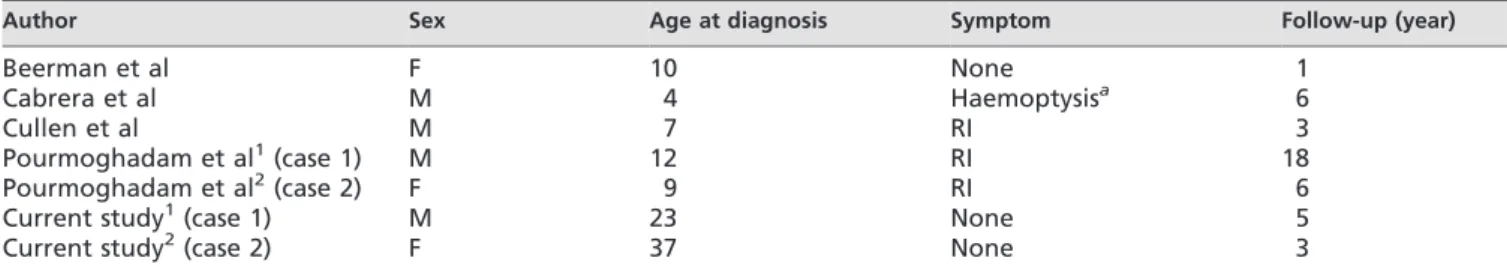
average period of 6.8 years (range 1–18 years) [2]. The clinical characteristics of those six patients have been summarised in Table 1, along with those of our patients. As in our adult cases, one of them (female, 10 years old) was asymptomatic [7]. Another (male, 4 years old) had been asymptomatic until he developed sudden haemop-tysis; the boy was subsequently followed up without surgery for at least 6 years [8]. Therefore, it seems that the time to develop symptomatic pulmonary hyperten-sion is variable among patients with unilateral pulmonary vein atresia, perhaps determined by the balance between the systemic vessels that supply and drain the affected lung, which would dictate the blood flow from the affected PA to the normal PA. The amount of lymphatic drainage may also play a role [2]. In the current case series, MDCT showed beaded thickening of the interlobular septa and nodularity along the fissure and pleura, which may represent systemic collaterals and engorged lympha-tic channels, and hypertrophied bronchial arteries. However, MDCT failed to show the severity of pulmon-ary hypertension or presence of venous drainage through the bronchial vein, which we felt were necessary to make a management decision. Here, although measurements of right and left PA pressures and their differences are available from catheter angiogram [9], quantification of systemic pulmonary shunt flow may be unreliable because of severe turbulence and heterogeneity of PA blood flow (Figure 2c). Likewise, four-dimensional MRI, despite its power in showing temporal change in contrast enhancement of the lung, may be unsuitable for this purpose. First-pass radionuclide angiography may serve this purpose better than catheter angiography or MRI because of its ability to calculate the amount of left-to-right shunt from the time–activity curve obtained from the unaffected lung.
A third management option in PV atresia, besides follow-up and pneumonectomy, is coil embolisation of the systemic arterial collaterals. In the article by Heyneman et al [4] this procedure controlled haemoptysis in one of the three adult patients, who became asymptomatic afterwards. After treating right PV atresia with transseptal percutaneous transluminal angioplasty and stent implan-tation, Ussia et al [5] performed embolisation of systemic pulmonary collateral arising from the descending thoracic aorta. Interestingly, coil embolisation or surgical ligation of systemic arterial collateral has been performed for management of pulmonary artery hypertension in chil-dren with a similar disorder, i.e. Scimitar syndrome [10].
In our series, chest radiographs did not allow correct differentiation between unilateral PA atresia and the Scimitar variant. However, using MDCT, a prompt
diagnosis was made, with exclusion of both normal and rudimentary pulmonary veins. To understand the haemodynamics of PA from MDCT findings, it helps to compare the densities of PAs with those of other systemic or pulmonary vessels. In the female patient, the contrast enhancement of the affected PA was increased compared with the contralateral counterpart, whereas in the male patient, as well as in the cases reported by Mataciunas et al [1] and Pourmoghadam et al [2], the density difference was in the opposite direction. In fact, the density of PA can be either increased or decreased in the affected lung — they simply parallel the contrast enhancement of systemic arteries.
In one of our patients, MDCT showed cyst formation in the affected lung. Such findings have not been previously described in patients with isolated PV atresia. However, since similar findings have been reported in paediatric cases of PV atresia associated with cardiac anomaly [11], as well as in bilateral Scimitar syndrome [12], we believe that they are not incidental findings but a radiological manifestation of this condition, representing destruction or underdevelopment of capillary networks at the alveolar level owing to inadequate pulmonary arterial blood supply or pressure damage from the high-pressure systemic collaterals.
Conclusion
We report two asymptomatic adult cases of isolated unilateral pulmonary vein atresia with pulmonary cysts. MDCT was the main diagnostic investigation in our patients, which provided accurate anatomical data for a correct diagnosis but no functional data necessary for a management decision. Unilateral pulmonary vein atresia encompasses a broad spectrum of clinical severity, and management may require a more individualised approach in patients with no or mild symptoms.
References
1. Mataciunas M, Gumbiene L, Cibiras S, Tarutis V, Tamosiunas AE. CT angiography of mildly symptomatic, isolated, unilateral right pulmonary vein atresia. Pediatr Radiol 2009;39:1087–90.
2. Pourmoghadam KK, Moore JW, Khan M, Geary EM, Madan N, Wolfson BJ, et al. Congenital unilateral pulmon-ary venous atresia: definitive diagnosis and treatment. Pediatr Cardiol 2003;24:73–9.
3. Tissot C, Corbelli R, Aggoun Y, Beghetti M, da Cruz E. Bronchoscopic diagnosis of asymptomatic unilateral
Table 1. Age and clinical characteristics of follow-up patients with isolated unilateral pulmonary vein atresia (modified from Pourmoghadam et al)
Author Sex Age at diagnosis Symptom Follow-up (year)
Beerman et al F 10 None 1
Cabrera et al M 4 Haemoptysisa 6
Cullen et al M 7 RI 3
Pourmoghadam et al1(case 1) M 12 RI 18 Pourmoghadam et al2(case 2) F 9 RI 6 Current study1(case 1) M 23 None 5 Current study2(case 2) F 37 None 3
pulmonary vein atresia in an infant. Pediatr Cardiol 2008; 29:976–9.
4. Heyneman LE, Nolan RL, Harrison JK, McAdams HP. Congenital unilateral pulmonary vein atresia: Radiologic findings in three adult patients. AJR Am J Roentgenol 2001;177:681–5.
5. Ussia GP, Marasini M, Rimini A, Pongiglione G. Atresia of right pulmonary veins with intact atrial septum and major aorto-pulmonary collateral treated with percutaneous stent implantation and embolization. J Interv Cardiol 2004;17:183–7. 6. Wiebe S, Maclusky I, Manson D, Holowka S, Yoo SJ. Hemoptysis: A rare cause can be related to a bronchial varix due to pulmonary venous obstruction. Pediatr Radiol 2003;33:884–6.
7. Beerman LB, Oh KS, Park SC, Freed MD, Sondheimer HM, Fricker FJ, et al. Unilateral pulmonary vein atresia:
Clinical and radiographic spectrum. Pediatr Cardiol 1983;4: 105–12.
8. Cabrera A, Vazquez C, Lekuona I. Isolated atresia of the left pulmonary veins. Int J Cardiol 1985;7:298–302.
9. Cullen S, Deasy PF, Tempany E, Duff DF. Isolated pulmonary vein atresia. Br Heart J 1990;63:350–4.
10. Uthaman B, Abushaban L, Al-Qbandi M, Rathinasamy J. The impact of interruption of anomalous systemic arterial supply on scimitar syndrome presenting during infancy. Catheter Cardiovasc Interv 2008;71:671–8.
11. Kim WS, Yeon KM, Kim I, Han MC, Chi JG. Radiological evaluation of pulmonary vein obstruction including two examinations by magnetic resonance imaging. Pediatr Radiol 1993;23:6–11.
12. Kabbani M, Haider N, Abu-Sulaiman R. Bilateral scimitar syndrome. Cardiol Young 2004;14:447–9.