1. INTRODUCTION
Graphene (G)/semiconductor hybrid nanomaterials have demonstrated high potential in enhancing the photocatalytic efficiency of semiconductor nanomaterials [1-10]. There are various materials that show photocatalytic properties, among which, TiO
2is one of the most widely used heterogeneous photocatalysts [11]. It has excellent physical and chemical stability, electronic and optical properties, and is inexpensive.
However, TiO
2suffers from a large band gap (3.2 eV for anatase TiO
2) which requires activation by UV light, and a fast electron-hole recombination rate which reduces its photocatalytic efficiency [11,12].
G-TiO
2hybrid nanomaterials in the literature are mostly prepared by hydrothermal, solvothermal and hydrolysis
✉ Jaehyeung Park; [email protected] Copyright ©2019 KIEEME. All rights reserved.
This is an Open-Access article distributed under the terms of the Creative Commons Attribution Non-Commercial License (http://creativecommons.org/licenses/by-nc/3.0) which permits unrestricted non-commercial use, distribution, and reproduction in any medium, provided the original work is properly cited.
reactions [2,3,13-15]. In these cases, graphene oxide (GO) instead of pristine graphene was used. The hybrid materials were prepared by reducing GO in the presence of a TiO
2precursor [titanium (IV) isopropoxide, titanium (IV) fluoride, titanium (IV) butoxide], resulting in the deposition of TiO
2particles on the surface of reduced GO. Because reduced GO was used, oxygen-containing species remained and could not be removed completely even after reduction. These defects disrupt the conjugated structure and the electron conduction of graphene [16]. To overcome this issue, we propose to synthesize G-TiO
2photocatalysts using pristine graphene and pre-made TiO
2nanoparticles. The goal is to develop high performance G-TiO
2hybrid nanomaterials with enhanced photocatalytic properties under visible light.
In this hybrid nanomaterials, graphene (G) acts as an electron acceptor to ensure fast charge transfer. Effective charge separation can therefore be achieved to slow down the electron-hole recombination, and as a result, the photocatalytic activity of TiO
2is enhanced [2,8,17].
Moreover, because graphene can absorb light in the visible region, the photo-excitation wavelength can be extended from
Photocatalytic Performance of
Graphene-TiO 2 Hybrid Nanomaterials Under Visible Light
Jaehyeung Park
Division of Advanced Materials Engineering, Dong-Eui University, Busan 47340, Korea (Received January 3, 2019; Revised January 22, 2019; Accepted January 22, 2019)
Abstract: This study describes the development of graphene-TiO2 conjugates for the enhancement of the photocatalytic efficiency of TiO2. Graphene-based hybrid nanomaterials have attracted considerable attention because of the unique and advantageous properties of graphene. In the proposed hybrid nanomaterial, graphene serves as an electron acceptor to ensure fast charge transfer. Effective charge separation can, therefore, be achieved to slow down electron-hole recombination. This results in an enhancement of the photocatalytic activity of TiO2. In addition, increased adsorption and interactions with the adsorbed reagents also lead to an improvement in the photocatalytic activity of graphene-TiO2 hybrid nanomaterials.
The acquired result is encouraging in that the photocatalytic activity of TiO2 was initiated using visible light (630 nm) instead of the typical UV light.
Keywords: Graphene, TiO2, Photocatalyst, Hybrid nanomaterial
Regular Paper 161
J. Korean Inst. Electr. Electron. Mater. Eng.
Vol. 32, No. 2, pp. 161-164 March 2019 DOI: https://doi.org/10.4313/JKEM.2019.32.2.161 ISSN 1226-7945(Print), 2288-3258(Online)
J. Korean Inst. Electr. Electron. Mater. Eng., Vol. 32, No. 2, pp. 161-164, March 2019: J. Park 162
UV to visible (400~700 nm) [18]. An absorbed photon promotes an electron from graphene, then the electron transfer from graphene into TiO
2. Consequently, the photocatalytic reactions can be carried out using the visible light instead of UV. In addition, the large surface area of graphene will increase the concentration of adsorbed reagents as well as the surface interactions [1]. The increased adsorption and interactions with the adsorbed reagents lead to enhanced photocatalytic activities of G-TiO
2hybrid nanomaterials [2-5].
2. METHOD FOR EXPERIMENT 2.1 Materials
Methyl pentafluorobenzoate (>97%), sodium azide, graphite flakes (Sigma), AEROXIDE® TiO
2P25, acetone, ethyl ether, methanol, triethylamine (>99%), N-methyl-2- pyrrolidone (NMP), hydrochloric acid (37%) were purchased from Sigma-Aldrich, and were used as received without any further purification.
2.2 Preparation of PFPA-COOH
4-Azido-2,3,5,6-tetrafluorobenzoic acid (PFPA-COOH) was synthesized following the previously reported protocol [19-21].
Sodium azide was added into solution of methyl 2,3,4,5,6- pentafluorobenzoate in acetone/water (8:3) mixture and then refluxed for 8 h. The azide replaced F in the para-position through a nucleophilic aromatic substitution reaction. The resulting was then hydrolyzed to PFPA-COOH by stirring with 10% NaOH in methanol overnight at room temperature. The solution was neutralized by adding 1 M HCl into the solution until reached pH 3. The compound was extracted with chloroform followed by drying under sodium sulfate.
After filtration, the solvent was then removed under vacuum to obtain PFPA-COOH as a pale yellow solid.
2.3 Preparation of G-TiO
2hybrid nanomaterials
The TiO
2particles were functionalized with PFPA-COOH and then covalently conjugated on FLG flakes via UV irradiation for 30 min [22]. For detail, TiO
2nanoparticles
were placed in a solution of PFPA-COOH in methanol and the solution was stirred at room temperature for overnight. Unmodified TiO
2nanoparticles were removed by centrifuged by repeating 3 times. Liquid phase exfoliated few layer graphene were used in this research. A suspension of G flakes in NMP was mixed with a suspension of PFPA- TiO
2. The mixture was irradiated with 450 W medium pressure Hg lamp (Ace Glass Inc., Vineland, NJ, USA) for 30 min while stirring. A 280 nm long-path optical filter was placed on top of the samples during irradiation.
The resulting reaction mixture was then washed and centrifuged for 3 times.
3. RESULTS AND DISCUSSION
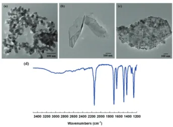
Figure 1 shows the transmission electron microscopy (TEM) images and IR spectrum of (a) PFPF-TiO
2, (b) G, (c) TiO
2modified G, and (d) PFPA-TiO
2. The average TiO
2nanoparticles size were 21 nm, measured by TEM.
In the IR spectra (Fig. 1(d)), the strong absorption at 2,130 and 1,700 cm
-1due to asymmetric stretch of the azido group and carbonyl group stretching indicating the TiO
2nanoparticles have been successfully functionalized with PFPA. After conjugation, nanoparticles were clearly visible on the graphene sheet (Fig. 1(c)).
The particles were placed throughout the sheet although possibility exists that the edges could fold over the basal plane. Control experiments using unfunctionalized TiO
2Fig. 1. TEM images of (a) PFPA-TiO2, (b) graphene flake, (c) G-TiO2, and IR spectra of (d) PFPA-TiO2.
전기전자재료학회논문지, 제32권 제2호 pp. 161-164, 2019년 3월: 박재형 163
nanoparticles were carried out under the same experimental conditions and almost no particles were observed on graphene flakes [22].
The UV-Vis spectrum of G-TiO
2shows a red-shifted absorption compared to TiO
2, which is absent in TiO
2and strong absorption in the visible light range (400~700 nm) (Fig. 2). The red-shift of λ
maxmeans the narrowing of the band gap after graphene conjugation. The extended light absorption in the visible region extends the photo excitation wavelength to 400~700 nm, which is a more efficient utilization of the solar spectrum to achieve photocatalysis.
The singlet oxygen sensor green (SOSG, Invitrogen) was used to characterize the photocatalystic activity of the materials. Singlet oxygen is generated when the excited electrons from the photocatalyst are trapped by oxygen in the aqueous solution [23,24]. In the presence of singlet oxygen, SOSG emits a green fluorescence, and the fluorescence intensity increases with the concentration of the singlet oxygen [25].
The experiment was carried out by mixing 10 µg of graphene, TiO
2, or G-TiO
2with 2.0 μM SOSG in pH 7.4 PBS buffer, and irradiating the sample with a LED lamp (UHP-Mic-LED-630, Prizmatix) at 630 nm. Fluorescence (525 nm, at 510 nm excitation) was measured every 10 min and the data are reported as I/I
0, where I is the fluorescence intensity and I
0is the initial fluorescence intensity before irradiation (Fig. 3). The fluorescence intensity increased with irradiation time for SOSG due to the degradation of SOSG in solution [26]. Addition of TiO
2or graphene gave the same result as the SOSG alone. When
G-TiO
2was added, however, the fluorescence intensity increased much more than those of TiO
2or FLG (Figure 4).
The result is encouraging that the photocatalytic activity of TiO
2was initiated using the visible light (630 nm) instead of UV.
4. CONCLUSION
In summary, we have successfully synthesized G-TiO
2hybrid nanomaterial. The materials prepared in this research synthesized by covalently attaching P25 TiO
2nanoparticles onto pristine graphene using the PFPA-mediated coupling chemistry. The mechanism of functionalization of PFPA chemistry on graphene has been proved in our previous study [9,22]. This hybrid nanomaterial showed enhanced photocatalytic activity under the visible light (630 nm) irradiation. The enhanced activities can be attributed by the strong interactions between graphene and the conjugated materials. We believe that the G-TiO
2hybrid nanomaterial prepared in this study should pave the way for the fabrication of high performance graphene-based photocatalyst that can be activated using visible light.
Fig. 2. UV-vis spectra of (a) TiO2, (b) TiO2-PFPA, and (c) G-TiO2.
Fig. 3. (a) Photocatalysis mechanism and (b) reaction of SOSG with singlet oxygen. The endoperoxide of SOSG product is highly fluorescent (excitation/emission maxima ~504/525 nm).
Fig. 4. Photocatalytic activity measurement, measured as I/I0 (excitation 630 nm).
J. Korean Inst. Electr. Electron. Mater. Eng., Vol. 32, No. 2, pp. 161-164, March 2019: J. Park 164
ORCID
Jaehyeung Park https://orcid.org/0000-0002-8255-5493
ACKNOWLEDGMENTS
This work was supported by Dong-eui University Grant (201802810001) and the Ministry of Trade, Industry &
Energy (MOTIE), Korea Institute for Advancement of Technology (KIAT) through the Encouragement Program for The Industries of Economic Cooperation Region (P0000828).
REFERENCES
[1] P. V. Kamat, J. Phys. Chem. Lett., 2, 242 (2011). [DOI: https://
doi.org/10.1021/jz101639v]
[2] H. Zhang, X. Lv, Y. Li, Y. Wang, and J. Li, ACS Nano, 4, 380 (2009). [DOI: https://doi.org/10.1021/nn901221k]
[3] Y. Zhang, Z. R. Tang, X. Fu, and Y. J. Xu, ACS Nano, 5, 7426 (2011). [DOI: https://doi.org/10.1021/nn202519j]
[4] R. Long, N. J. English, and O. V. Prezhdo, J. Am. Chem. Soc., 134, 14238 (2012). [DOI: https://doi.org/10.1021/ja3063953]
[5] S. D. Perera, R. G. Mariano, K. Vu, N. Nour, O. Seitz, Y.
Chabal, and K. J. Balkus Jr, ACS Catal., 2, 949 (2012).
[DOI: https://doi.org/10.1021/cs200621c]
[6] B. Qiu, M. Xing, and J. Zhang, J. Am. Chem. Soc., 136, 5852 (2014). [DOI: https://doi.org/10.1021/ja500873u]
[7] R. Mo, Z. Lei, K. Sun, and D. Rooney, Adv. Mater., 26, 2084 (2014). [DOI: https://doi.org/10.1002/adma.201304338]
[8] I. V. Lightcap and P. V. Kamat, J. Am. Chem. Soc., 134, 7109 (2012). [DOI: https://doi.org/10.1021/ja3012929]
[9] J. Park, T. Jin, C. Liu, G. Li, and M. Yan, ACS Omega, 1, 351 (2016). [DOI: https://doi.org/10.1021/acsomega.6b00113]
[10] J. Park and M. Yan, Nanotechnol. Rev., 5, 417 (2016). [DOI:
https://doi.org/10.1515/ntrev-2015-0043]
[11] A. L. Linsebigler, G. Lu, and J. T. Yates, Chem. Rev., 95, 735 (1995). [DOI: https://doi.org/10.1021/cr00035a013]
[12] K. Hashimoto, H. Irie, and A. Fujishima, Jpn. J. Appl. Phys., 44, 8269 (2005). [DOI: https://doi.org/10.1143/JJAP.44.8269]
[13] J. S. Lee, K. H. You, and C. B. Park, Adv. Mater., 24, 1084 (2012). [DOI: https://doi.org/10.1002/adma.201104110]
[14] Y. B. Tang, C. S. Lee, J. Xu, Z. T. Liu, Z. H. Chen, Z.
He, Y. L. Cao, G. Yuan, H. Song, L. Chen, L. Luo, H. M.
Cheng, W. J. Zhang, I. Bello, and S. T. Lee, ACS Nano, 4, 3482 (2010). [DOI: https://doi.org/10.1021/nn100449w]
[15] C. Nethravathi and M. Rajamathi, Carbon, 46, 1994 (2008).
[DOI: https://doi.org/10.1016/j.carbon.2008.08.013]
[16] C. Gómez-Navarro, R. T. Weitz, A. M. Bittner, M. Scolari, A. Mews, M. Burghard, and K. Kern, Nano Lett., 7, 3499 (2007). [DOI: https://doi.org/10.1021/nl072090c]
[17] G. Williams, B. Seger, and P. V. Kamat, ACS Nano, 2, 1487 (2008). [DOI: https://doi.org/10.1021/nn800251f]
[18] C. Chen, W. Cai, M. Long, B. Zhou, Y. Wu, D. Wu, and Y. Feng, ACS Nano, 4, 6425 (2010). [DOI: https://doi.org/
10.1021/nn102130m]
[19] L. Liu and M. Yan, Angew. Chem. Int. Ed., 45, 6207 (2006).
[DOI: https://doi.org/10.1002/anie.200602097]
[20] H. Wang, J. Ren, A. Hlaing, and M. Yan, J. Colloid Interface Sci., 354, 160 (2011). [DOI: https://doi.org/10.1016/
j.jcis.2010.10.018]
[21] T. Kubo, X. Wang, Q. Tong, and M. Yan, Langmuir, 27, 9372 (2011). [DOI: https://doi.org/10.1021/la201324h]
[22] J. Park, H.S.N. Jayawardena, X. Chen, K. W. Jayawardana, M. Sundhoro, E. Ada, and M. Yan, Chem. Commun., 51, 2882 (2015). [DOI: https://doi.org/10.1039/C4CC07936A]
[23] G. Munuera, A. Navio, and V. Rives-Arnau, J. Chem. Soc., Faraday Trans. 1, 77, 2747 (1981). [DOI: https://doi.org/
10.1039/f19817702747]
[24] S. Wang, R. Gao, F. Zhou, and M. Selke, J. Mater. Chem., 14, 487 (2004). [DOI: https://doi.org/10.1039/b311429e]
[25] X. Ragàs, A. Jiménez-Banzo, D. Sánchez-García, X. Batllori, and S. Nonell, Chem. Commun., 0, 2920 (2009). [DOI: https://
doi.org/10.1039/b822776d]
[26] A. Gollmer, J. Arnbjerg, F. H. Blaikie, B. W. Pedersen, T.
Breitenbach, K. Daasbjerg, M. Glasius, and P. R. Ogilby, Photochem. Photobiol., 87, 671 (2011). [DOI: https://doi.org/
10.1111/j.1751-1097.2011.00900.x]