Vol. 28, No. 11 (2018)
620
Enhanced Self-Cleaning Performance of Ag-F-Codoped TiO
2/SiO
2Thin Films
Byeong-Min Kim and Jung-Sik Kim
†Department of Materials Science and Engineering, University of Seoul, Seoul 02504, Republic of Korea (Received October 2, 2018 : Revised October 2, 2018 : Accepted October 23, 2018)
Abstract
Highly self-cleaning thin films of TiO2-SiO2 co-doped with Ag and F are prepared by the sol-gel method. The as- prepared thin films consist of bottom SiO2 and top TiO2 layers which are modified by doping with F, Ag and F-Ag elements.XRD analysis confirms that the prepared thin film is a crystalline anatase phase. UV-vis spectra show that the light absorption of Ag-F-TiO2/SiO2 thin films is tuned in the visible region. The self-cleaning properties of the prepared films are evaluated by a water contact angle measurement under UV light irradiation. The photocatalytic performances of the thin films are studied using methylene blue dye under both UV and visible light irradiation. The Ag-F-TiO2/SiO2 thin films exhibit higher photocatalytic activity under both UV and visible light compared with other samples of pure TiO2, Ag-doped TiO2, and F-doped TiO2 films.
Key words
photocatalyst, titanium dioxide, Ag-F-TiO2 film, self-cleaning, sol-gel process.1. Introduction
Titanium dioxide(TiO
2) has become the most promising candidate for extensive application in the field of self- cleaning, photocatalysis, phototovoltaic, pigment, antibac- terial and hydrogen production due to its extra ordinary properties like chemical-physical stability, thermal and electrical properties, low cost and nontoxicity.
1,2)To extend its applicability in self-cleaning surfaces, many researchers have been developing various ways to apply TiO
2coating on different substrates.
3,4)Generally, TiO
2and SiO
2are used as high and low refractive index materials, re- spectively. TiO
2-SiO
2based coatings are found to be applicable for coating various substrates due to their high, photoactivity, strong redox property, high thermal stability, high chemical durability, low thermal expansion coefficient, versatile refractive index and hydrophilicity.
5,6)Recently, modified TiO
2has attracted a great attention, because it could result in a higher photocatalytic activity compared with undoped TiO
2. Among various dopant species, Ag and F elements doped in TiO
2showed enhancing the photocatalytic activity towards degradation of organic compounds. Ag is an extremely attractive noble metal due to its low cost, high efficiency, antimicrobial
property, high oxygen adsorption reactivity and ease of preparation and its remarkable catalytic activity.
7)The deposition of Ag into TiO
2has been of considerable interest for both mechanistic and applicability reasons because Ag nanoparticles can act as electron traps sup- porting electron-hole separation.
8,9)Visible light absorption by silver surface plasmons is considered for electron transfer to TiO
2surface and thus it can be activated by visible light.
Similarly, researchers have found that the doping of nitrogen or fluorine is an effective way to enhance the photocatalytic activity of TiO
2.
10)Compared with N doping, F-doping can improve photocatalytic activity under both UV and visible light irradiations. F-doping converted Ti
4+to Ti
3+by charge compensation, and the existence of Ti
3+could increase the photogenerated charge carrier separation rate, and enhance the photocatalytic activity.
11)The previous investigation have also indicated that surface fluorination can generate more free OH radicals and creates oxygen vacancies.
12)Also, upon F- doping in TiO
2, F centers are formed which helps to narrowing the band gap of TiO
2.
13)Several work has been reported on Ag or F doped TiO
2thin film photocatalysts
14,15)while very few studies ad-
†Corresponding author
E-Mail : [email protected] (J.-S. Kim, Univ. of Seoul)
©Materials Research Society of Korea, All rights reserved.
This is an Open-Access article distributed under the terms of the Creative Commons Attribution Non-Commercial License (http://creative- commons.org/licenses/by-nc/3.0) which permits unrestricted non-commercial use, distribution, and reproduction in any medium, provided the original work is properly cited.
dressed self-cleaning and photocatalytic effect of Ag and F codoped TiO
2thin films. Lin et al prepared F-doped TiO
2loaded with Ag(Ag-F-TiO
2) powder by sol-gel process combined with photoreduction method and investigated the effects of Ag loading on the photocatalytic activities of F-TiO
2.
7)Therefore, it is necessary to understand photocatalytic performance of Ag and F co-doped TiO
2thin films.
In the present work, we have synthesized Ag-F co- doped TiO
2/SiO
2thin film photocatalyst by a sol-gel process. The effect of Ag and F doping on the self- cleaning and photocatalytic performance of prepared thin films were evaluated by measuring the water contact angle and photocatalytic degradation of methylene blue, respectively.
2. Experimental 2.1 Raw materials
Tetraethyl orthosilicate(TEOS, Si[OC
2H
5]
4) and titanium tetraisopropoxide(Ans.) (TTIP, Ti[OCH(CH
3)
2]
4) were pur- chased form Aldrich. Ethyl alcohol(EtOH, C
2H
5OH), Nitric acid (HNO
3, 64~66 %), n-butyl alcohol(n-butanol, CH
3[CH
2]
3OH), silver nitrate(AgNO
3, 99.8 %), and ammonium fluoride(NH
4F, 97 %) were purchased form Duksan. Hy- drochloric acid(HCl, 36.5~38 %) and isopropyl alcohol (IPA, C
3H
8O) was purchased from J. T. Baker and Dong- Woo Fine-Chem, respectively.
2.2 Preparation of TiO
2/SiO
2-based thin films The thin films of Ag, F and Ag-F co-doped TiO
2/SiO
2double layer were prepared by a sol-gel method. Initially, SiO
2thin film was prepared according to a previous report.
16)SiO
2-sol was aged for 2 days and then used to coat the glass substrate using a spin coater. After spin coating, the SiO
2-coated glass substrate was annealed at 400
oC for 1hr.
The TiO
2sol was prepared by mixing titanium tetra- isopropoxide with ethanol, n-butanol, deionized water, and HCl. Similarly, Ag-TiO
2, F-TiO
2and Ag-F-TiO
2sol were prepared using precursors such as AgNO
3and NH
4F. TiO
2sol, Ag-TiO
2sol, F-TiO
2sol and Ag-F-TiO
2sol were aged for 12hr, then each sol was spin coated on
a glass substrate using a spin coater and annealed at 500
oC for 1 hr. Table 1 represents the molar ratio of chemical precursors used to prepare sols. The prepared thin films of TiO
2/SiO
2, Ag-TiO
2/SiO
2, F-TiO
2/SiO
2and Ag-F-TiO
2/SiO
2were denoted as TS, ATS, FTS, and AFTS, respectively.
2.3 Materials characterization
The XRD patterns of thin films were acquired on an X-ray diffraction(New D8-Advance, Bruker-AXS) with a 2 θ range of 20~80
o. The optical properties of the thin films were studied on a UV-Vis spectrometer(S-4100, SCINCO). The surface morphology of the samples was investigated using a scanning electron microscopy(Field Emission S-4700, Hitachi) and transmission electron microscopy(TECNAI F20 G2, FEI). The XPS measure- ment was carried out on an X-ray photoelectron spec- troscopy(VG ESCALAB 220i, Thermo Scientific). Surface hydrophilicity of the films was quantified from measure- ments of the water contact angle. Experiments were performed at room temperature using a goniometer (Surface tech, GSTD, Korea) attached to a camera. Water contact angles were measured on the thin film surface by dropping water droplets(2 µL) at pH 5.7. The photo- induced hydrophilicity of the films was measured using a UV lamp(Daytime CFL 20W Black-light, Korea). The peak wavelength of a UV-lamp was 365 nm.
2.4 Photocatalytic reaction measurement
The photocatalytic activity was evaluated for degrad- ation of methylene blue(MB) solution under UV and visible light irradiation. The thin films(2.5 × 2.5 cm
2) were dipped into 5 mL MB solution(2ppm). Prior to the photocatalytic reaction was carried out, the thin film was kept in the dark under magnetic stirring for 1 h for the saturation adsorption of MB on the coatings. UV lamp (Day time CFL 20W Black light, Korea) and incandescent bulb (NB-220-C 200W, Lanxi China) with ultraviolet light filter(Kenko zeta UV L41, Japan) were used for UV and visible light irradiation, respectively. The concentration of MB aqueous solution was determined every 15 min with a UV-Vis spectrometer(S-4100, SCINCO).
Table 1. Molar ratios of chemical precursors.
Coating materials Molar ratio
SiO2 TEOS : H2O : HCl : IPA = 1 : 4 : 0.03 : 20
TiO2
(
TS) TTIP : EtOH : H2O : HCl : n-butanol = 1 : 20 : 1 : 0.35 : 1Ag-TiO2
(
ATS) TTIP : EtOH : H2O : HNO3: n-butanol : AgNO3= 1 : 20 : 1 : 0.35 : 1 : 0.03 F-TiO2(
FTS) TTIP : EtOH : H2O : HNO3: n-butanol : NH4F = 1 : 20 : 1 : 0.35 : 1 : 0.04Ag-F-TiO2
(
AFTS) TTIP : EtOH : H2O : HNO3: n-butanol : AgNO3: NH4F = 1 : 20 : 1 : 0.35 : 1 : 0.03 : 0.043. Results and Discussion 3.1 XRD analysis
The XRD patterns of thin films are shown in Fig. 1.
The obtained diffraction patterns are crystalline in nature and clearly matched with the anatase phase TiO
2(JCPDS 21-1272). No diffraction patterns of rutile or brookite were detected in these samples. In case of Ag containing samples(Ag-TiO
2and Ag-F-TiO
2), no any peak of silver was detected in the diffraction patterns. This may be due to the low concentration of silver below the detection limit. Also, F-doping did not cause any shift in peak position of TiO
2phase because the ionic radius of fluorine atom(0.133 nm) is almost similar to the substituted oxygen atom(0.132 nm).
13)The crystallite size of the samples
were estimated using the Debye-Scherrer equation. The crystallite sizes calculated for TS, ATS, FTS and AFTS were about 18.99 nm, 18.88 nm, 19.08 nm and 19.10 nm, respectively. Our XRD results are in good agreement with previous reports and it was found that the Ag and F doping in TiO
2does not affects crystal structure of TiO
2thin films.
17)3.2 Optical properties
The optical absorption spectra of the thin films are shown in Fig. 2. The optical absorption of TiO
2thin film was in the UV region. ATS thin film showed strong absorption peak at 401 nm which is due to the dipole resonance of silver nanoparticles.
18,19)No any significant shift was observed for F-doped TiO
2thin films. Previous reports suggest that the optical properties of TiO
2are not affected by F-doping.
20)The absorption edge of AFTS samples shows significant red shift in the visible light region. This can be attributed to the surface plasmon resonance effect of silver nanoparticles and F centers generated by substituted oxygen vacancies with F dopant.
The UV-Vis transmittance spectra of the thin films are shown in Fig. 3. The transmittance values observed for thin film samples were about 75 %, 72 %, 69 %, and 65 % for TS, ATS, FTS, and AFTS sample, respectively.
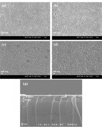
3.3 Morphological properties
SEM images of all thin film samples are shown in Fig.
4. Almost spherical particles of few nanometer in size were detected in all thin film samples. As shown in images, F and Ag containing samples also shows uniform porous structure of the thin film. Fig. 3(e) shows cross- sectional view of a representative AFTS sample. The thickness of SiO
2and TiO
2layer were found to be 215 nm and 74 nm, respectively.
Fig. 2. UV-Vis absorbance spectra of TiO2, Ag-TiO2, F-TiO2 and Ag- F-TiO2 thin films.
Fig. 1. XRD patterns of TiO2, Ag-TiO2, F-TiO2 and Ag-F-TiO2 thin films.
Fig. 3. UV-vis transmittance spectra of TiO2, Ag-TiO2, F-TiO2 and Ag-F-TiO2 thin films.
Fig. 5 shows TEM images of ATS and AFTS samples.
TEM images clearly shows that the Ag NPs are uniformly dispersed on the surface of TiO
2. The size of Ag and TiO
2NPs were found to be 2 nm and 12 nm, respectively.
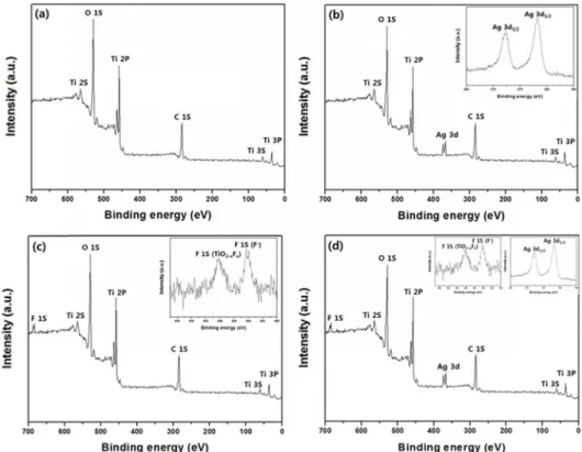
3.4 XPS analysis
The chemical state of Ti, O, F, and Ag were analyzed by XPS measurement as shown in Fig. 6. For TiO
2, Ti 2p
3/2and Ti 2p
1/2peaks are detected at 458.49 eV and 464.28 eV, respectively. The splitting between the Ti 2p
1/2and Ti 2p
3/2is 5.79 eV, indicating a normal state of Ti
4+in the sample.
21)The peak observed at 530 eV is assigned to lattice oxygen of TiO
2. Fig. 6(b) shows the XPS of ATS sample in which the Ag 3d
5/2and Ag 3d
3/2binding energy regions are found at 367.8 and 373.8 eV, respectively. The corresponding spin energy separation is 6.0 eV. This is the characteristic of metallic Ag, con- firming that the Ag present in the metallic state.
22)The XPS of FTS and AFTS is shown in Fig. 6(c-d). The binding energy peak at 684.3 eV indicates that the fluorine is present in the thin films sample in the form of TiF
4and/or physically adsorbed F
−on TiO
2. Similarly, the peak at 688 eV could assigned to nonstoichiometric solid solution of F in TiO
2of the TiO
2-xF
xtype.
23)Fig. 7 shows high resolution XPS spectra of O1s configuration for TS, ATS, FTS, and AFTS samples. The Ti-O peak intensity was in the order of AFTS < ATS < FTS < TS, while the surface hydroxyl peak intensity was in the order of TS < FTS < ATS < AFTS. The higher hydroxyl density was found for AFTS sample which might be beneficial to trap holes reaching the surface region of the film. This leads to the enhancement of both the charge transfer efficiency and photocatalytic activity.
3.5 Photo-induced hydrophilicity
Hydrophilicity of the prepared thin films were studied using water contact angle measurements under UV light irradiation. Fig. 8 shows the contact angles of all thin film samples. Before UV light irradiation, the nature of thin films was hydrophobic and the water contact angles of all thin films were in the range of 21-24
o. After UV light irradiation, the water contact angles decreased to about 4
owithin 5 min of UV irradiation. All thin film samples showed super hydrophilicity upon UV light irradiation.
3.6 Photocatalytic activity
In order to evaluate the self-cleaning activity of the films and to give an example of potential applications, photocatalytic activity of the thin films was evaluated by photocatalytic degradation of methylene blue aqueous solution under UV and visible light irradiation. The concentration of MB solution under dark conditions (without light) didn’t change. Also, in presence of light irradiation and without thin films, the concentration of MB solution remains constant. Therefore, the degradation of MB solution can be occurred by both light irradiation and photocatalytic thin film.
Fig. 9 and Fig. 10 show the MB degradation profiles for all samples(TS, ATS, FTS, and AFTS coating films) under UV and visible light irradiation, respectively. Upon visible light irradiation, pure TiO
2sample did not show any photo-activity, but other samples of ATS, FTS and AFTS showed photocatalytic activities because of formed F centers
13)and surface plasmon resonance effect of Ag dopant.
24)It is shown that the photo-activity of AFTS thin film under UV as well as visible light was higher
Fig. 4. SEM surface images of (a) TS, (b) ATS, (c) FTS, (d) AFTS,and (e) cross section of AFTS films.
Fig. 5. TEM images of (a) ATS and (b) AFTS films.
than that of other samples. In agreement with previous researches, the incorporation of Ag on F-TiO
2is a prom- ising way to enhance the photocatalytic performance.
25)The F-TiO
2thin film showed lower photocatalytic activity than the Ag-TiO
2thin film under UV and visible light irradiation. The photocatalytic activity was high in
Fig. 6. Survey XPS spectra of (a) TS, (b) ATS, (c) FTS, and (d) AFTS films(Inset: high-resolution XPS spectra of Ag 3d and F 1S).Fig. 7. High-resolution XPS spectra of O 1S for (a) TS, (b) ATS, (c) FTS, and (d) AFTS films.
the order of AFTS > TS > FTS > ATS under UV and AFTS > ATS > FTS > TS under visible light irradiation, respectively.
The enhanced photocatalytic activity of the AFTS thin film may be due to the difference in Fermi level of Ag and F-TiO
2. As the Fermi level of Ag is lower than that of F-TiO
2, the photo-induced electrons can transfer to the Ag particles loaded on the surface of TiO
2. Then, the transferred electrons are trapped by Ag and separated effectively. Ultimately, more reactive oxygen species are generated due to the photo-generated charge carriers and enhance the photocatalytic activity.
26)In addition, the surface plasmon resonance of Ag particles on F-TiO
2is excited by visible light, enhancing the surface electron excitation and photo-generated charge carrier separation.
The combined effect of Ag played an important role to transfer electrons from the sample surface to adsorbed O
2, and to inject photo-excited electrons into the F-TiO
2conduction band, producing separated electrons and holes, which can react with adsorbates.
It has been reported that in the F doped TiO
2, doped F can convert Ti
4+to Ti
3+by charge compensation.
27)Upon light irradiation, photo-generated electrons accumulate at the lower lying surface state of Ti
3+, while holes accumulate at the valence band of TiO
2. The photo- generated electrons could easily diffuse after doping fluorine, and then captured by Ag.
28)The hydroxyl density of AFTS was higher than that of other samples, so the AFTS sample is efficient to trap holes reaching the surface region of the thin film, which ultimately enhance photocatalytic activity by promoting charge transportation.
4. Conclusions
TiO
2/SiO
2thin films doped with F, Ag and Ag-F were
successfully prepared by a sol-gel method, followed by spin coating. Optical analysis confirmed that the Ag-F- TiO
2/SiO
2coating film showed good visible light absorption. All prepared thin films of (Ag-TiO
2, F-TiO
2, and Ag-F-TiO
2)/SiO
2showed excellent self-cleaning and photocatalytic properties. Self-cleaning performance of Ag-F-TiO
2/SiO
2thin film was higher than other samples under UV and visible light. This higher self-cleaning performance of Ag-F-TiO
2/SiO
2film was mainly attributed to the synergetic effect of F and Ag dopant, enhancing the formation of oxygen vacancies and the separation of electron-hole pairs.
Fig. 8. Contact angles of photocatalysts(TiO2, Ag-TiO2, F-TiO2, and Ag-F-TiO2)/SiO2 under UV light irradiation.
Fig. 10. Comparisons of photo-decomposition for MB solution with photocatalysts(TiO2, Ag-TiO2, F-TiO2, and Ag-F-TiO2)/SiO2 under visible light irradiation.
Fig. 9. Comparisons of photo-decomposition for MB solution with photocatalysts(TiO2, Ag-TiO2, F-TiO2, Ag-F-TiO2)/SiO2 under UV light irradiation.
Acknowledgement
This research work was supported by a grant(17- CTAP-C133057-01) from Infrastructure and transportation technology promotion research program funded by Min- istry of Land, Infrastructure, and Transport of the Korean government.
References
1. S. Banerjee, D. D. Dionysiou and S. C. Pillai, Appl.
Catal. B Environ. 176-177, 396 (2015).
2. H. M. Yadav, J.-S. Kim and S. H. Pawar, Korean J.
Chem. Eng., 33, 1989 (2016).
3. W.-J. Lee, Y.-B. Choi and D.-S. Bae, Korean J. Mater.
Res., 27, 289 (2017).
4. L. Zhou, S. Yan, B. Tian, J. Zhang and M. Anpo, Mater.
Lett., 60, 396 (2006).
5. H. M. Yadav, T. V. Kolekar, S. H. Pawar and J.-S. Kim, J. Mater. Sci.: Mater. Med. 27: 57, 1 (2016).
6. M. Houmard, G. Berthomé, J. C. Joud and M. Langlet, Surf. Sci., 605, 456 (2011).
7. X. Lin, F. Rong, D. Fu and C. Yuan, Powder Technol., 219, 173 (2012).
8. B.-M. Kim and J.-S. Kim, Korean J. Mater. Res., 26, 73 (2016).
9. E. Albiter, M. A. Valenzuela, S. Alfaro, G. Valverde- Aguilar and F. M. Martinez-Pallares, J. Saudi Chem. Soc., 19, 563 (2015).
10. A. M. Asiri, M. S. Al-Amoudi, S. A. Bazaid, A. A.
Adam, K. A. Alamry and S. Anandan, J. Saudi Chem.
Soc., 18, 155 (2014).
11. J. C. Yu, J. Yu, W. Ho, Z. Jiang and L. Zhang, Chem.
Mater., 14, 3808 (2002).
12. D. Li, H. Haneda, N. K. Labhsetwar, S. Hishita and N.
Ohashi, Chem. Phys. Lett., 401, 579 (2005).
13. D. Li, H. Haneda, S. Hishita, N. Ohashi and N. K.
Labhsetwar, J. Fluor. Chem., 126, 69 (2005).
14. D. J. R. Gutierrez, N. R. Mathews and S. S. Martinez, J. Photochem. Photobiol. A: Chem., 262, 57 (2013).
15. J. Xu, Y. Ao, D. Fu and C. Yuan, Appl. Surf. Sci., 254, 3033 (2008).
16. B.-M. Kim, H. M. Yadav, J.-S. Kim, J. Coat. Technol.
Res., 13, 905 (2016).
17. S. C. Chan and M. A. Barteau, Top. Catal., 54, 378 (2011).
18. I. Pastoriza-Santos and L. M. Liz-Marzán, Nano Lett., 2, 903 (2002).
19. S. P. Deshmukh, R. K. Dhokale, H. M. Yadav, S. N.
Achary, S. D. Delekar, Appl. Surf. Sci., 273, 676 (2013).
20. T. Yamaki, T. Umebayashi, T. Sumita, S. Yamamoto, M.
Maekawa, A. Kawasuso and H. Itoh, Nucl. Instrum.
Methods Phys. Res., Sect. B, 206, 254 (2003).
21. D. K. Kim and W. Y. Maeng, Korean J. Mater. Res., 26, 271 (2016).
22. X. Yang, Y. Wang, L. Xu, X. Yu and Y. Guo, J. Phys.
Chem. C, 112, 11481 (2008).
23. C. Trapalis, N. Todorova, T. Giannakopoulou, G.
Romanos, T. Vaimakis and J. Yu, Int. J. Photoenergy, 2008, 534038 (2008).
24. H. M. Sung-Suh, J. R. Choi, H. J. Hah, S. M. Koo and Y. C. Bae, J. Photochem. Photobiol. A: Chem., 163, 37 (2004).
25. X. Lin, F. Rong, D. Fu and C. Yuan, Powder Technol., 219, 173 (2012).
26. M. Jakob, H. Levanon and P. V. Kamat, Nano Lett., 3, 353 (2003).
27. Y. Wu, H. Liu, J. Zhang and F. Chen, J. Phys. Chem. C, 113, 14689 (2009).
28. S. N. Subbarao, Y. H. Yun, R. Kershaw and K. Dwight, Inorg. Chem., 18, 488 (1979).