Journal of Bacteriology and Virology 2006. Vol. 36, No. 2 p.89 – 98
Coxsackievirus B3 H3와 10A1 감염에 의한 마우스 심장 내에서의 유전자 변이 관찰
가톨릭대학교 생명공학부 분자바이러스연구실
1, 성균관대학교 의과대학 순환기내과
2, 국립보건연구원 간염 폴리오 연구팀
3남재환
1*·임병관
2·조영주
1·김대선
1·김연정
1·정수영
1·지영미
3·전은석
2Host Gene Profiling of Coxsackievirus B3 H3- and 10A1-infected Mouse Heart
Jae-Hwan Nam1*, Byung-Kwan Lim2, Young-Joo Cho1, Dae-Sun Kim1, Yeun-Jung Kim1, Soo-Young Chung1, Young-Mee Jee3 and Eun-Seok Jeon2
1
Department of Biotechnology, The Catholic University of Korea, 43-1 Yeokgok Dong, Wonmi-Ku, Bucheon, 420-743, Korea;
2Department of Medicine, Sunkyunkwan University School of Medicine, Cardiac and
Vascular Center, Samsung Medical Center, 50 Il-Won Dong, Kangnam-Ku, Seoul 135-710, Korea;
3
Division of Hepatitis and Poliovirus, Department of Virology, National Institute of Health, 5 Nokbun Dong, Eunpyung Ku, Seoul, 136-701, Korea
Received : March 28, 2006 Accepted : May 19, 2006
Coxsackievirus B3 (CVB3) is a non-enveloped virus that has a single-stranded RNA genome. CVB3 induces myocarditis, and ultimately, dilated cardiomyopathy. A myocarditis variant of CVB3 (CVB3 H3) and its antibody-escape mutant (CVB3 10A1) were studied previously; H3 was found to induce myocarditis and 10A1 was found to be attenuated in infected mice. Although amino acid residue 165, located in a puff region of VP2, was found to be altered (i.e., the H3 asparagine was altered to aspartate in 10A1), the detailed mechanism of attenuation was not clearly elucidated. Here, DNA microarray technology was used to monitor changes in mRNA levels of infected mouse hearts after CVB3 H3 and 10A1 infection. This tool was used to elucidate the pathogenic mechanisms of viral infection by understanding virus-host interactions. We identified several genes, including protein tyrosine kinases, Ddr2 and Ptk2, as well as Clqb and Crry, involved in complement reactions, which may be involved in these viral processes. Thus, gene profiling can provide an opportunity to understand host immune responses to viral infection for gene therapy and may contribute to the identification of the target gene that is modified during treatment of viral myocarditis.
Key Words: Coxsackievirus B3 H3, 10A1, cDNA microarray, Gene profile
서 론
콕사키바이러스 B (Coxsackievirus B, CVB)의 혈청형 (sero- type)은 6개가 있으며 (CVB1 - 6) 이들은 전형적인 human enteroviruses (Picornaviridae)에 속한다. 이 바이러스의 게놈
은 일반적으로 단일가닥 (single-stranded) positive RNA이며 약 7400 base 정도의 길이를 가지고 있다 (34). 심근염 (Myo- carditis) 및 확장성 심근증 (Dilated cardiomyopathy, DCM) 환 자 심장의 약 5~50% 정도에서 바이러스 게놈이 확인되어 CVB3가 심근에 존재함이 밝혀졌다 (23). 또한 CVB3가 사람 및 생쥐를 감염 하면 급성기에는 심근염을 유발하고 만성화 되면 확장성 심근증이 유발 되는 것이 실험적으로 증명 되어 CVB3가 심장 특이적 질환을 유발하는 것으로 확인되었다 (31). 현재 인구 10만 명당 5~8명이 CVB3 감염에 의해 확
89
*교신저자: 남재환. 420-743, 경기도 부천시 원미구 역곡2동 산 43-1, 가톨릭대학교 생명공학부 분자바이러스연구실
Phone: 02-2164-4852, Fax: 02-2164-4917, e-mail: jhnam@catholic.ac.kr
장성 심근증에 걸릴 위험이 있으며 특히 유아기 때 감염되 면 사망률이 8%에 이르는 것으로 보고되고 있다 (34).
바이러스 심근염은 감염 후 병리학적 상태에 따라 3 기간 으로 나누고 있다. 바이러스의 증식이 활발한 급성기 (acute phase), 바이러스에 의해 유발된 면역기전에 의해 바이러스 에 감염된 심근세포의 손상이 발생하는 아급성기 (subacute phase), 바이러스 증식 없이 심근손상이 장기적으로 진행되 는 만성기 (chronic phase)로 구분하고 있다 (2,3). 급성기는 활발한 바이러스의 증식으로 심근손상이 일어나는 시기로 심근에서의 바이러스 역가 (titer)가 높고 심근괴사 (necrosis) 가 관찰되나 염증세포의 침윤은 거의 관찰되지 않는다 (27, 33). 배양한 심실세포에 바이러스를 감염 시키면 심근세포의 고사 (apoptosis)를 관찰할 수 있어 심근염 초기의 세포손상 의 기전은 주로 바이러스 증식에 의한 직접적인 세포독성 (direct cytotoxicity) 때문으로 알려져 있다 (3,15). 이 시기에 는 일부 cytokine 유전자 발현이 심장조직에서 관찰된다. 아 급성기는 감염 후 4일에서 14일에 이르는 기간으로 염증세 포의 침윤이 관찰되며 혈청 중화항체가 가장 높은 시기이 다. 또한 이시기에는 IL-1β, TNF-α, IL-2, INF-γ와 같은 cyto- kine들이 증가하고 (20,22,25) 염증세포들에 의해 nitric oxide (NO)가 증가하게 되어 (10,14,21) 감염 후 자가 면역 활성화 에 의해 만성기에서 지속적인 심근손상을 유발할 수 있다.
바이러스 감염 후 자가 면역반응이 증가 되고 이것으로 인 한 심근증 발병의 원인 규명을 위해 많은 연구가 진행 중에 있으며 특히 Th1과 Th2 면역반응의 상호 균형 (balance), 즉 사이토카인의 상호 균형이 질환의 진행에 매우 중요함이 확인 되었다 (12,29). 이러한 연구 결과들은 바이러스를 감염 시킨 마우스 모델을 이용해 얻어졌다. 실험에 마우스를 이용 하는 이유는 사용 가능한 많은 inbred strain이 있으며, 유전 적 정보가 잘 밝혀져 있고, 특정 유전자 주입 쥐의 개발과 사용이 용이하다는 장점이 있어 췌장염 (pancreatitis), 수막염 (meningitis), 마비 (paralysis), 급 · 만성 심근염과 확장성 심 근증 등의 모델들로 사용되고 있다 (2,26). 자가 면역 심근 염 모델의 경우 바이러스 감수성이 있는 Balb/c 마우스에 coxsackievirus (CVB3)나 cytomegalovirus (CMV)와 같은 전혀 다른 바이러스를 감염시켜 실험한 예가 있으며 두 모델에서 모두 감염 후 7~14일에 급성 심근염이 발생되고 28일 후에 는 만성 심근염이 유발되었으며 IgM과 IgG를 포함한 다량 의 자가 항체가 심근 미오신 (myosin)에 대해서 만들어 지는 것이 확인되어 바이러스에 의한 만성 자가 면역 질환이 유 도됨이 증명되었다 (11,16,19, 28).
CVB3에는 임상적으로 심각한 심근염을 유발하는 주 (myocarditic strain, cardiovirulent strain)와 경미한 급성열성질 환이나 무균성 수막염 등을 유발하는 약독화 된 주 (non-
cardiovirulent strain)로 나눌 수 있다. 이러한 약독화는 주로 바이러스 캡시드 단백질 (capsid protein)이나 5'UTR (untran- slated region) 부분의 염기서열 치환이 중요한 영향을 미치는 것으로 확인 되었다 (7,9). 특히 CVB3의 Woodruff 주는 마우 스에서 심근염을 유발하기 때문에 주로 심장 질환 연구에 사 용되고 있으며 (34), 이 주의 플라크 분리주 (plaque purified strain)인 H3주는 감염된 마우스 심장에서 주로 분리되는 등 심장 특이적인 심근염 균주로 보고되어 있다 (31). 또한 H3 주의 항체회피바이러스 (antibody escaping virus)주인 10A1주 는 오직 VP2 부분의 165번째 아미노산인 asparagine(N)이 aspartate(D)으로 변하였으나 H3에 비해 약독화 되어 있음이 보고되었다 (18,31). 이러한 약독화의 기작은 정확히 알려져 있지 않으나 N165D 부분이 VP2에서 항원 중화 부분 (anti- genic neutralization site)으로 알려진 puff region에 위치하기 때문에 아마도 이러한 변화가 바이러스와 세포 표면의 수용 체 (receptor)와의 결합을 변화시켰거나 혹은 심근염을 유발 하는 바이러스의 능력을 변화 시켰을 것으로 추정하고 있다 (18). 그러나 두 바이러스의 분자 병리학적 차이가 완전히 이 해되어 있지는 않은 실정이다
본 연구에서는 바이러스 감염 시 생쥐 심장에서 일어나는 다양한 숙주세포 유전자의 변이를 추적하기 위해 CVB3의 H3 바이러스주 및 H3에 대한 약독화 주인 10A1을 마우스 에 감염 시킨 후 시간대별로 심장을 적출한 후 바이러스 감 염에 의해 영향을 받는 심장 유전자의 mRNA 증감 변이 (profile)를 high-through put 기술인 cDNA microarray를 이용 하여 조사하였다. 이러한 조사는 CVB3에 의한 심장 질환의 유발 기작을 바이러스 감염에 영향을 받거나 영향을 주는 숙 주세포의 측면에서 이해할 수 있게 해주며 또한 CVB3 H3와 10A1주의 약독화 기작에 대한 이해의 단서를 제공해 줄 것 으로 기대된다.
재료 및 방법
1. 바이러스 및 세포
바이러스 배양 및 역가 확인에 사용한 자궁경부암세포주 인 HeLa-UVM는 Dulbecco's Modified Eagles Medium (DMEM, Invitrogen-GIBCO, Rockville, MD, USA)에 불활화 시킨 fetal bovine serum (Invitrogen-GIBCO, Rockville, MD, USA) 10%를 첨가하여 37℃, CO2 배양기에서 배양 하였다. 사용한 HeLa- UVM (본 논문에서는 HeLa 세포주로 명명)는 Sally Huber (University of Vermont, USA)로부터 입수 하였다. 사용된 CVB3 H3 및 10A1주는 90% 가량 자란 HeLa 세포에 10 moi로 감염 시켜 37℃에서 16시간 감염 시킨 후 3회에 걸쳐 냉동과 해동을 반복하여 세포 lysate 및 배양액을 함께 원심
분리한 후 상층액을 분할하여 -80℃에 사용할 때까지 보관 하였다. 보관된 바이러스 stock은 HeLa 세포주를 이용한 plaque assay를 실시하여 바이러스 역가를 결정하였다.
2. 바이러스성 심근염 생쥐 모델
4주령 된 Balb/c 생쥐 수컷을 이용하여 실험하였다. 바이 러스에 감염시키기 1주일 전에 생쥐 공급 업체 (오리엔트바 이오, 성남, 경기도, 대한민국)로부터 실험에 사용할 생쥐를 수령하여 동물실에서 안정화 시켰다. 이 후 H3 및 10A1 바 이러스주를 마리 당 1×10⁴ pfu로 복강을 통해서 감염 시켰 다. 감염 후 7일, 13일째에 바이러스가 감염된 생쥐를 안락사 하여 즉시 심장을 적출해서 액체질소를 이용하여 급속 냉동 하여 사용 시까지 -80℃에 보관하였다. 나머지 H3와 10A1이 감염된 마우스들 각 20마리는 15일 동안 사망률을 확인하 였다.
3. 마우스 심장 염색
CVB3 감염에 의해 심장에서 일어나는 염증 (inflammation) 반응을 확인하기 위해 Evans-blue dye를 사용하여 염색하였 다. 전체적인 방법은 김 등의 논문과 동일하다 (17). 간단히 방법을 서술하면 심장을 적출하기 12시간 전에 복강으로 10 µl의 Evans-blue dye (10 mg/ml in Phosphate-buffered Saline)를 주사한다. 이 후 심장을 적출하고 냉동 절편을 만들어 형광 현미경 아래에서 관찰하면 염증이 일어난 부분은 진한 빨간 색으로 표시된다.
4. 심장 total RNA 분리
바이러스가 감염된 마우스의 심장을 적절한 시기에 적출
하고 무게를 잰 후 적출된 심장을 액체질소에 넣어 급속 냉 동하였다. 냉동된 마우스 심장을 막자사발에 넣고 액체질소 를 조금씩 부으면서 곱게 갈았다. 여기에 TRIsol (Invitrogen- Life Technology, Gaithersburg, MD, USA) 용액을 넣고 제조사 의 방법대로 total RNA를 정제하고 spectrophotometer를 이용 하여 정량한 후 -80℃에 사용 전까지 보관하였다.
5. cDNA microarray 기술
cDNA microarray는 약 7400개의 유전자를 가지고 있는 chip을 (주)디지털지노믹스(서울, 대한민국)의 Twin-Chip을 구입하여 사용하였다. 바이러스가 감염된 생쥐의 심장에서 추출된 total RNA는 Cy3 dye (감염되지 않은 생쥐 심장의 RNA)와 Cy5 dye (감염된 마우스 심장 RNA) (GE Healthcare, Waukesha, WI, USA)를 사용하여 역전사 (reverse transcription) 하여 fluorescein-labeled cDNA를 준비하였다. Cy3 및 Cy5로 표시된 cDNA를 섞어 chip 위에서 42℃에서 16시간 동안 처 리하였다. Chip을 세척 후 ScanArray Life (Perkin-Elmer Life Sciences, Wellesly, MA, USA)를 사용하여 scanning하여 얻은 이미지를 GenePix software (Axon In- struments, Sunnyvale, CA, USA)를 사용하여 gene expression ratio (감염 대 비감염)를 분석하였다.
6. cDNA microarray 데이터 분석
각 cDNA microarray spot의 형광 정도 (fluorescence inten- sity)는 local median background subtraction에 의해 계산되었 다. Robust scatter-plot smoother LOWESS 기능을 유전자 발현 의 intensity-dependent normalization을 위해 이용하였다. Scatter plot 분석은 Microsoft Excel 2000을 이용하여 분석 하였다.
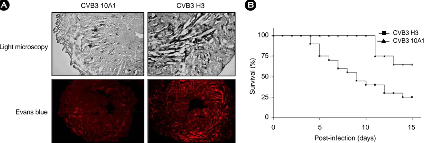
Figure 1. Characterization of CVB3 H3 and 10A1 infected mouse. (A) Histology of myocardial injury after CVB3 H3 and 10A1 infection. Injury to myocytes was identified with Evans Blue dye, and appears as a red image. Analysed hearts were harvested at 7 days after challenge. (B) Survival curve after CVB3 H3 and 10A1 challenge in mice. Each group contained 20 mice. The survival curve is shown as percentage survival against time post-infection.
CVB3 10A1 CVB3 H3
Light microscopy
Evans blue
A B
100
75
50
25
0
Survival (%)
0 5 10 15 Post-infection (days)
CVB3 H3 CVB3 10A1
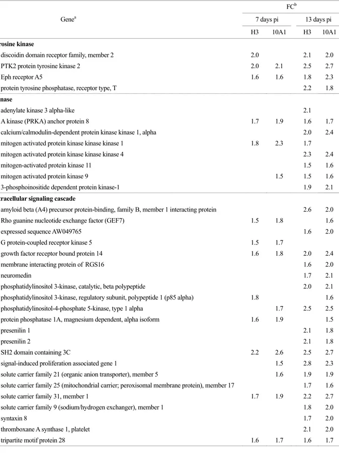
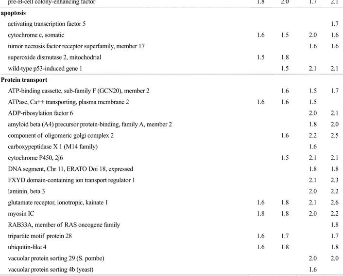
Table 1. Functional grouping of mRNAs of infected mouse heart that change more than 1.5 fold after CVB3 H3 and CVB3 10A1 infection FCb
7 days pi 13 days pi Genea
H3 10A1 H3 10A1
tyrosine kinase
discoidin domain receptor family, member 2 2.0 2.1 2.0
PTK2 protein tyrosine kinase 2 2.0 2.1 2.5 2.7
Eph receptor A5 1.6 1.6 1.8 2.3
protein tyrosine phosphatase, receptor type, T 2.2 1.8
Kinase
adenylate kinase 3 alpha-like 2.1
A kinase (PRKA) anchor protein 8 1.7 1.9 1.6 1.7
calcium/calmodulin-dependent protein kinase kinase 1, alpha 2.0 2.4 mitogen activated protein kinase kinase kinase 1 1.8 2.3 1.7
mitogen activated protein kinase kinase kinase 4 2.3 2.4
mitogen-activated protein kinase 11 1.5 1.6
mitogen activated protein kinase 9 1.5 1.5 1.6
3-phosphoinositide dependent protein kinase-1 1.9 2.1
intracellular signaling cascade
amyloid beta (A4) precursor protein-binding, family B, member 1 interacting protein 2.6 2.0
Rho guanine nucleotide exchange factor (GEF7) 1.5 1.8 1.6
expressed sequence AW049765 1.6 2.0
G protein-coupled receptor kinase 5 1.5 1.7
growth factor receptor bound protein 14 1.6 1.8 2.0 2.4
membrane interacting protein of RGS16 1.6 2.0
neuromedin 1.7 2.1
phosphatidylinositol 3-kinase, catalytic, beta polypeptide 2.0 2.1 phosphatidylinositol 3-kinase, regulatory subunit, polypeptide 1 (p85 alpha) 1.8 1.6 phosphatidylinositol-4-phosphate 5-kinase, type 1 alpha 1.7 2.5 2.5 protein phosphatase 1A, magnesium dependent, alpha isoform 1.6 1.9 1.5
presenilin 1 2.1 1.8
presenilin 2 2.1 1.8
SH2 domain containing 3C 2.2 2.6 2.5 2.7
signal-induced proliferation associated gene 1 1.5 2.8 2.3
solute carrier family 21 (organic anion transporter), member 5 1.6 1.9 1.9 solute carrier family 25 (mitochondrial carrier; peroxisomal membrane protein), member 17 1.7 1.6
solute carrier family 31, member 1 1.7 1.9 2.2 2.7
solute carrier family 9 (sodium/hydrogen exchanger), member 1 1.8 2.0
syntaxin 8 1.7 2.0
thromboxane A synthase 1, platelet 2.1 2.0
tripartite motif protein 28 1.6 1.7 1.6 1.7
Table 1. continued
FCb
7 days pi 13 days pi Genea
H3 10A1 H3 10A1
immune response
complement component 1, q subcomponent, beta polypeptide 1.7 2.0 2.4 2.9
chemokine (C-C motif) ligand 25 2.5 2.6
CD8 antigen, alpha chain 1.8 1.7
complement component factor h 2.1 2.0
complement receptor related protein 1.7 1.8
colony stimulating factor 2 receptor, beta 1, low-affinity 1.5 1.9
interferon (alpha and beta) receptor 1 1.8 1.7
interferon gamma receptor 1.6 1.7
lymphocyte antigen 6 complex, locus A 1.7
N-myc (and STAT) interactor 1.9 2.1
pre-B-cell colony-enhancing factor 1.8 2.0 1.7 2.1
apoptosis
activating transcription factor 5 1.7
cytochrome c, somatic 1.6 1.5 2.0 1.6
tumor necrosis factor receptor superfamily, member 17 1.6 1.6
superoxide dismutase 2, mitochodrial 1.5 1.8
wild-type p53-induced gene 1 1.5 2.1 2.1
Protein transport
ATP-binding cassette, sub-family F (GCN20), member 2 1.6 1.5 1.7
ATPase, Ca++ transporting, plasma membrane 2 1.6 1.6 1.5
ADP-ribosylation factor 6 2.0 2.1
amyloid beta (A4) precursor protein-binding, family A, member 2 1.8 2.0
component of oligomeric golgi complex 2 1.6 2.2 2.5
carboxypeptidase X 1 (M14 family) 1.6
cytochrome P450, 2j6 1.5 2.1 2.1
DNA segment, Chr 11, ERATO Doi 18, expressed 1.8 1.8
FXYD domain-containing ion transport regulator 1 2.1 2.3
laminin, beta 3 2.0 2.2
glutamate receptor, ionotropic, kainate 1 1.6 1.8 2.1 2.6
myosin IC 1.8 1.8 2.0 2.2
RAB33A, member of RAS oncogene family 1.8
tripartite motif protein 28 1.6 1.7 1.7
ubiquitin-like 4 1.6 1.8 1.8
vacuolar protein sorting 29 (S. pombe) 2.0 2.0
vacuolar protein sorting 4b (yeast) 1.6
유전자의 logarithmic gene expression ration이 +1.5보다 크거 나 -1.5보다 작으면 의미 있는 유전자 발현의 차이로 고려했 다 (즉 최소한 3배 이상 유전자 발현이 차이가 나는 것을 분 석 대상으로 삼았다).
결 과
1. CVB3 H3와 10A1의 병리학적 차이
Knowlton 등의 논문 (18)에 의하면 CVB3 H3와 10A1주 가 감염된 마우스 심장의 심근염 정도 (myocardial score)는 각각 2.08±1.00과 0.33±0.49이고, 바이러스 역가는 조직 100 mg 당 5.32±0.13 및 4.92±0.13 log pfu임을 보여주었다. 즉 CVB H3주가 10A1주 보다 심장 염증은 더 심하게 그리고 바이러스 증식도 더 많이 되고 있음을 보여주고 있다. 본 연 구에서도 CVB3 H3와 10A1을 1×10⁴ pfu로 마우스에 감염 시켜 7일 후 Evans blue 염색을 통해 CVB3 H3와 10A1의 염 증 정도를 비교하였다 (Fig. 1A). CVB3 H3이 감염된 마우스 심장에서 10A1이 감염된 마우스 심장 보다 더 심한 염증반 응을 확인하여 이전 논문 (18)과 동일한 결과를 얻었다. 또한 두 바이러스주를 1×10⁴ pfu로 감염 시킨 마우스 각 20마리 의 생존율을 비교한 결과 CVB3 H3가 감염된 마우스는 감염 15일 후에 25% 정도의 생존율을 보여주었으나 10A1이 감염 된 마우스는 60% 정도의 생존율을 보여주었다 (Fig. 1B).
2. cDNA microarray 분석
CVB3 H3와 10A1을 각각 1×10⁴ pfu로 4주령 된 수컷 Balb/C 마우스 복강에 감염 시켰다. 감염 후 7일째와 13일 째에 마우스 심장을 적출하여 total RNA를 정제한 후 cDNA microarray 실험에 사용하였다. 감염 후 7일째와 13일째 마우 스를 사용한 이유는 7일째 마우스 심장은 급성기의 전형적 인 상태를 보여주고 있으며, 13일째 마우스는 만성기 시작의 마우스 심장 상태를 보여주기 때문에 두 시기로 나누어 유전
자 분석 실험을 수행하였다.
cDNA microarray 실험 데이터 분석은 데이타의 normali- zation을 위해 intensity/location-dependent normalization 방법 을 이용하였다 (36). 확보된 결과 중 LOWESS (f=0.2)를 이 용하여 normalization 한 결과인 Global M 값을 사용하여 유 전자 발현을 분석하였다. M 값은 log2 (Cy5/Cy3)의 식으로 계산된 것으로 이 값이 1이면 감염된 마우스 심장에서 그 유 전자의 발현이 2배 높고, -1이면 감염되지 않은 마우스 심장 에서 2배 높음을 의미한다. 즉 바이러스 감염에 의해 그 유 전자의 발현이 2배 감소하였음을 의미한다. 또한 분석된 데 이터들은 raw 데이터에서 signal intensity 값이 10 이상인 것 만 선별하여 비교하였다 (즉 signal intensity가 크다는 의미는 기술적 변이에 의한 차이가 적음을 의미한다). Global M 값이 최소한 1.5 이상 차이가 나는 것이 실제 mRNA 양의 차이를 잘 반영할 것으로 추측되어 본 연구에서 보여준 모든 데이터 (Table 1, 2, 3)는 증가 (up-regulation)된 세포 인자 (cellular factor)들 중 global M 값이 1.5 이상인 것만을 대상으로 하 여 분석하였다. 발현양이 감소 (down-regulation)되는 세포 인 자 (cellular factor)들은 분석 대상에서 제외하였다. 그 이유는 CVB3가 숙주세포에 감염되면 바이러스의 2A 단백질이 host cap-dependent 전사 기작에 매우 중요한 eIF4G를 자르게 된 다 (5). 따라서 일정 시간이 지나면 바이러스가 감염된 숙주 세포 내의 유전자 발현은 자연적으로 감소하게 되어 결국 apoptosis 등이 일어나서 감염된 세포가 사라지게 된다. 따라 서 바이러스가 감염된 후 유전자 발현이 감소하는 유전자 보 다 증가하는 유전자가 바이러스 감염에 좀 더 의미 있는 영 향을 미칠 것으로 생각하였다. 본 연구에서 분석한 Table 1, 2, 3은 모두 바이러스 감염에 의해 증가되는 cellular factor들 만을 정리하였으며 global M값은 1.5 이상, signal intensity는 10.0 이상의 유전자들만을 대상으로 하여 정리하였다.
Table 1은 유전자 기능을 중심으로 시간대별로 감염된 마 우스 심장에서 나타나는 cellular factor들의 profile을 정리 하 Table 1. continued
FCb
7 days pi 13 days pi Genea
H3 10A1 H3 10A1
G-protein coupled receptor protein signaling pathway
coagulation factor II (thrombin) receptor-like 3 1.6
membrane interacting protein of RGS16 2.0
prostaglandin I receptor (IP) 1.8 2.0
regulator of G-protein signaling 11 1.5 1.6
a Description of gene encoding mRNA, b Global M Value by data normalization
였으며, Table 2와 3은 각각 바이러스 감염 후 7일째와 13일 째 CVB3 H3와 10A1에서 공통적으로 증가하거나 각각 따로 증가한 cellular factor들의 유전자 심볼을 정리하였다. 이러한 심볼은 각 클론에 해당하는 유전자 이름이다.
고 찰
Coxsackievirus는 뉴욕주 Coxsackie의 허드슨강 근처의 마 을에서 열병을 앓고 있는 어린이로부터 최초로 분리되었으며 Table 2. mRNAs of infected mouse heart that change more than 1.5 fold at 7 day post-infection
Gene Symbola
Induction in both CVB3 H3 and CVB3 10A1 infected mouse heart
Akap8, Alas1, Arhgef7, Atp2b2, Bdnf, C1qb, C230035B04, Cog2, Comt, Crry, Csf2rb1, Cstb, Cycs, D130006K24Rik, D17Wsu94e, Dcx,Ddr2, Elovl1, Epha5, Evi2b, Fxyd1, Gba2,Gprk5, Grb14, Grcc2F, Grik1, Gtf2ird1, Hnrpd, Jdap1-pending, LOC223754, Lpin1, Map3k1, Mpg, Mrpl23, Mrps14, Myo1c, Ntan1, Pbef-pending, Pclo, Ppm1a, Ptk2, Rfc2, Sh2d3c, Slc30a4, Slc31a1, Snap25bp, Sod2, Spg4, Surb7, Tfam, Tmpo, Tnfsf12, Trim28, Tspyl, Ub14, Uros, Wrn, Zfh4-pending
Induction in only CVB3 10A1 infected mouse heart
Abcf2, Acas2, Adam8, Arcn1, B630003E05Rik, Bat4, Cav2, Cnih, Cpd1-pending, Cyp2j6, D4Wsu125e, D5Buc26e, Deb1, E430018M07Rik, Eif2s2, Entpd1, Es2el, Esp, Gfi1, Gnpat, Inpp5e, Klf15, Litaf, Mapk9, MGC25248, MGC28597, Mllt3, Mnt, Mpv17, Mrps22, Ndr1, Nktr, Nr2f6, Nudt5, Panx1, Pip5k1a, Rab14, Rpo1-1, Scd1, Sdf2, Sipa1, Slc21a5, Sorbs1, Spp1, Sptlc2, Suv39h1, Tagln2, Tcfeb, Umpk, Uty, Wig1, Zap3-pending
Induction in only CVB3 H3
infected mouse heart G1rzfp-per, Gyg1, Hn1, Lamr1, Mtap4, Osp94, Pik3r1
a Gene name of clone encoding mRNA
Table 3. mRNAs of infected mouse heart that change more than 1.5 fold at 13 day post-infection Gene Symbola
Induction in both CVB3 H3 and CVB3 10A1 infected mouse heart
Abcf2, Acas2, Aco1, Actn4, Adam8, Akap8, Alas1, Amfr, Apacd-pending, Apba2, Apbb1ip-pending, Apc7-pending, Arf6, Asah2, Asb8, Atf5, AW049765, B630002E05Rik, Bace, Bat4, BC026401, Brd4, C130052I12Rik, C1qb, Camkk1, Caskin2-pending, Ccl25, Cd8a, Cfh, Cited1, Clstn1, Clybl, Cnot8, Cog2, Comt, Cops3, Cpd1-pending, Crry, Crybb1, Ctsh, Cycs, Cyp2j6, D11Ertd18e, D130006K24Rik, D17Wsu94e, D730049H07Rik, Dcx, Ddr2, Deb1, Dnajb9, E430007K15Rik, E430018M07Rik, Elovl1, Entpd1, Epha5, Es2el, Evi2b, Expi, Fgfr2, Fibp, Fosb, Fxyd1, G0s2, G1rzfp-pending, Gas41-pending, Gcat, Gfi1, Gh, Gnpat, Gpi1, Grb14, Grcc2f, Grik1, Gtf2ird1, Gus, Gzma, H2afy, Hip1r, Hirip5, Hnrpd, Hs1bp3-pending, Ifnar1, Ifngr, Itpr5, Lamb3, Lamr1, Lcat, Lcp1, Litaf, LOC207352, LOC216543, LOC227619, LOC23373, LOC270153, LOC270153, Lpin1, Ly6a, Madh1, Map3k4, Mapk11, Mapk9, MGC25248, MGC28597, MGC40859, Minpp1, Mir16-pending, Mnt, Mpg, Mpv17, Mrpl12, Mrpl17, Mrps14, Mrps22, Mrps30, Ms4a4d, Mta1, Myo1c, Myo7a, N4wbp4-pending, Ncl, Ndr1, Nkx2-2, Nmi, Nmu, Nthl1, Osp94, Ovca2-pendingm Panx1, Papss1, Pbef-pending, Pcm1, Pdpk1, Pik5k1a, Plscr3, Pou2f2, Prodh, Psen1, Psen2, Pstpip1, Ptgir, Ptk2, Ptprt, Pxf, Rab14, Rab33a, Rangnrf-pending, Rfc2, Rgs11, Rnasep2-pending, Rpl8, Sc5d, Sh2d3c, Sipa1, Slc221a5, Slc25a17, Slc31a, Slc9a1, Smap-pending, Snap25bp, Sorbs1, Spg4, Spp1, Srprb, Ssbp3, Stx8, Surb7, Suv39h1, Tagln2, Tbx2, Tbxas1, Tmpo, Tnfrsf17, Trim28, Tslpr, Tspyl, Vamp4, Vps29, Wdr10, Wig1, Xpo1, Yars, Zap3-pending, Zfpm2
Induction in only CVB3 10A1 infected mouse heart
Al181838, Al317223, Al317395, Al645590, Al848594, Arhgef7, Ass1, Atp2a2, AW060733, B430109J1, Bdnf, C130038E, C1qtnf5, Camk2b, Cnih, Cstb, D16Ertd36, D5Buc26e, D6Ertd253, Epb4.1l3, Fbxo16, Galns, Gba2, Gng10, Klf1a, Klf15, LOC22375, MGC27915, Mllt3, Mtap4, Ngef, Nktr, Notch1, Npr3, Nr2f6, Nudel-pending, Pclc, Pik3r1, Pirb, Ppp1cb, Ppp3ca, Prkacb, Prkcm, Rad51l1, Rbbp9m Rnase1, Rpo1-1, Rsu1, Scd1, Scotin-pending, Skz1-pending, Slc22a4, Slc25a11, Sox5, Sox6, Sptlc2, Stra6, Stx6, Surf4, Tfam, Tnfsf9, Trim16, U3-55k-pending, Ubl4, Uty, YEAST3-1, Zap3-pending
Induction in only CVB3 H3
infected mouse heart Adh1, Al848594, Ak3l, Akt2, Atp2b2, C230035B04, Cpxm, D3Ucla1, F2rl3, Glud, Map3k1, Ncl, Nr2f6, Nudc, Rce1, Stx4a, Uros, Vps4b, Wwp2-pending, Zak-pending, Zfp52
a Gene name of clone encoding mRNA
(24), 영아마우스에 감염 시켰을 때 이완된 마비증세 (flaccid paralysis)를 유발하는 것을 A type (CVA)으로, 유발하지 않는 것을 B type (CVB)으로 구분하였다 (4,24). CVB를 감염 시 켜 마우스에서 유발되는 심근염은 임상적으로 관찰되는 심 근염과 병리소견이 유사하여 바이러스성 심근염의 병리학적 연구에 많이 이용되고 있다. 이러한 실험 결과 CVB3 감염 초기 급성기에 심근에서 발생하는 병리학적 변화는 주로 대 식세포 (macrophage), 자연살해세포 (natural killer cell) 등에 의한 심근세포 손상에 의한 것이고, 바이러스 감염 후 2주 정도에 일어나는 현상은 주로 T 림프구가 관여하는 것으로 알려져 있다 (2,3). 급성 바이러스성 심근염이나 확장성 심 근증으로 진행하는 것에 대한 병리학적 기전은 초기에는 바 이러스를 제거하기 위한 숙주의 면역기전으로 심근염이 진 행되고 이후 심장조직에 바이러스가 없는 상태에서 관찰되 는 지속적인 염증반응으로 알려져 있다 (1).
본 연구에서 사용된 CVB3 10A1은 약독화 바이러스주로서 CVB3 H3에서 유래되었으며 VP2 부분의 165번째 아미노산 인 asparagine(N)이 aspartate(D)로 변하였다. 또한 CVB3 H3 에 비해 바이러스 감염 후 마우스의 사망률이 현저히 낮은 특징을 보이고 있다 (18). 본 연구는 이러한 CVB3 10A1과 H3 바이러스주의 차이를 이용하여 두 바이러스주가 감염된 마우스 심장세포 내에서 바이러스 감염에 영향을 받는 (혹은 영향을 미치는) cellular factor들의 mRNA level의 차이를 조 사하여 coxsackievirus의 병리학적 질환 유발 기전의 이해를 돕고자 하였다.
Ddr2 (discoidin domain receptor family, member 2)는 H3와 10A1 바이러스가 감염된 마우스 심장 모두에서 증가 (up- regulation) 되었다 (Table 1). 이 유전자는 transmembrane rece- ptor protein tyrosine kinase signaling pathway의 기능을 하고 있다 (32). 실제로 receptor tyrosine kinase (PTK)는 세포의 성 장 등에 매우 중요한 역할을 하며 세포 내의 신호 전달에 가 장 중요한 역할을 하고 있다고 알려져 있다 (32). 따라서 바 이러스 감염에 의한 PTK family인 Ddr2의 증가는 바이러스 성장 혹은 바이러스에 대한 방어 기작의 시작에 매우 중요할 것으로 기대된다. 이외에도 바이러스 감염에 의해 증가된 다 른 PTK 관련 유전자로는 Ptk2 (PTK2 protein tyrosine kinase 2)가 있다 (Table 1) 이 유전자는 integrin-mediated signaling pathway에 중요한 역할을 하고 있으며, cytoplasmic protein tyrosine kinase로서 FAK family에 속한다 (13). 이 유전자의 활성 (activation)은 특정 펩타이드나 세포의 extracellular matrix 와의 접촉에 의해 유도되는 세포의 성장이나 intracellular signal transduction pathway의 초기 단계에서 매우 중요한 역 할을 하고 있다 (35). 따라서 바이러스 감염에 의해 이 유전 자가 증가 된다는 것은 바이러스 감염에 의해 숙주세포 내로
의 신호 전달에 이 유전자가 특정 역할을 할 수 있을 것이라 는 것을 의미 한다. 이외에도 바이러스 감염에 의해 증가되 는 다른 PTK 관련 세포 인자 (cellular factor)로는 Epha5 (Eph receptor A5)가 있다 (Table 1, 2 및 3).
Pik3rl (phosphatidylinositol 3-kinase, regulatory subunit, poly- peptide 1, p85 alpha)는 감염 7일째에는 H3가, 13일째에서는 10A1이 감염된 심장에서만 증가 된다 (Table 1, 2 및 3). 감 염 후 13일째 H3가 감염된 마우스 심장에서는 Pik3rl이 직접 증가 되지는 않지만 유사한 신호 전달 기작을 보여주고 있는 Pdpk1 (3-phosphoinositide dependent protein kinase-1), Pik3cb (phosphatidylinositol-3-kinase, catalytic, beta polypeptide), Pip5k1a (phosphatidylinositol-4-phosphate 5-kinase, type 1 alpha)가 증가 된다 (Table 1, 2 및 3).
Clqb (complement component 1, q subcomponent, beta poly- peptide)와 Crry (complement receptor related protein)는 모두 감염 후 7일째와 13일째에서 증가 되고 있다 (Table 1, 2 및 3). 이들 유전자는 주로 보체를 활성화 시키는 신호 전달 체 계에 속하고 있으며 (8), 따라서 CVB 감염 후 보체에 의한 자연 면역 체계 (innate immune system)가 매우 중요한 역할 을 하고 있음을 시사하고 있다.
Cycs (cytochrome C, somatic)는 감염 후 7일과 13일째에서 CVB3 H3와 10A1 바이러스가 감염된 마우스 심장에서 모 두 증가 되고 있다 (Table 1, 2 및 3). 이 유전자는 cytochrome c를 encoding 하고 있으며 cytochrom c는 apoptosis의 시작 에 매우 중요한 역할을 하고 있다 (6). 즉 이미 알려진 것 처럼 anti-apoptotic member를 억제 (suppression) 하거나 pro- apoptotic member를 활성화 (activation) 시키면 미토콘드리아 막의 투과성이 변해 cytochrome c가 세포질로 나오게 되고 나온 cytochrome c는 Apaf-1과 결합하여 apoptosome을 형성 하여 caspase-9을 활성화 시킴으로서 apoptosis가 유도 된다 (6). 이러한 사실은 CVB3의 감염 시 apoptosis가 심장의 파 괴에 직접적인 역할을 한다는 발표와 일치하며 (3,15) 따라 서 CVB3가 심장에 감염되면 Cycs가 induction 되어 apopto- sis를 유발하고 이는 CVB3에 의한 심장의 파괴 기작에 일정 역할을 할 것으로 추정되어 진다.
이러한 유전자 외에도 Sod2 (superoxide dismutase 2, mito- chodrial)는 감염 후 7일째 CVB3 H3와 10A1가 감염된 마우 스 심장에서 증가 된다 (Table 1과 2). 일반적으로 superoxide metabolism은 세포 내에 생성된 radical을 제거하여 세포에 독성을 주는 기작을 억제 한다 (30). 따라서 이 유전자는 CVB3 감염에 의해 생성된 free radical을 제거하여 숙주를 보호하기 위해 감염 초기에 증가된 것으로 추정되어 지며, 이러한 현상을 기반으로 하여 CVB3 감염에 대한 방어 유전 자를 선별할 수 있을 것이다.
본 연구는 CVB3 감염에 의해 증가 되는 다양한 세포 인 자 (cellular factor)들을 확인 하였으며, 이를 통해 바이러스 감염에 의해 숙주 안에서 일어나는 다양한 병리학적 기작을 분자 수준에서 이해할 수 있는 기반을 제공 하였다. 또한 이 러한 유전자에 대한 좀 더 자세한 연구를 통해 CVB3 감염 에 의해 유도되는 심근염 및 확장성 심근증을 억제할 수 있 는 치료용 유전자 타켓의 선별을 기대 할 수 있을 것이다.
감사의 글
저자 중 남재환과 임병관은 실험 수행에 있어서 제1저자 로서 동일하게 기여하였음. 본 연구는 보건복지부 보건의료 진흥사업의 지원 (A050768) 및 2006년도 질병관리본부 용역 사업의 지원으로 수행되었음.
참 고 문 헌
1) 전은석: 심근염과 심근증의 원인. 순환기 23(부록): 208 -216, 1993.
2) 전은석: 심근염의 동물 model. 순환기 21(1): 53-56, 1997.
3) 전은석: 바이러스 심근염과 심근증의 병인기전. 생화학 뉴스 22(1): 36-44, 2002.
4) Carpernter CM, Boak RA: Coxsackie viruses; a review of pathologic, epidemiologic, diagnostic and etiologic observa- tions. Calif Med. 77(2): 127-130, 1952.
5) Carthy CM, Granville DJ, Watson KA, Anderson DR, Wilson JE, Yang D, Hunt DW, McManus BM: Caspase activation and specific cleavage of substrates after coxsackie- virus B3-induced cytopathic effect in HeLa cells. J Virol 72:
7669-7675, 1998.
6) Cho SG, Choi EJ: Apoptotic signaling pathways: caspases and stress-activated protein kinases. J Biochem Mol Biol.
35(1): 24-27, 2002.
7) Dan M, Chantler JK: A genetically engineered attenuated coxsackievirus B3 strain protects mice against lethal infection.
J Virol 79(14): 9285-9295, 2005.
8) Gasque P: Complement: a unique innate immune sensor for danger signals. Mol Immunol 1(11): 1089-1098, 2004.
9) Gutierrez AL, Denova-Ocampo M, Racaniello VR, del Angel RM: Attenuating mutations in the poliovirus 5' untran- slated region alter its interaction with polypyrimidine tract- binding protein. J Virol 71(5): 3826-3833, 1997.
10) Hiraoka Y, Kishimoto C, Takada H, Sasayama S: Role of oxygen derived free radicals in the pathogenesis of coxsackie- virus B3 in mice. Cardiovasc Res 27: 957-961, 1993.
11) Huber SA, Job LP, Auld KR, Woodruff JF: Sex-related differences in the rapid production of cytotoxic spleen cells active against uninfected myofibers during coxsackievirus B3 infection. J Immunol 126: 1336-1340, 1981.
12) Huber SA, Born W, O'Brien R: Dual functions of murine gammadelta cells in inflammation and autoimmunity in coxsackievirus B3-induced myocarditis: role of Vgamma1+
and Vgamma4+ cells. Microbes Infect 7(3): 537-543, 2005.
13) Isakov N, Biesinger B: Lck protein tyrosine kinase is a key regulator of T-cell activation and a target for signal interven- tion by Herpesvirus saimiri and other viral gene products. Eur J Biochem. 267(12): 3413-3421, 2000.
14) Ishiyama S, Hiroe M, Nishikawa T, Abe S, Shimojo T, Ito H, Ozasa S, Yamakawa K, Matsuzaki M, Mohammed MU, Nakazawa H, Kasajima T, Marumo F: Nitric oxide contri- butes to the progression of myocardial damage in experimental autoimmune myocarditis in rats.Circulation. 95(2): 489-496, 1997.
15) Jeon ES, Sheng Z, Knowlton KU: Coxsackievirus induces myocytes apoptosis. Circulation 94(suppl): 1-356, 1996.
16) Khatib R, Chason JL, Silberberg BK, Lerner AM: Age- dependent pathogenecity of group B coxsacieviruses in Swiss- Webster mice. J Infect Dis 141: 394-403, 1980.
17) Kim JY, Jeon ES, Lim BK, Kim SM, Chung SK, Kim JM, Park SI, Jo I, Nam JH: Immunogenicity of a DNA vaccine for coxsackievirus B3 in mice: protective effects of capsid proteins against viral challenge. Vaccine 23(14): 1672-1679, 2005.
18) Knowlton KU, Jeon ES, Berkley N, Wessely R, Huber S: A mutation in the puff region of VP2 attenuates the myocarditic phenotype of an infectious cDNA of the Woodruff variant of coxsackievirus B3. J Virol 70(11): 7811-7818, 1996
19) Lawson CM, O'Donoghue HL, Reed WD: Mouse cytome- galovirus infection induces antibodies which cross-react with virus and cardiac myosin: a model for the study of molecular mimicry in the pathogenesis of viral myocarditis. Immunology 275: 513-519, 1992.
20) Levine B, Kalman J, Mayer L, Fillit HM, Packer M:
Elevated circulating levels of tumor necrosis factor in severe chronic heart failure. N Engl J Med 223: 236-241, 1990.
21) Lowenstein CJ, Hill SL, Lafond-Walker A, Wu J, Allen G, Landavere M, Rose NR, Herskowita A: Nitric oxide inhi- bits viral replication in murine myocarditis. J Clin Invest 97:
1837-1843, 1996.
22) Matsumori A, Yamada T, Suzuki H, Matoba Y, Sasayama S: Increased circulating cytokines in patients with myocarditis and cardiomyopathy. Br Heart J 72: 561-566, 1994.
23) Maisch B, Ristić AD, Hufnagel G, Pankuweit S: Patho- physiology of viral myocarditis: The role of the humoral immune response. Cardiovasc Pathol 11: 112-122, 2002.
24) McLean DM: Coxsackieviruses and echoviruses. Am J Med Sci. 251(3): 351-368, 1966.
25) McMurray J, Abdullah I, Dargie HJ, Shapiro D: Increased concentration of tumor necrosis factor in cachectic patients with severe chronic heart failure. Br Heart J 66: 356-358, 1991.
26) Mena I, Perry CM, Harkins S, Rodriguez F, Gebhard J, Whitton JL: The role of B lymphocytes in coxsackievirus B3 infection. Am J Pathol 155: 1205, 1999.
27) Pontes-de-Carvalho L, Santana CC, Soares MB, Oliveira GG, Cunha-Neto E, Ribeiro-dos-Santos R: Experimental chronic chagas' disease myocarditis is an autoimmune disease preventable by induction of immunological tolerance to myo- cardial. J Autoimmun 18: 131-138, 2002.
28) Rose NR: Myocarditis from infection to autoimmunity. The Immunologist 64: 67-75, 1996.
29) Shioi T, Matsumori A, Sasayama S: Persistent expression of
cytokine in the chronic stage of viral myocarditis in mice.
Circulation 94(11): 2930-2937, 1996.
30) Toyozaki T, Hiroe M, Tanaka M, Nagata S, Ohwada H, Marumo F: Levels of soluble Fas ligand in myocarditis. Am J Cardiol 82(2): 246-248, 1998.
31) Van Houten N, Bouchard PE, Moraska A, Huber S: Selec- tion of an attenuated coxsackievius B3 variant, using a mono- clonal antibody reactive to myocyte antigen. J Virology 65:
1286-1290, 1991.
32) Vogel W: Discoidin domain receptors: structural relations and functional implications. FASEB J. 13 Suppl: 77-82, 1999.
33) Wilson FM, Miranda QR, Chason JL, Lerner M: Residual pathologic changes following murine coxsackie A and B myocarditis. Am J Pathol 55: 253-265, 1969.
34) Woodruff JF: Viral myocarditis: A review. Am J Pathol 101:
424, 1980.
35) Xiong WC, Mei L: Roles of FAK family kinases in nervous system. Front Biosci 8: 676-682, 2003.
36) Yang YH, Dudoit S, Luu P, Lin DM, Peng V, Ngai J, Speed TP: Normalization for cDNA microarray data: a robust com- posite method addressing single and multiple slide systematic variation. Nucleic Acids Res 30(4): e15, 2002.