J Korean Soc Radiol 2015;73(2):100-104 http://dx.doi.org/10.3348/jksr.2015.73.2.100
INTRODUCTION
Inflammatory pseudotumor was first detected in the lung and described by Brunn in 1939. It was so named by Umiker and Iverson in 1954 because of its tendency to mimic a malig- nant process, clinically and radiologically (1). Inflammatory pseudotumor commonly involves the lung and the orbit, but it can occur in nearly every site in the body (2). Inflammatory pseudotumor has been reported in various sites in the abdo- men, including the liver. Abdominal inflammatory pseudotu- mor should be included in the differential diagnosis of any soft- tissue mass within the abdomen and viscera (1). There are va- rious causes of inflammatory pseudotumor including secondary infection (2).
Perihepatitis is defined as inflammation of the peritoneal cap- sule of the liver and is classically described as being associated
with pelvic inflammatory disease (PID) (the so-called Fitz-Hugh- Curtis syndrome). Fitz-Hugh-Curtis syndrome is thought to re- sult from the peritoneal spread of infection from the pelvic cavi- ty (3).
We report a case of inflammatory pseudotumor of the liver and omentum caused by PID, which shows the intraperitoneal spreading mechanism in Fitz-Hugh-Curtis syndrome.
CASE REPORT
A 47-year-old woman was admitted to our hospital for right sided abdominal pain. Physical examination showed a tender abdomen and slight rigidity in the right upper and lower quad- rants. Body temperature was measured as 38.5°C.
Laboratory data of liver function parameters, such as aspar- tate aminotransferase (12 IU/L, normal range: 12–35 IU/L), al-
Inflammatory Pseudotumor in the Liver and Right Omentum Caused by Pelvic Inflammatory Disease: A Case Report
골반염에 의해 발생한 간과 그물막의 염증성 거짓종양: 증례 보고
Hyukjun Byun, MD, Seong Hoon Kim, MD*
Department of Radiology, Daegu Fatima Hospital, Daegu, Korea
Inflammatory pseudotumor can develop in any part of the human body. It is one of the most important tumor-mimicking lesions that require differential diagnosis.
There are various causes of inflammatory pseudotumor, one of which is infection and its resultant inflammation. Pelvic inflammatory disease (PID) often causes peri- hepatitis, which is called Fitz-Hugh-Curtis syndrome. In Fitz-Hugh-Curtis syndrome, bacteria spread along the right paracolic gutter, causing inflammation of the right upper quadrant peritoneal surfaces and the right lobe of the liver. We experienced a case of PID with accompanying inflammatory pseudotumor in the liver and the right omentum. This case identically correlates with the known intraperitoneal spreading pathway involved in Fitz-Hugh-Curtis syndrome, and hence, we present this case report.
Index terms
Inflammatory Pseudotumor Pelvic Inflammatory Disease Fitz-Hugh-Curtis Syndrome
Received January 28, 2015 Revised March 4, 2015 Accepted April 21, 2015
*Corresponding author: Seong Hoon Kim, MD Department of Radiology, Daegu Fatima Hospital, 99 Ayang-ro, Dong-gu, Daegu 701-724, Korea.
Tel. 82-53-940-7165 Fax. 82-53-954-7417 E-mail: [email protected]
This is an Open Access article distributed under the terms of the Creative Commons Attribution Non-Commercial License (http://creativecommons.org/licenses/by-nc/3.0) which permits unrestricted non-commercial use, distri- bution, and reproduction in any medium, provided the original work is properly cited.
anine aminotransferase (12 IU/L, normal range: 7–35 IU/L), to- tal bilirubin (0.15 mg/dL, normal range: 0.1–1.2 mg/dL) were within the normal range. Erythrocyte sedimentation rate (37 mm/hr, normal range: 0–20 mm/hr) and C-reactive protein (CRP, 8.57 mg/dL, normal range: 0.0–0.5 mg/dL) were elevated with leukocytosis [white blood cell (WBC) count 14220/mm3, normal range: 4400–11000/mm3].
On abdominal ultrasound image, a 4 cm sized hypoechoic mass was detected in the right paracolic gutter (Fig. 1A). Also, a heterogenous hypoechoic lesion in liver segment #6 was ob- served (Fig. 1B). Bilateral tubo-ovarian abscesses were detected, with the left side being more severely affected.
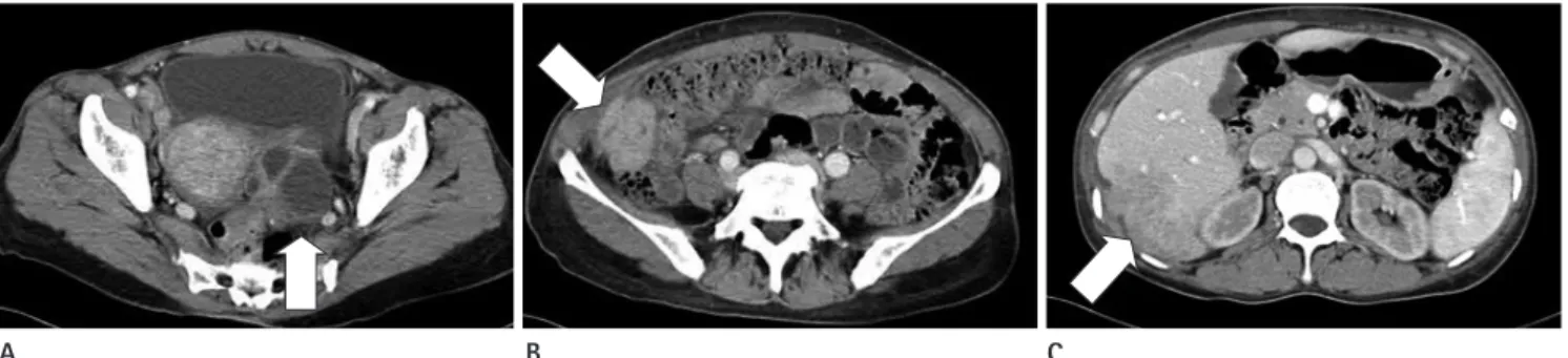
In serial abdominal computed tomography scan (multidetec- tor row CT, SOMATOM Sensation 64, Siemens Healthcare, Forchheim, Germany), bilateral tubo-ovarian abscesses with PID (Fig. 2A), a well-enhanced mass in the right paracolic gutter (Fig.
2B), and a heterogenously enhanced lesion in liver segment #6 with capsular retraction and enhancement were noted (Fig. 2C) [CT parameters: 120 kVp, 160 mAs, collimation 64 × 0.6 mm
(with z-flying focal spot), and slice thickness 5 mm, Protocol:
The images were taken in precontrast, arterial, portal, and delay- ed phases. Precontrast phase image was taken in the initial scan.
Then, 1.5 mL per kg of contrast media was administered via in- travenous bolus injection. Through bolus tracking, arterial phase image was taken when the Hounsfield unit (HU) value in the abdominal aorta exceeded 100 HU. Portal phase image was taken 50 seconds after bolus tracking, and the delayed phase image was taken 160 seconds after bolus tracking. The scan time for each phase was 6 seconds].
In positron emission tomography CT scan, intensely increased fluorodeoxyglucose uptake was noted in bilateral tubo-ovarian abscesses [maximum standardized uptake value (SUVmax): 8.16], right omental mass (SUVmax: 10.15) (Fig. 3), and mass-like le- sion in liver segment #6 (SUVmax: 9.23).
We performed percutaneous ultrasound-guided biopsy for the mass in liver segment #6 (Fig. 4A) and the right omental mass (Fig. 4B). The images showed infiltration of lymphoplasma cells and some lymphocytes without cytologic atypia. The masses were
Fig. 1. Abdominal ultrasound shows a 4 cm sized hypoechoic mass (A, arrow) in the right paracolic gutter. A heterogenous hypoechoic lesion (B, arrow) is detected in liver segment #6.
A B
Fig. 2. Serial abdominal CT scan shows bilateral tubo-ovarian abscesses (A, arrow) with PID and a well-enhanced mass (B, arrow) in the right paracolic gutter. A heterogenously enhanced lesion (C, arrow) in liver segment #6 with capsular retraction and enhancement is noted.
PID = pelvic inflammatory disease
A B C
pathologically confirmed to be inflammatory pseudotumors.
The patient underwent a two-week course of intravenous an- tibiotic treatment. The right sided abdominal pain and tender- ness subsided, and the laboratory data also improved (WBC count 9700/mm3, CRP 0.26 mg/dL). The patient was discharged and she was lost to follow-up.
DISCUSSION
Inflammatory pseudotumors have been described under var- ious names due to the variable sites of involvement; plasma cell granuloma (heart and lung), inflammatory myofibroblastic tu- mor (lung), inflammatory myofibrohistiocytic proliferation, his- tiocytoma, xanthoma, fibroxanthoma, fibrous xanthoma, xan- thogranuloma, xanthomatous pseudotumor, plasma cell-histio- cytoma complex (lung), plasmacytoma, solitary mast cell gr- anulomas, and inflammatory fibrosarcoma (urinary bladder) (1, 2).
Inflammatory pseudotumor may also occur in the abdominal cavity, which is an important mimicker of intraperitoneal tu- mor. Differentiation from malignant tumors can often be chal- lenging because the radiologic findings of inflammatory pseu- dotumors are rather nonspecific. Abdominal inflammatory pseudotumor should be considered in the differential diagnosis of any soft-tissue mass within the abdomen and viscera. Percu- taneous biopsy is the most reliable method for differential diag- nosis, and it helps us to avoid unnecessary exploratory laparot- omy or a hepatectomy in case of an uncertain diagnosis (1, 2).
The causes of inflammatory pseudotumor are not known.
Some authors claim, on the basis of multiple evidence, that it is a low-grade fibrosarcoma with inflammatory cells. The propen- sity of inflammatory pseudotumors to be locally aggressive, to frequently be multifocal, and to progress occasionally to a true malignant tumor supports this idea. In some cases, inflamma- tory pseudotumor is thought to result from inflammation fol- lowing minor trauma or surgery, or to be associated with other malignancy (1, 3).
There appears to be a subset of inflammatory pseudotumors that occur secondary to infection (1). Organisms found in asso- ciation with inflammatory pseudotumor include mycobacteria associated with spindle cell tumor; Epstein-Barr virus found in splenic and nodal pseudotumors; actinomycetes and nocardiae found in hepatic and pulmonary pseudotumors, respectively;
and mycoplasma in pulmonary pseudotumors (1, 4). There have been case reports of inflammatory pseudotumor associated with infections caused by other organisms, including Mycobac- terium avium-intracellulare complex, Corynebacterium equi,
Fig. 4. Pathologic analysis of the mass-like lesion in liver segment #6 (A) and the right omental mass (B) shows infiltration of lymphoplasma cells and lymphocytes (hematoxylin and eosin staining, × 400).
A B
Fig. 3. Positron emission tomography CT reveals an intensely in- creased fluorodeoxyglucose uptake in the right omental mass (arrow).
Escherichia coli, Klebsiella, Bacillus sphaericus, Pseudomonas, Helicobacter pylori, and Coxiella burnetti (1, 3-6).
PID refers to infection and resultant inflammation of the up- per female genital tract, including the endometrium, fallopian tubes, and ovaries. PID is the result of ascending infection from the vagina and cervix; the most common organisms are Neisse- ria gonorrhoeae and Chlamydia trachomatis. Polymicrobial in- fection can occur in 30–40% of cases; tuberculosis and actino- mycosis occur much less frequently (7).
In advanced PID patients, tubo-ovarian abscess and perihep- atitis can develop. Perihepatitis is classically described as being associated with PID, the so-called Fitz-Hugh-Curtis syndrome (8). In Fitz-Hugh-Curtis syndrome, bacteria spread by means of direct extension along the right paracolic gutter or through the lymphatic system, causing inflammation of the right upper qu- adrant peritoneal surfaces and the right lobe of the liver (7-9).
Inflammation of the liver capsule is not visible on ultrasound (7). In dynamic CT, Fitz-Hugh-Curtis syndrome has been re- ported to manifest as intense enhancement along the anterior surface of the liver (7, 9). The capsular enhancement seen in ear- ly-phase images may reflect increased blood flow in the inflam- ed hepatic capsule. On enhanced MR, dynamic postcontrast images show subcapsular and periportal areas of hypervascu- larity in the arterial phase. These areas are isointense relative to the rest of the hepatic parenchyma with delayed postcontrast se- quences (7).
In this case report, the patient complained of lower and right sided abdominal pain with fever along with laboratory findings that suggested an infection. CT scan of the patient after admis- sion confirmed the diagnosis of PID. The patient was referred to the gynecologist and colposcopy showed prominent cervical motion tenderness. CT scan also showed multiple mass-like le- sions in the right omentum and liver segment #6. These lesions were pathologically confirmed as inflammatory pseudotumors.
As already mentioned, the causes of inflammatory pseudotu- mor are not fully known, and they are rather vague and diverse.
An infection with subsequent inflammation can be one of the causes. In this case, PID was confirmed through symptoms, signs, laboratory findings, and radiologic findings, although the pathogen responsible for PID was not clearly identified (10).
An inflammatory pseudotumor was found in the right omen- tum and liver segment #6, which corresponds to the intraperito-
neal spreading pathway involved in Fitz-Hugh-Curtis syndrome.
Tubo-ovarian abscess in the left adnexa appears to be a cystic lesion, whereas the lesions in the right omentum and the liver ap- pear to be of solid nature; therefore, it is debatable as to whether these lesions have a common origin. But it is known that inflam- matory lesions are observed in various forms during the course of their progression. Adnexal lesions, especially, are tubular st- ructures which can appear as cystic lesions (like tubo-ovarian ab- scess) (7). In addition, the patient had no previous trauma or op- eration history, comorbid malignancy, or infection other than PID. Therefore, this case serves as evidence supporting the known intraperitoneal spreading mechanism in Fitz-Hugh-Curtis syn- drome, through a series of consequences such as PID and in- flammatory pseudotumor.
REFERENCES
1. Narla LD, Newman B, Spottswood SS, Narla S, Kolli R. In- flammatory pseudotumor. Radiographics 2003;23:719-729 2. Patnana M, Sevrukov AB, Elsayes KM, Viswanathan C, Lub-
ner M, Menias CO. Inflammatory pseudotumor: the great mimicker. AJR Am J Roentgenol 2012;198:W217-W227 3. Sanders BM, West KW, Gingalewski C, Engum S, Davis M,
Grosfeld JL. Inflammatory pseudotumor of the alimentary tract: clinical and surgical experience. J Pediatr Surg 2001;
36:169-173
4. Dehner LP. The enigmatic inflammatory pseudotumours:
the current state of our understanding, or misunderstand- ing. J Pathol 2000;192:277-279
5. Wood C, Nickoloff BJ, Todes-Taylor NR. Pseudotumor re- sulting from atypical mycobacterial infection: a “histoid”
variety of Mycobacterium avium-intracellulare complex in- fection. Am J Clin Pathol 1985;83:524-527
6. Lévy S, Sauvanet A, Diebold MD, Marcus C, Da Costa N, Thiéfin G. Spontaneous regression of an inflammatory pseu- dotumor of the liver presenting as an obstructing malignant biliary tumor. Gastrointest Endosc 2001;53:371-374 7. Rezvani M, Shaaban AM. Fallopian tube disease in the non-
pregnant patient. Radiographics 2011;31:527-548 8. Lee JW, Kim S, Kwack SW, Kim CW, Moon TY, Lee SH, et al.
Hepatic capsular and subcapsular pathologic conditions:
demonstration with CT and MR imaging. Radiographics
2008;28:1307-1323
9. Sam JW, Jacobs JE, Birnbaum BA. Spectrum of CT findings in acute pyogenic pelvic inflammatory disease. Radiograph-
ics 2002;22:1327-1334
10. Berek JS. Berek & Novak’s Gynecology, 14th ed. Philadel- phia: Lippincott Williams & Wilkins, 2007:549-551
골반염에 의해 발생한 간과 그물막의 염증성 거짓종양: 증례 보고
변혁준 · 김성훈*
염증성 거짓종양은 인체 어디에서나 관찰될 수 있고 이는 종양과의 감별이 반드시 필요한 중요한 병변이다. 염증성 거짓종 양이 생기는 원인은 다양하며, 그 중 대표적인 것이 감염 및 그와 동반된 염증이다. 골반염 환자에서 우측 부대장홈을 통해 세균이 전파되어 간주위염이 유발되는 경우가 있으며, 이를 피츠-휴-커티스 증후군(Fitz-Hugh-Curtis syndrome)이라 한 다. 우리는 골반염 환자에서 간과 우측 그물막에 염증성 거짓종양이 발생한 증례를 경험하였으며, 이것이 피츠-휴-커티스 증후군의 전파경로와 일치하기에 소개하고자 한다.
대구파티마병원 영상의학과