– 99 –
대한근전도∙전기진단의학회지 16(2):99~102, 2014 J Korean EMG Electrodiagn Med
Address reprint requests to Jee Young Kim, M.D.
Department of Neurology, Myongji hospital
55, Hwasu-ro 14beon-gil, Deogyang-gu, Goyang-si, Gyeonggi-do, Korea TEL: 82-31-810-5446, FAX: 82-31-969-0500, E-mail: [email protected]
투고일: 2014년 10월 13일, 1차 수정일: 2014년 11월 26일, 2차 수정일: 2014년 11월 28일, 최종 수정일: 2014년 12월 2일, 게재확정일: 2014년 12월 2일
INTRODUCTION
Guillain-Barré syndrome (GBS) is typically character- ized by acute onset of ascending paralysis and hypo- or areflexia. However, some cases presented regional weak- ness and these regional variants expanded the clinical profile of GBS. In 1986, Ropper described 3 patients who had acutely progressive, predominant oropharyn- geal, neck and shoulder weakness with normal or slight- ly reduced strength in legs. He called this regional vari- ant “pharyngeal-cervical-brachial weakness (PCB)”.1
However, because of its rarity, the pathomechanism and electrophysiological features are poorly understood. We report a patient who experienced acute, predominant bibrachial weakness and had reversible conduction failure.
CASE REPORT
A 42-year-old man visited our hospital due to mild upper limb weakness and right calf pain for 2-3 days.
His medical history was unremarkable including upper respiratory infection, gastroenteritis, vaccination, or
길랑바레증후군의 양상지아형 1예
1명지병원 신경과 , 2이화여자대학교 의학전문대학원 신경과학교실
윤도영
1∙박두용
1∙한현정
1∙박기덕
2∙김지영
1– Abstract –
A Case with Bibrachial Variant of Guillain-Barré Syndrome
Do Young Yoon, M.D.
1, Doo Yong Park, M.D.
1, Hyun Jung Han, M.D.
1, Kee Duk Park, M.D.
2, Jee Young Kim, M.D.
11Department of Neurology, Myongji Hospital, Goyang
2Department of Neurology, School of Medicine, Ewha Womans University, Seoul
The bibrachial variant of Guillain-Barré syndrome (GBS) is characterized by rapidly progressive and pre- dominant weakness of both upper extremities with hyporeflexia or areflexia. Because the bibracial variant is rare, the pathomechanism and disease course has not been clarified. We report a 42-year-old man with rapidly progressive weakness of both arms. His electrophysiological study showed partial conduction blocks of motor nerves normalized without development of excessive temporal dispersion within 3 weeks.
This case suggests that the main pathophysiological mechanism is reversible conduction block and it may be in the continuous spectrum of axonal motor GBS subtype.
Key Words: Guillain-Barré syndrome, pharyngeal-cervical-brachial variant, acute motor axonal neu- ropathy
윤도영∙박두용∙한현정∙박기덕∙김지영
toxin or drug exposure. In neurologic examination, the strength of both shoulder abductors on the Medical Research Council (MRC) was grade 4+/5. Two day after admission, his arm weakness was dramatically aggravat- ed, and strength was 3/5 in right arm and 2~3/5 in left arm. There was a slight limitation of left eye abduction and mild weakness of right ankle dorsiflexion of MRC grade 4+/5. There was no nystagmus, blepharoptosis and pupillary abnormality. Dysphagia or dysarthria were not observed, and the strength of other leg muscles and neck muscles was normal. Tendon reflexes were decreased in upper and lower limbs. Sensation to all modalities was preserved. Ataxia or autonomic dysfunction was absent.
Routine laboratory tests and arterial blood gas analysis were within the normal range. Anti-GQ1b and anti-GM1 antibody were negative. Also other serologic markers for paraproteimia, hepatitis B and C, HIV were negative.
Brain and cervical spine magnetic resonance imaging (MRI) was unremarkable. The cerebrospinal fluid con- tained 0 cell/L with a protein level of 37.5 mg/dl on day 2.
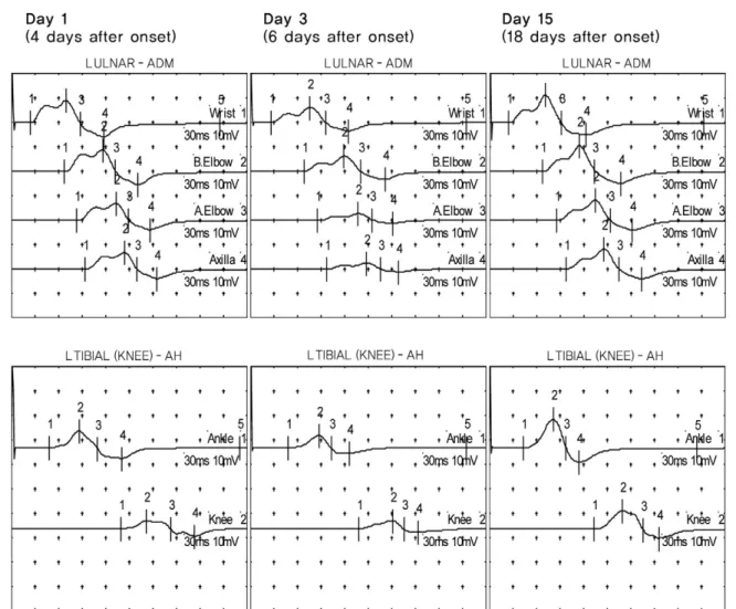
On day 1, nerve conduction study (NCS) demonstrated
partial motor conduction block (CB) in left tibial and ulnar nerves (Table 1 and Figure 1). And no potential was observed in right peroneal nerve. On day 3, NCS showed motor CB in left ulnar nerve and increased ter- minal latency (TL) in median, ulnar and left peroneal nerves (Table 1 and Figure 1). To rule out neuromuscu- lar junction disorder, repetitive nerve stimulation test (RNST) of facial, spinal accessory and ulnar nerves, recording orbicularis oculi, trapezius, flexor carpi ulnaris and abductor digiti minimi muscles, was performed. In low- frequency RNST, there was no abnormal decre- ment. A decrement greater than 10% was considered abnormal, in accordance with the suggestion by the AAEM Quality Assurance Committee.2
A diagnosis of GBS was made. On days 4 through 8, he received intravenous immunoglobulin (0.4 g/kg daily). On day 6, he noticed improvement of upper limb weakness and muscle strength was MRC grade 4. On day 8, mus- cle strength in both shoulder extensors improved to grade 4+. On day 15, TL was normalized and CB disap- peared in left ulnar nerve (Table 1 and Fig. 1). Also, his weakness was completely recovered.
– 100 – Table 1. Serial motor nerve conduction study
Day 1 Day3 Day 15 2014.10.07
(4 days after onset) (6 days after onset) (18 days after onset) (1 month after onset)
Right Left Right Left Right Left Right Left
Median nerve
TL msec (Amp mV) 3.6 (9.3) 3.6 (12.5) 3.9 (6.1) 3.8 (8.8) 3.4 (13.2) 3.6 (16.7) 3.1 (16.8) 3.3 (19.3) W-E m/sec (Amp mV) 51.0 (9.0) 50.9 (12.2) 53.3 (5.8) 56.6 (7.4) 54.0 (12.3) 61.4 (15.6) 54.9 (16.7) 58.7 (18.5) E-Ax m/sec (Amp mV) 57.8 (9.8) 66.7 (11.5) 59.5 (5.4) 59.5 (7.1) 60.5 (11.8) 66.7 (14.9) 64.0 (16.6) 60.3 (17.7)
F-wave (msec) 30.5 31.1 30.1 NP 29.1 30.4 28.4 29.2
Ulnar nerve
TL msec (Amp mV) 2.4 (15.2) 2.4 (15.2) 2.5 (11.3) 2.7 (10.1) 2.3 (16.7) 2.4 (16.9) 2.3 (16.6) 2.1 (17.3) W-E m/sec (Amp mV) 60.0 (14.7) 56.8 (14.7) 60.5 (10.9) 61.9 (10.1) 58.6 (15.8) 56.8 (15.9) 59.3 (16.5) 56.5 (16.9) E-E m/sec (Amp mV) 71.4 (14.5) 62.5 (11.2) 62.5 (9.8) 54.1 (4.5) 60.6 (15.5) 57.1 (12.7) 64.0 (16.4) 61.9 (16.8) E-Ax m/sec (Amp mV) 80.0 (14.4) 70.0 (11.1) 72.0 (9.4) 66.7 (4.2) 64.5 (15.0) 66.7 (12.6) 66.8 (16.1) 66.5 (16.7)
F-wave (msec) 29.7 30.6 31.7 NP 30.3 31.3 29.5 30
Peroneal nerve
TL msec (Amp mV) np 4.7 (4.7) np 5.9 (3.0) np 5.4 (4.3) 4.7 (0.3) 4.5 (4.8)
K-A m/sec (Amp mV) np 43.8 (4.0) np 41.5 (2.5) np 48.6 (4.2) 44.3 (0.3) 48.0 (4.5)
F-wave (msec) np 49.4 np 53.8 np 51.6 np 49.1
Posterior Tibial nerve
TL msec (Amp mV) 4.9 (15.4) 4.8 (11.8) 4.9 (9.5) 4.8 (7.8) 4.5 (19.0) 4.3 (18.2) 4.5 (19.4) 4.2 (19.7) K-A m/sec (Amp mV) 44.3 (11.5) 45.7 (6.8) 46.4 (7.9) 46.4 (5.3) 49.4 (13.6) 48.4 (11.9) 52.0 (15.7) 47.7 (16.2)
F-wave (msec) 49.4 49.5 49.4 51.1 49.3 50.5 48.9 48.9
H-reflex (msec) 29.8 29.6 33.7 34.3 31.1 31.3 30.4 32.2
TL: terminal latency, Amp: Amplitude, W-E: wrist-elbow, E-E: below elbow-above elbow, E-Ax: elbow-axilla, K-A: knee-ankle, NP: no potential
NCS was performed for median nerve recording abductor pollicis brevis muscle, ulnar nerve recording abductor digiti minimi mus- cle, peroneal nerve recording extensor digitorum brevis muscle, tibial nerve recording abductor hallucis brevis muscle.
DISCUSSION
This case presented acute onset, progressive weakness predominant in both upper arms and hyporeflexia. We thought that this case could belong to PCB variant of GBS. However, in contrast to earlier reports,1,3-5 the strength of his pharyngeal and cervical muscle was pre- served. On day 3, mild external ophthalmopelgia was observed and we thought that this was a case of bibrachial variant overlapping Fisher syndrome (FS), clinically. Recently, Gürsoy et al. reported a patient with an overlap of the PCB variant of GBS and Miller-Fisher syndrome.6 In this case, antiganglioside antibodies against GQ1b, GD1a, and GD1b were found.6In a large retrospective analysis of 100 patients with PCB, Nagashima et al. reported that 26 patients were diag- nosed with FS overlap.7And they found anti-GT1a IgG antibodies in 51% of 100 patients with PCB and identi-
fied that anti-GQ1b antibodies were positive in 73% of PCB-FS overlap patients.7However, anti-GQ1b or GT1a antibodies were not found in our patient.
On day 1 (4 days after onset), NCS showed partial CB in left ulnar and left tibial nerves and on day 3, revealed prolonged TL in median, ulnar and deep peroneal nerves and CB in left ulnar nerve. Sensory NCS was normal. On day 15 (18 days after onset), TL was normalized and CB was resolved completely. F-waves reappeared with nor- mal latency. Sensory NCS was preserved. These features suggest that the main pathophysiological mechanism of this case was reversible conduction failure of motor fibers. Reversible conduction failure was described in some cases with acute motor axonal neuropathy (AMAN) and acute motor conduction block neuropathy (AMCBN).8,9Mostly, the patients with reversible con- duction failure showed fast recovery. Cappaso et al.
reported 2 cases with acute symmetric weakness.9 In
길랑바레증후군의 양상지아형 1예
– 101 –
Fig. 1. Serial recordings of motor nerve conduction study. Upper tracings: left ulnar nerve motor conductions recorded from abductor digiti minimi muscle. Lower tracings: left tibial motor conductions recorded from abductor hallucis bre- vis muscle.
윤도영∙박두용∙한현정∙박기덕∙김지영
– 102 – electrophysiological study, early partial motor CB in forearm segments was observed, but CB was resolved within 2-5 weeks without excessive temporal dispersion of proximal motor responses.9Hiraga et al. investigated differences of recovery patterns between acute inflam- matory demyelinating neuropathy and AMAN patient in 97 GBS patients.10In this study, 19 AMAN patients had improvement within 4 weeks.10 It is supposed that reversible conduction failure of motor fibers is attributed to immune-mediated reversible conduction impairment at the axolemma of nodes of Ranvier.9,10Our patient showed clinical improvement within 3 weeks.
The main points to consider in this case are as follows.
(1) Our patient presented predominant bibrachial weak- ness-“bibrachial variant” of GBS; (2) the main mecha- nism of his weakness was reversible conduction failure;
(3) this variant may be in the continuous spectrum of axonal GBS subtypes (AMAN, AMCBN).
REFERENCES
1. Ropper AH: Unusual clinical variants and signs in Guil- lain-Barré syndrome. Arch Neurol 1986:43:1150-1152 2. AAEM Quality Assurance Committee. Literature review
of the usefulness of repetitive nerve stimulation and single fiber EMG in the electrodiagnostic evaluation of patients with suspected myasthenia gravis or Lambert-Eaton myas- thenic syndrome. Muscle Nerve 2001:24:1239-1247 3. Wakerley BR, Yuki N: Pharyngeal-cervical-brachial vari-
ant of Guillain-Barré syndrome. J Neurol Neurosurg Psy-
chiatry 2014:85:339-344
4. Arai M, Susuki K, Koga M: Axonal pharyngeal-cervical- brachial variant of Guillain-Barré syndrome without anti- GT1a IgG antibody. Muscle Nerve 2003:28:246-250 5. Capasso M, Notturno F, Manzoli C, Yuki N, Uncini A:
Reversible conduction failure in pharyngeal-cervical- brachial variant of Guillain-Barré syndrome. Muscle Nerve 2010:42:608-612
6. Gürsoy AE, Kolukısa M, Babacan-Yıldız G, Altıntaç Ö, Yaman A, Asil T: Reversible conduction failure in overlap of Miller Fisher syndrome and pharyngeal-cervical- brachial variant of Guillain-Barré syndrome in the spec- trum of nodo-paranodopathies. J Clin Neurosci 2014:21:
1269-1271
7. Nagashima T, Koga M, Odaka M, Hirata K, Yuki N: Con- tinuous spectrum of pharyngeal-cervical-brachial variant of Guillain-Barré syndrome. Arch Neurol 2007:64:1519- 1523
8. Kuwabara S, Yuki N, Koga M, Hattori T, Matsuura D, Miyake M et al.: IgG anti-GM1 antibody is associated with reversible conduction failure and axonal degeneration in Guillain-Barré syndrome. Ann Neurol 1998:44:202-208 9. Capasso M, Caporale CM, Pomilio F, Gandolfi P, Lugaresi
A, Uncini A: Acute motor conduction block neuropathy:
another Guillain-Barré syndrome variant. Neurology 2003:
61:617-622
10. Hiraga A, Mori M, Ogawara K, Kojima S, Kanesaka T, Misawa S et al. Recovery patterns and long term prognosis for axonal Guillain-Barré syndrome. J Neurol Neurosurg Psychiatry 2005:76:719-722
s