• 검색 결과가 없습니다.

Particularly, paresis of the wrist or finger extensor muscles is more common because of anti­synergic effect of upper limb,

N/A
N/A
Protected

Academic year: 2021

Share "Particularly, paresis of the wrist or finger extensor muscles is more common because of anti­synergic effect of upper limb,"

Copied!
5
0
0

로드 중.... (전체 텍스트 보기)

전체 글

(1)

Introduction

Stroke is the 2nd most common cause of mortality in the world, and it is also the most common cause of morbidity among all the neurological diseases.

Therefore, prevention, early diagnosis and treatment of complications after stroke are important.

1,2

Motor weakness of limb is common complaint after stroke.

Particularly, paresis of the wrist or finger extensor muscles is more common because of anti­synergic effect of upper limb,

3,4

which may be called as pseudoradial nerve palsy.

5

Stroke patients may also have polyneuropathies and entrapment neuropathies because of long stay in bed due to paresis.

6

It could lead to pain, paresthesia and loss of function of parts innervated by the nerves. If there are another causes

뇌졸중 환자의 마비 측에 동반된 급성 요골신경병증: 증례보고

정필우

1

, 김기훈

2

, 박병규

2

, 김동휘

2

1금천 재활요양병원 재활의학과, 2고려대학교 의과대학 재활의학교실

A Case of Acute Radial Neuropathy in the Affected Side of a Patient with Stroke

Pill Woo Choung

1

, Ki Hoon Kim

2

, Byung Kyu Park

2

, Dong Hwee Kim

2

1

Department of Physical Medicine & Rehabilitation, Kumcheon Su Hospital,

2

Department of Physical Medicine & Rehabilitation, College of Medicine, Korea University, Seoul, Korea

Received November 21, 2016

Revised (1st) January 23, 2017, (2nd) May 4, 2017 Accepted May 10, 2017

Corresponding Author: Dong Hwee Kim

Department of Physical Medicine & Rehabilitation, Korea University Ansan Hospital, Korea University College of Medicine, 123 Jeokgeum-ro, Danwon-gu, Ansan 15355, Korea

Tel: 82-31-412-5330, Fax: 82-31-412-5344, E-mail: rmkdh@korea.ac.kr

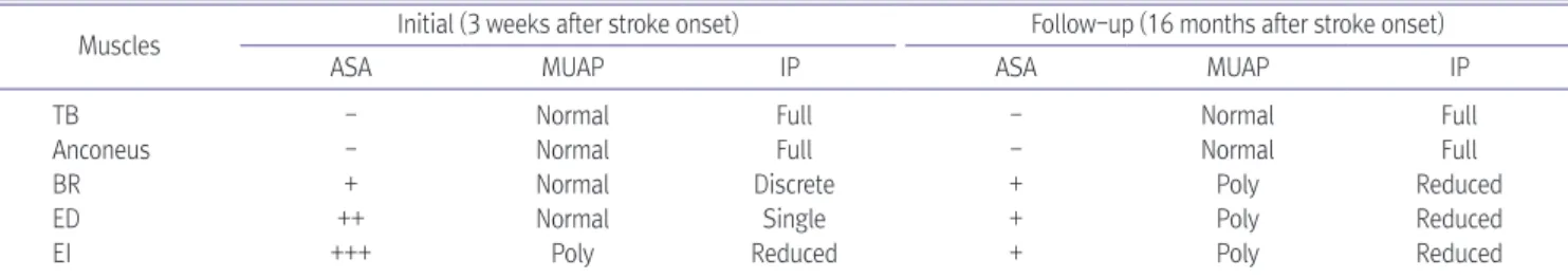
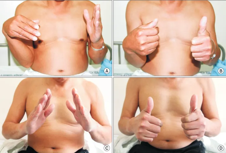
Motor weakness of limbs and trunk are common complaint after stroke. If there are other concurrent causes of motor weakness like focal entrapment neuropathy in paretic limb, it is hard to find them because of weakness due to stroke. A man with the left basal ganglia hemorrhage complained of motor weakness of the right upper extremity, especially wrist and finger extensors. Motor nerve conduction study revealed the right radial neuropathy with definite conduction block between 13 cm and 15 cm proximal to the lateral epicondyle. Ultrasonography showed swelling of right radial nerve at the same lesion. After dexamethasone injection around the right spiral groove, electrodiagnostic findings and clinical feature were significantly improved. In case stroke patients with hemiplegia show persistent motor and sensory impairment in the specific peripheral nerve territory despite of recovery, concomitant neuropathy on the affected side should be considered.

Key Words: stroke, radial nerve, wrist drop, electrodiagnois, ultrasonography

Copyright © by Korean Association of EMG Electrodiagnostic Medicine

This is an Open Ac cess article distributed under the terms of the Creative Commons Attribution Non-Commercial License (http://creativecommons.org/licenses/by-nc/4.0) which permits unrestricted non-commercial use, distribution, and reproduction in any medium, provided the original work is properly cited.

ISSN 1229-6066 https://doi.org/10.18214/jkaem.2017.19.1.10 J Korean Assoc EMG Electrodiagn Med 19(1):10-14, 2017

J Korean Assoc

Electrodiagn Med EMG

(2)

of motor weakness like focal entrapment neuropathy in paretic limb, it is hard to be found because of underlying weakness. In stroke patients, entrapment neuropathies are frequently developed in the median, ulnar and peroneal nerves.

7

The radial neuropathy rarely occurs, the case reports about radial neuropathy in stroke patients are absent. This case report demon­

strated acute radial neuropathy around spiral groove in paretic limb after stroke that was confirmed with electromyographic and ultrasonographic studies and had nearly full recovery following steroid injection.

Case Report

Fifty one­year­old man, who was diagnosed as intracranial hemorrhage in the left basal ganglia one month ago, complained of the right side motor weakness, especially in the upper extremity. The patient had a history of hypertension for one year.

Fifteen days after onset, the manual muscle tests of the right upper extremity revealed grossly grade 1 to 2, while the muscle strength of the right lower extremity was normal. Sensory examination which was performed on palmar side of the right hand and flexor

Table 1. Serial Nerve Conduction Studies of Right Radial Motor Nerve

Nerve Stimulation site Recording site Initial (3 weeks after stroke onset) Follow-up (16 months after stroke onset) Latency (msec) Amplitude (mV) Latency (msec) Amplitude (mV)

Radial Forearm EI 2.2 6.6 2.2 7.4

LE+5 5.3 6.3 4.9 7.1

LE+15 0 7.1 5.3

Radial (inching test) LE+5 EI 4.9 6.9 5.0 7.1

LE+7 5.3 6.8 5.3 7.2

LE+9 5.5 6.8 5.6 7.1

LE+11 5.9 6.8 6.0 6.9

LE+13 6.2 5.6 6.3 6.9

LE+15 0 7.8 4.4

LE+5: 5 cm proximal to lateral epicondyle (LE), LE+7: 7 cm proximal to LE, LE+11: 11 cm proximal to LE, LE+13: 13 cm proximal to LE, LE+15: 15 cm proximal to LE, EI: extensor indicis

A

B C

Fig. 1. Nerve conduction study of the radial nerve (A; right, B; left) showed complete block in the right arm. Inching test of the right radial

motor nerve revealed the complete conduction block between 13 cm and 15 cm proximal to the lateral epicondyle (C).

(3)

part of the right forearm was intact, and deep tendon

reflexes were hyperactive on the right side. Pathologic reflexes were absent. During rehabilitation program, the proximal part of right side motor weakness was improved to grade 4. However, physical examination demonstrated discrepancy between flexor and extensor of the right wrist and finger strength: trace in extensor and good in flexor. Also, detailed sensory examination

demonstrated hypesthesia on the right superficial radial sensory nerve territory.

The electrodiagnostic study was performed to find out focal entrapment peripheral neuropathy, including the radial neuropathy. The right radial motor segmental conduction study revealed complete conduction block in the arm segment (Table 1, Fig. 1). Inching test of the right radial motor nerve around arm demonstrated

Table 2. Serial Needle Electromyographic Examinations

Muscles Initial (3 weeks after stroke onset) Follow-up (16 months after stroke onset)

ASA MUAP IP ASA MUAP IP

TB - Normal Full - Normal Full

Anconeus - Normal Full - Normal Full

BR + Normal Discrete + Poly Reduced

ED ++ Normal Single + Poly Reduced

EI +++ Poly Reduced + Poly Reduced

TB: triceps brachii, BR: brachioradialis, ED: extensor digitorum, EI: extensor indicis, ASA: abnormal spontaneous activities such as fibrillation potentials or positive sharp waves, MUAP: motor unit action potentials, IP: interference patterns, Poly: polyphasic motor unit action potentials

A B

C D

LE+14, C A=13 mm S

2

LE+14, C A=10 mm S

2

LE+15, C A=17 mm S

2

LE+15, C A=10 mm S

2

Fig. 2. Transverse ultrasonograms of

right radial nerve (A and B) demon-

strated swelling of the right radial nerve

around 15 cm proximal to the lateral

epicondyle (LE) compared with the left

radial nerve (C and D). Arrows indicate

radial nerve. LE+14, 14 cm proximal to

LE; LE+15, 15 cm proximal to LE; CSA,

cross-sectional area.

(4)

the complete conduction block between 13 cm and 15 cm proximal to the lateral epicondyle. Needle electromyography showed abnormal spontaneous activities and polyphasic motor unit action potentials with single to reduced interference patterns in the right radial nerve innervated muscles distal to the anconeus (Table 2). Ultrasonographic examination showed focal swelling of right radial nerve at 15 cm proximal to the lateral epicondyle compared with the left: 17 mm

2

in the right; 10 mm

2

in the left (Fig. 2B). Dexamethasone injection under ultrasonographic guidance was performed around the radial nerve at the level of 15 cm proximal to lateral epicondyle. Three months later, the muscle strength of wrist and finger extensors improved to grade 4, and motion of the right hand was grossly similar compared to unaffected side (Fig. 3). Follow­up electrodiagnostic study revealed interval improvement

of the right radial neuropathy (Tables 1, 2).

Discussion

The radial neuropathy can be confused with stroke due to apparent intrinsic hand muscle weakness. In this case, motor recovery of the hemiparetic limbs were accomplished except the wrist and finger extensors and hypesthesia was remained in the radial nerve distribution. Because of persistent weakness of the particular muscle group and sensory change in the radial nerve distribution despite recovery of the motor and sensory impairment, the concomitant focal neuropathy such as radial neuropathy would be suspected and electrodiagnostic and ultrasonographic examinations were performed. The previous studies about entrapment neuropathy in patients with acute

A B

C D

Fig. 3. Weakness of the right wrist and finger extension (A) and the right thumb extension (B) is observed 3 weeks after stroke onset. Muscle

strength of the right wrist and fingers demonstrates nearly full recovery 16 months after stroke onset (C and D).

(5)

or chronic stroke demonstrated that median or ulnar

nerve was usually involved and the entrapment neuropathy may be an important cause for morbidity in patients with stroke.

8,9

However, there is no report about focal radial nerve neuropathy in stroke patients.

The focal neuropathy in the radial nerve can occur during unconsciousness after acute stroke, or during profound sleep, when the arm is draped over a chair or a hard space for a prolonged period. Delayed diagnosis of the concomitant neuropathy in patients with stroke could miss the early appropriate treatment, which lead to permanent nerve injury and functional impairment.

In usual electrodiagnostic study, the conduction block between stimulating points of radial nerve could localize the lesion. Furthermore, we performed short segment study at 2­cm intervals from 5 cm to 15 cm proximal to lateral epicondyle and localized the focal lesion of radial nerve.

Also, the ultrasonography could be used to evaluate the morphology of the radial nerve or space­occupying mass or structure around the lesion which was detected by electrodiagnostic examination. Ultrasound images of the radial neuropathies shows hypoechoic swelling of radial nerve.

10

The use of ultrasonography allows direct visualization of anatomical structures and can find the accurate lesion where the nerve was swollen.

The steroid injection around the swollen nerve could improve the symptoms. Ultrasonography­guided steroid injection is helpful to accurately localize the injectate around the nerve and to potentially reduce the risk of neurovascular injury. The effect of steroid injection is not yet clarified in entrapment neuropathy. In acute experimental neuropathy with conduction block, local steroid injection demonstrated significant increase of the recovery rates of amplitude, but no significant increase of conduction velocity.

11

In our case, it was unclear that clinical and electrophysiological recovery would be related with natural course or local steroid effect. However, steroid perineural injection would be

helpful to relive sensory symptoms such as tingling or pain.

In conclusion, if stroke patients with hemiplegia show discrepancy of the motor weakness between the flexor and extensor muscles of same parts or hypesthesia in the specific peripheral nerve territory, focal neuropathy on the affected side such as radial neuropathy should be considered. And to evaluate and manage the peripheral neuropathy, the electrodiagnostic and ultrasonography studies are useful.

References

1. The World Health Report 2000: Health systems improving performance. Geneva, Switzerland: WHO, 2000.

2. Kalra L, Yu G, Wilson K, Roots P: Medical complications during stroke rehabilitation. Stroke 1995: 26: 990­994 3. Johnston KC, Li JY, Lyden PD, Hanson SK, Feasby TE, Adams

RJ, et al: Medical and neurological complications of ischemic stroke: experience from the RANTTAS trial. RANTTAS Investigators. Stroke 1998: 29: 447­453

4. Davenport RJ, Dennis MS, Wellwood I, Warlow CP:

Complications after acute stroke. Stroke 1996: 27: 415­420 5. Tahir H, Daruwalla V, Meisel J, Kodsi SE: Pseudoradial nerve

palsy caused by acute ischemic stroke. J Investig Med High Impact Case Rep 2016: 4: 1­4

6. Katirji MB, Wilbourn AJ: Common peroneal mononeuropathy: a clinical and electrophysiologic study of 116 lesions. Neurology 1988: 38: 1723­1728

7. Rempel DM, Diao E: Entrapment neuropathies: pathophy­

siology and pathogenesis. Journal of Electromyography and Kinesiology 2004: 14: 71­75

8. Kabayel L, Balci K, Turgut N, Kabayel DD: Development of entrapment neuropathies in acute stroke patients. Acta Neurol Scand 2009: 15: 53­58

9. hunkar R, Balci K: Entrament neuropathies in chronic stroke patients. J Clin Neurophysiol 2012: 29: 96­100

10. Jacobson JA, Fessell DP, Lobo Lda G, Yang LJ: Entrapment neuropathies I: upper limb (carpal tunnel excluded). Seminars in Musculoskeletal Radiology 2010: 14: 473­486

11. Hong CZ, Cheng BB, Liu A, Yu J: Local steroid injection:

effect on the recovery of nerve conduction in experimental

neuropathy. Arch Phys Med Rehabil 1990: 71: 42­45

수치

Table 1. Serial Nerve Conduction Studies of Right Radial Motor Nerve
Table 2. Serial Needle Electromyographic Examinations
Fig. 3.	Weakness	of	the	right	wrist	and	finger	extension	(A)	and	the	right	thumb	extension	(B)	is	observed	3	weeks	after	stroke	onset.	Muscle	 strength	of	the	right	wrist	and	fingers	demonstrates	nearly	full	recovery	16	months	after	stroke	onset	(C	and	D).

참조

관련 문서

【판결요지】[1] [다수의견] 동일인의 소유에 속하는 토지 및 그 지상 건물에 관하여 공동저 당권이 설정된 후 그 지상 건물이 철거되고 새로 건물이 신축된 경우에는

1) 신경전도 속도검사 (Nerve conduction velocity test).. 162. Electrodiagnostic

1 John Owen, Justification by Faith Alone, in The Works of John Owen, ed. John Bolt, trans. Scott Clark, "Do This and Live: Christ's Active Obedience as the

The greater tubercle is palpable on the line from the lateral epicondyle of the distal humerus in the direction of the humeral longitudianl axis and just below the acromion

As a result of the thermal conduction analysis, a cooling zone is formed on the surface of the welding metal along the arc by a cooling medium, compared

The purpose of this study was to analyze the impaction pattern of the impacted mandibular third molar and the relationship with the inferior alveolar nerve

The effect of the stress management program on the stress coping and perceived stress of the patients with finger replantation. Unpublished master's

It is the purpose of this study to investigate the anti microbacterial Activity and anti inflammatory Effects of water extract Rhododendron brachycarpum