• 검색 결과가 없습니다.

Elbow Healthcare System for Flexion and Extension Abnormality of Elbow

N/A
N/A
Protected

Academic year: 2021

Share "Elbow Healthcare System for Flexion and Extension Abnormality of Elbow"

Copied!
6
0
0

로드 중.... (전체 텍스트 보기)

전체 글

(1)

Elbow Healthcare System for Flexion and Extension Abnormality of Elbow

1)

Seong-Yoon Shin*

Abstract

In a broad sense, the healthcare system refers to the conventional medical service for the field of treatment. From head to toe, our whole body corresponds to the subject of the medical service. In this paper, we discuss the abnormality of flexion and extension in general elbow disease. Flexion and extension refer to flexing and extending of the arm while it is set to be at 90 degrees. In this case, if the angle of the arm is remarkably small or is accompanied with pain, there is an abnormality that occurs in the elbow. We tested the flexion and extension of the elbow for 100 people in their 50s and calculated the number of people for each case. Afterwards, we classified people with abnormalities in flexion and extension and presented the respective treatment methods. In this paper, a system was constructed for the treatment of musculoskeletal disorders

▸Keyword: Healthcare System, Elbow Disease, Flexion and Extension, Musculoskeletal Disorders

I. Introduction

In a broad sense, healthcare refers to including all of treatment, prevention, and the course of health management, and in a narrow sense, it refers to remote medical treatment and health counseling. If healthcare and IT technology are combined, many people can use medical services more freely. In this paper, we describe the method of treating elbow disease from the perspective of the musculoskeletal system from the viewpoint of the healthcare system. We will start with a study on the flexion and extension of the elbow.

Flexion of the elbow provides important functions such as pulling, lifting, and eating food. The elbow extension also allows the user to perform actions such as throwing or stretching. Related papers were published [1] that measured variability of maximum elbow joint torque calculated during periodic elbow flexion and extension.

The purpose of [2] was to determine the cerebral measurement reliability of manual testing of elbow flexor muscle spasms using the modified Ashworth scale. In this

study, we extended the patient's elbow to the maximum extension of the maximum arm length at the maximum flexion position, with the forearm gripped away.

The goal of [3] was to develop an animal model of post-traumatic elbow build-up and to assess the possibility of studying the cause of PTJS(post-traumatic joint stiffness). After sacrifice, the flexion-extension mechanical joint test showed reduced range of motion and increased stiffness of the injured fixation limb relative to control and pseudo-animals with functional effects correlated with degree of impairment.

Subluxation or dislocation of the ulnar nerve in the elbow flexion is one of the causes of ulnar nerve roots, which causes internal elbow pain, numbness and movement disorders of the ulnar side of the hand.

However, some studies have used palpated or ultrasonography to demonstrate that flexion causes ulnar nerve displacement in healthy elbows[4][5].

The goal of [6] was to evaluate the location and signal

∙First Author: Seong-Yoon Shin, Corresponding Author: Seong-Yoon Shin

*Seong-Yoon Shin (s3397220@kunsan.ac.kr), School of Computer Information & Communication Engineering, Kunsan National University

∙Received: 2018. 09. 17, Revised: 2018. 10. 17, Accepted: 2018. 10. 23.

(2)

intensity of ulnar nerve in healthy elbow grooves with healthy elbows and extended flexion, with additional analysis of neurological location of healthy elbow and ulnar neuropathy.

Triceps play an important role during elbow flexion/extension exercises. Therefore, it is necessary to investigate the possibility of motion change by applying a vibration stimulus to the triceps to provide cognitive support to the upper extremities. In [7], a vibration stimulus was added to the deltoids during one continuous bending motion of the elbow angle. From the experimental results, an interesting feature of the motion change occurring in the triceps by vibration stimulation was obtained.

In [8], it can propose a protocol that can objectively evaluate upper limb function. Applying to push-ups implies that loss of elbow flexion affects more movements than loss of elbow extension

Therefore, many studies have been conducted regarding the flexion and extension of the elbow. We will examine the contents of related research to this extent and investigate the structure of the elbow, diseases, and treatments related to flexion and extension.

Next, we will look at the research related to the Healthcare System.

To provide a more convenient service and medical environment, [9] proposes a cyber-physical system for patient-oriented medical applications and services called Health-CPS, which is built on cloud and big data analysis technology.

With the development of information and communication technology, IOT (Internet of Things) appeared. [10] At first, the key security requirements of modern healthcare systems based on the Body Sensor Network(BSN) are highlighted. Next, we proposed a secure IoT-based healthcare system using the BSN-Care BSN that can efficiently meet these requirements.

The healthcare systems are rapidly adopting large amounts of data, driven by record keeping, compliance and regulatory requirements, and patient care. The advances in healthcare system will rapidly enlarge the size of the health records that are accessible electronically. Concurrently, fast progress has been made in clinical analytics. The proposed knowledge system[11]

is developed based on variety of databases such as Electronic Health Record (EHR), Medical Imaging Data, Unstructured Clinical Notes and Genetic Data. The

proposed methodology asynchronously communicates with different data sources and produces many alternative decisions to the doctor

[12] describes the architecture of the Patient Centered Research Results Institute (PCORI) for the clinical data research network of the Healthcare Management System (SCILHS, http://www.SCILHS.org).

II. Structure of Elbow

The structure of the elbow is shown in Fig. 1. The description of the detailed structure is as follows.

Fig. 1. Structure of Elbow

The ulna is the longer, larger, and medial of the lower arm bones. Many muscles in the arms and forearms attach to the ulna and perform arm, hand and wrist movements. The movement of the ulna is essential for everyday functions such as throwing a ball and driving a car.

The radius is slightly shorter than the sides of the two arm bones. It is on the thumb side of the forearm and rotates to allow the wrist to rotate. Several muscles in the arms and forearms are inserted in the inferior and inferior radius and provide motion to the upper extremities. This movement is essential for everyday tasks such as writing, drawing, and throwing a ball.

The humerus is the largest bone in the arm and the

only bone in the upper arm. There are many powerful

muscles fixed to the humerus that manipulate the forearm

of the shoulder and the forearms of the elbow. The

(3)

movement of the humerus is essential for all the various activities of the arm, such as throwing, lifting and writing.

The joint capsule of the elbow has thickened ulnar and radial ligaments. The front of the capsule is reinforced with fibers coming from the muscles of the upper arm.

Like all synovial joints, a thin layer of soft articular cartilage covers the tip of the bone forming the elbow joint.

There is a joint cavity filled with synovial fluid between the joint surfaces. The joint can be partially or completely subdivided by a joint disc known as meniscus.

At the elbow joint, the end of the bone is covered with articular cartilage. Articular cartilage is a soft and smooth material. It protects the bone tip from friction when the elbows snap together as they move. Articular cartilage is soft enough to act as a shock absorber. If you are not injured, it will be hard enough to last forever.

Ⅲ. Flexion and Extension of Elbow

3.1 Elbow Flexion

Elbow flexion is an elbow test procedure for the forearm tunnel syndrome test. The cubital tunnel is an important pathway for the ulnar nerve when it passes behind the elbow. Admission and trauma are the main causes of bilateral tunnel syndrome, and sensations and tingling symptoms in this area may only appear in a few years.

The patient should be awake and be cooperative in elbow flexion testing. Elbow flexion is shown in Fig. 2(a).

1) The patient makes the elbow 90 degrees.

2) The bottom of the hand should face upward.

3) The patient should have his / her elbow fully bent by raising his / her hand.

4) The examiner instructs the patient to remain in position for a maximum of 1 to 3 minutes.

5) The inspector examines the patient for a few minutes. He can apply gentle pressure to increase elbow flexion.

6) At this time, the angle should be 130 ~ 145 degrees.

7) If the angle is too small or accompanied by pain, it is called cubital tunnel syndrome.

(a) (b)

Fig. 2. Structure of Elbow

3.2 Elbow Extension

If there is an exudate or a bodily fracture, the entire extension of the elbow is blocked. According to several studies, elbow expansion testing is a fast and reliable test that excludes potential fractures.

The patient should be awake and be cooperative in the elbow dilation test. Elbow extension is shown in Fig. 2(b).

1) The patient makes an elbow 90 degrees.

2) The bottom of the hand should face up.

3) The patient pulls his / her hand down completely.

4) The examiner instructs the patient to hold the posture for a maximum of 1 to 3 minutes.

5) The inspector examines the patient for a few minutes. He can apply gentle pressure to increase elbow flexion.

6) At this time, the angle should be 0 ~ -5 degrees.

7) If the angle is too small or painful, it is called potential fractures.

Ⅳ. Therapy

4.1. Elbow Flexion-Cubital Tunnel Syndrome First, if there is an abnormality in the flexion and cubital tunnel syndrome is displayed, there are non-surgical treatment and surgical treatment options available.

In non-surgical treatment methods, the first priority is

to change habits so as to avoid excessive flexion of the

elbow joint in everyday life. Next, in order to avoid elbow

(4)

flexion during sleep, there is a nighttime splint method in which elbows are fixed in a stretched state. Patients can also consider splints to prevent the flexion of the elbow joint additionally during daytime, physical therapy, etc.

There are also avoiding posture in which the ulnar nerve is pressed in everyday life, stretching the elbow joint during sleeping, avoiding excessive elbow flexion, not leaning the elbow on the desk while talking on the phone, etc. There are also topical steroid injections, nonsteroidal analgesic anti-inflammatory drugs, and vitamin B6 dosage regimens for drug treatment. Implementing steroid injection treatment directly on the passage of the ulnar nerve around the elbow joint may cause changes in the surrounding soft tissue in the future, and therefore, it should be avoided.

Even for initial lesions, surgery should be considered when there is no improvement in symptoms by the non-surgical treatment or when nerve compression is progressed. The surgical method is decided by taking into account the difficulty of surgery and the advantages and disadvantages of each method. The most important principle is to remove the structure that presses the nerve around the ulnar nerve, move the nerve in the medial upper and the posterior forward in order to prevent the nerve from being lengthened or being pressed while the elbow is flexed. Representative surgical treatments include simple decompression, decompression with anterior transposition of ulnar nerve, medial epicondylectomy, endoscopic decompression, etc.

4.2. Elbow Extension-Potential Fractures

An elbow fracture may occur when a shock is directly applied to the elbow or the arm is twisted. Sprains, deformation, or dislocation may occur simultaneously with fracture. X-rays are used to inspect fractures and dislocations. Sometimes computed tomography (CT) inspection can be used to provide more details.

Here we will look at splice protection. If the bone fragments are not displaced, fractures may be treated with splints to fix the elbows in the healing process. Your doctor will often take an x-ray to see if your bones have not changed during the healing process.

Splints are usually worn for about 5-6 weeks before smooth movements begin. If the fracture changes during this time, you may need to revisit the fracture.

Fractures that are out of position or unstable are more likely to require surgery. Surgery replaces and stabilizes

debris and removes bone fragments. Emergency surgery is necessary to treat wounds and bones in order to minimize the risk of infection when the fracture is open.

Surgery is usually required for the following fractures.

When the bone is in a different position away from its position, it is called a displaced fracture. A piece of broken bone that punctures the skin, called an open fracture.

Surgery for fractures typically involves placing the broken pieces of bone in place and keeping them immobile until they are stuck and cured.

Because of the increased risk of infection during surgery, open fractures should be operated as soon as possible. During surgery, the wound is completely removed and the bones are usually restored during the same operation.

Ⅴ. Experiment

The experiment was implemented in Visual C++ 2015 in Windows 7, and DB used MySQL Server 5.6 version. For the elbow’s healthcare system, first, personal information was entered, and the system received data for whether the subject felt pain and whether there was an abnormality in flexion or extension. Certainly, a situation where all three symptoms were accompanied was possible. Fig. 3 is the healthcare system inquiry screen of Seong-Yoon Shin’s elbow. This patient is diagnosed with cubital tunnel syndrome because a high value in the flexion of the elbow was displayed, and while not in the stage for surgery, physical therapy treatment for 3 days was ordered.

Fig.3. Inquiry Screen of Healthcare System

(5)

In this study, we conducted experiments with 100 general people in their 50s. Personal information, the flexion angle, the extension angle, and date of whether a person felt pain were entered. It didn’t matter even with all three types or with more than(equal) one. For reference values, flexion was set between 130 degrees and 145 degrees, and extension was set between 0 degrees and –5 degrees. The experimental result was shown in Table 1.

Total persons

Flexion angle

Extension

angle with

pain Note

Large Small Large Small Person

(Total persons:

88) 3

17 (Pain :

4) 4

23 (Pain :

2) 6

If accompanied with pain, flexion

and extension were both

treated.

Percent 3% 17%

(4%) 4% 23%

(2%) 6%

Table. 1. Experimental Results

In the experiment, in many cases, flexion the elbow and the extension angle were smaller than the reference values. However, the reason for this is that the age of the people is in the 50s, and it is the result of the bone being solidified to some extent. In the case of being accompanied with pain, the number was very few. In particular, in flexion, pain was a problem that could be corrected by physical therapy. However, for the pain caused in extension, we encouraged people to take a complete medical examination at the hospital because these were cases where they suffered a fracture when they were young or are currently suffering from a fracture.

Ⅵ. Conclusions

In this study, we presented a healthcare system dealing with abnormalities of flexion and extension in elbow disease. Flexing and extending the arm at a 90 degree angle were defined as flexion and extension, respectively, and when the angle of the arm was remarkably small or was accompanied with pain, we had the patient take a complete medical examination at the hospital because we were in doubt of a fracture of the elbow. Here, treatment methods were also easily determined and reported even when abnormality occurred. In addition, we presented a

healthcare system including patient input/output, angle measurement, and treatment for the elbow flexion and extension. We tested the elbow’s flexion and extension for 100 people in their 50s, and as a result, the angle was 3% in flexion, 17% (4% accompanied with pain) was small in flexion, the angle was large for 4% in extension and was shown to be 23% (2% accompanied with pain). Even when flexion was accompanied with pain, it was mostly resolved by physical therapy, but for extension, a fracture was suspected, certainly having recommended a complete medical examination to patients.

REFERENCES

[1] Ballaz L, Raison M, Detrembleur C, Gaudet G, Lemay M. “Joint torque variability and repeatability during cyclic flexion-extension of the elbow.” BMC sports science, medicine and rehabilitation. Vol. 11, No. 1, April 2016.

[2] Charalambous, Charalambos P. “Interrater reliability of a modified Ashworth scale of muscle spasticity,” Classic Papers in Orthopaedics. Springer, London, pp. 415-417, 2014.

[3] Lake, S. P., Castile, R. M., Borinsky, S., Dunham, C. L., Havlioglu, N. and Galatz, L. M. “Development and use of an animal model to study post-traumatic stiffness and contracture of the elbow,” J. Orthop. Res., Vol. 34, pp.

354–364, February 2016, doi:10.1002/jor.22981 [4] Van Den Berg PJ, Pompe SM, Beekman R, Visser LH.,

“Sonographic incidence of ulnar nerve (sub)luxation and its associated clinical and electrodiagnostic characteristics,” Muscle Nerve, Vol. 47, No. 6, pp. 849–

855, June 2013.

[5] Yang SN, Yoon JS, Kim SJ, Kang HJ, Kim SH., “Movement of the ulnar nerve at the elbow: a sonographic study,”

J Ultrasound Med, Vol. 32, No. 10, pp. 1747–1752, October 2013.

[6] Y. Kawahara, T. Yamaguchi, Y Honda, Y. Tomita, and M. Uetani, “The Ulnar Nerve at Elbow Extension and Flexion: Assessment of Position and Signal Intensity on MR Images,” Radiology, Vol. 280, No. 2, pp. 489-492, February 2016.

[7] K. Honda and K. Kiguchi, “A fundamental study on the effect of vibration stimulation on triceps brachii during elbow flexion motion for perception-assist,” 2016 International Symposium on Micro-NanoMechatronics and Human Science (MHS), pp. 1-3, November, 2016.

Nagoya, doi: 10.1109/MHS.2016.7824213

(6)

[8] Fradet, L., Liefhold, B., Rettig, O., Bruckner, T., Akbar, M., & Wolf, S. I., “Proposition of a protocol to evaluate upper-extremity functional deficits and compensation mechanisms: application to elbow contracture,” Journal of Orthopaedic Science, Vol. 20, No. 2, pp. 321-330, March 2015.

[9] Zhang, Y., Qiu, M., Tsai, C. W., Hassan, M. M., & Alamri, A., “Health-CPS: Healthcare cyber-physical system assisted by cloud and big data,” IEEE Systems Journal, Vol. 11, No. 1, pp. 88-95, March 2017.

[10] Gope, P., & Hwang, T. “BSN-Care: A secure IoT-based modern healthcare system using body sensor network,”

IEEE Sensors Journal, Vol.16, No. 5, pp.1368-1376, March 2016.

[11] Manogaran, G., Thota, C., Lopez, D., Vijayakumar, V., Abbas, K. M., & Sundarsekar, R., “Big data knowledge system in healthcare,” In Internet of things and big data technologies for next generation healthcare. pp.

133-157, Springer, Cham, January 2017.

[12] Mandl, K. D., Kohane, I. S., McFadden, D., Weber, G.

M., Natter, M., Mandel, J., ... & Adams, W. G., “,” Journal of the American Medical Informatics Association, Vol.

21, No. 4, pp. 615-620, May 2014.

Authors

Seong-Yoon Shin received his M.S. and

Ph.D degrees from the Dept. of Computer

Information Engineering of Kunsan

National University, Kunsan, Korea, in 1997

and 2003, respectively. From 2006 to the

present, he has been a professor in the

same department. His research interests include image

processing, computer vision, and virtual reality.

수치

Fig.  1.  Structure  of  Elbow
Fig.  2.  Structure  of  Elbow

참조

관련 문서

The index is calculated with the latest 5-year auction data of 400 selected Classic, Modern, and Contemporary Chinese painting artists from major auction houses..

The twin rod cylinders are suitable for applications that require the non rotation of the piston and resistance of flexion. A tandem cylinder can provide amplified output

– Head rotation to the left causes extension of left arm and leg and flexion of right arm and leg; head rotation to the right causes extension of right arm and leg and

- 노뼈머리는 위팔뼈 작은머리(capitulum of the humerus)와 자뼈의 노패임(ulnar radial notch)과 관절을 이룬다...

The mainstay therapy for excessive iron deposition in pa- tients with primary and secondary hemochromatosis is phlebotomy and iron chelation, respectively, which are de-

 The PL bundle is hidden behind the AM bundle and becomes visible during flexion as the femoral insertion moves

• For first language acquisition, there seems to be a critical period of the first five years, during which children must be exposed to rich input.. There is also

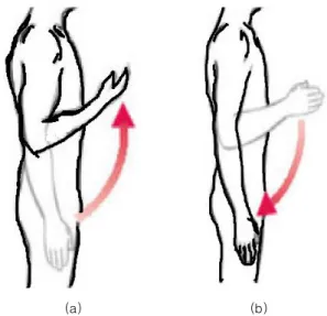
Figure 8.5b Movements allowed by synovial joints. (b) Angular movements: flexion, extension, and