Journal of
Preventive Medicine
& Public Health
248
Copyright © 2018 The Korean Society for Preventive Medicine J Prev Med Public Health 2018;51:248-256 • https://doi.org/10.3961/jpmph.18.112Effect of Uric Acid on the Development of Chronic Kidney Disease: The Korean Multi-Rural Communities Cohort
Study
Kwang Ho Mun1, Gyeong Im Yu1, Bo Youl Choi2, Mi Kyung Kim2, Min-Ho Shin3, Dong Hoon Shin1
1Department of Preventive Medicine, Keimyung University School of Medicine, Daegu, Korea; 2Department of Preventive Medicine, Hanyang University College of Medicine, Seoul, Korea; 3Department of Preventive Medicine, Chonnam National University Medical School, Gwangju, Korea
Original Article
Objectives: Several studies have investigated the effects of serum uric acid (SUA) levels on chronic kidney disease (CKD), with discrep- ant results. The effect of SUA levels on CKD development was studied in the Korean rural population.
Methods: A total of 9695 participants aged ≥40 years were recruited from 3 rural communities in Korea between 2005 and 2009. Of those participants, 5577 who participated in the follow-up and did not have cerebrovascular disease, myocardial infarction, cancer, or CKD at baseline were studied. The participants, of whom 2133 were men and 3444 were women, were grouped into 5 categories ac- cording to their quintile of SUA levels. An estimated glomerular filtration rate of <60 mL/min/1.73 m2 at the time of follow-up was considered to indicate newly developed CKD. The effects of SUA levels on CKD development after adjusting for potential confounders were assessed using Cox proportional hazard models.
Results: Among the 5577 participants, 9.4 and 11.0% of men and women developed CKD. The hazard ratio (HR) of CKD was higher in the highest quintile of SUA levels than in the third quintile in men (adjusted HR, 1.60; 95% confidence interval [CI], 1.02 to 2.51) and women (adjusted HR, 1.56; 95% CI, 1.14 to 2.15). Furthermore, CKD development was also more common in the lowest quintile of SUA levels than in the third quintile in men (adjusted HR, 1.83; 95% CI, 1.15 to 2.90). The effect of SUA was consistent in younger, obese, and hypertensive men.
Conclusions: Both high and low SUA levels were risk factors for CKD development in rural Korean men, while only high levels were a risk factor in their women counterparts.
Key words: Cohort studies, Chronic kidney disease, Uric acid, Korea
Received: May 21, 2018 Accepted: August 10, 2018 Corresponding author: Dong Hoon Shin, MD, PhD
Department of Preventive Medicine, Keimyung University School of Medicine, 1095 Dalgubeol-daero, Dalseo-gu, Daegu 42601, Korea E-mail: [email protected]
This is an Open Access article distributed under the terms of the Creative Commons Attribution Non-Commercial License (http://creativecommons.org/licenses/by- nc/4.0/) which permits unrestricted non-commercial use, distribution, and repro- duction in any medium, provided the original work is properly cited.
INTRODUCTION
Chronic kidney disease (CKD) is a major cause of mortality worldwide; more than 2 million patients receive renal replace-
pISSN 1975-8375 eISSN 2233-4521
ment therapy, and an estimated 10-fold higher number need therapy, but do not receive treatment [1]. In Korea, 80 674 pa- tients received kidney transplantation or underwent renal di- alysis in 2014 [2]. Furthermore, it has been found that 14 and 17% of patients who underwent peritoneal dialysis and hemo- dialysis, respectively, were bedridden or dependent in activi- ties of daily living, with various comorbidities such as cardio- vascular disease (CVD) and infections [2]. Several common risk factors that contribute to CKD development are diabetes mel- litus (DM), hypertension (HTN), obesity, elevated creatinine levels, and hyperuricemia [3-5]. Thus, controlling these risk factors is an important aspect of preventing CKD develop-
Preventive Medicine
& Public Health
ment.Uric acid, an end-product of the metabolism of purine nu-cleotides, occurs at higher levels in humans than in other mammals due to its loss during the uricase process [6]. In the 2007-2015 national health claims database, the prevalence of gout in 2015 was 13.57 per 1000 people in men and 1.58 in women, and the incidence per 1000 people was higher in men (3.21) than in women (0.67) [7].
High levels of serum uric acid (SUA) have both advantages and disadvantages for the human body. SUA is known to have a neuroprotective effect, lowering the risk of Parkinson disease and cognitive impairment [8,9]. Moreover, it also acts as an antioxidant in the serum [10]. However, SUA has a strong pro- oxidant effect at the cellular level and is known to be responsi- ble for cardiovascular mortality [11,12]. Furthermore, high lev- els of SUA have been reported to be responsible for the devel- opment of HTN and higher mortality [12,13].
Several reports have shown a relationship between SUA and CKD. In several studies, high SUA levels were found to contrib- ute to CKD development [4,14]. However, some studies have failed to show a significant contribution of SUA to CKD devel- opment [15,16]. Moreover, other studies of CKD patients have shown SUA-lowering therapy to be beneficial; however, a me- ta-analysis did not support those findings [17,18].
A few reports have shown that low SUA levels conferred an increased risk of CKD development and mortality. A study by Kanda et al. [19] showed that both high and low SUA levels contributed to the loss of kidney function in Japanese men.
One study on CKD patients reported a J-shaped relationship between mortality and SUA levels, with the lowest quintile of SUA as a risk factor [20]. Another study by Lee et al. [21] re- ported that low SUA levels (the lowest quintile) were a risk fac- tor for mortality in new-onset hemodialysis patients.
We analyzed data from the Multi-Rural Communities Cohort (MRCohort), consisting of rural inhabitants aged ≥40 years collected from 3 centers. The effects of various levels of SUA on CKD development were studied, and further studies on specific groups affected by SUA should be conducted.
METHODS Study Population
The MRCohort was established in 2004 as part of the Korean Genomic and Epidemiology Study, designed to assess the risk factors associated with CVD in the Korean population. The
study was conducted in 3 rural areas in Korea: Goryeong, Yangpyeong, and Namwon. Within these 3 rural areas, villages were chosen through multistage cluster sampling, and partici- pants aged ≥40 years were recruited.
As of 2009, a total of 9695 participants had been recruited to the cohort, as shown in the flow diagram (Figure 1). Partici- pants were followed up every 2-4 years. As of 2013, among the 9695 participants, 7020 had participants returned for fol- low-up.
Among the 9695 participants, 2675 who did not participate in the follow-up were excluded. A total of 781 participants with cancer, cerebrovascular disease, or myocardial infarction prior to enrollment were also excluded from the study. Partici- pants with missing self-reported information on lifestyle fac- tors including alcohol consumption, smoking, and exercise, and participants with missing laboratory data, including SUA and creatinine levels or other confounding variables (body mass index [BMI], fasting glucose level, and serum lipid levels) were not included in the study. Lastly, participants with an es- timated glomerular filtration rate (eGFR) equivalent to CKD stage 3 or higher (eGFR <60 mL/min/1.73 m2) at baseline were excluded from the study. Finally, 5577 participants were analyzed, of whom 2133 were men and 3444 were women.
This study was conducted with the approval of the ethics com- Figure 1. Flow diagram of the enrollment of study subjects.
Missing data include serum uric acid, creatinine, smoking, drinking, exercise, height, and weight variables. CVA, cere- brovascular disease; MI, myocardial infarction; CKD, chronic kidney disease.
Participants at the baseline (n=9695)
Participants (n=6239)
Participants (n=6155)
Participants for analysis (n=5577)
Participants excluded for follow-up loss (n=2675) Participants excluded for CVA, MI, cancer (n=781)
People excluded for missing data (n=84)
People excluded for baseline CKD (n=578)
250
mittee of Keimyung University in Korea (no. 40525-201803- HR-09-01).
Data Collection
Data were collected from 3 centers using a standardized questionnaire and examination procedures by trained inter- viewers and examiners. All interviewers and technicians were trained by the same trainers using a standardized protocol from the coordinating center.
The questionnaire included demographic, lifestyle, disease, and medication history information. The demographic infor- mation consisted of identification number, age, gender, edu- cational status, and marital status. The lifestyle factors were smoking, alcohol consumption, and exercise status. Partici- pants’ self-reported history of previous diseases such as HTN, diabetes, CVD, myocardial infarction, and cancer was also col- lected.
Anthropometric measurements were obtained by a trained examiner at each center using a standard protocol. Height was obtained using a standard height scale, and for weight mea- surements, the scale was zero-balanced before each measure- ment. BMI was computed as weight divided by height squared.
Participants were categorized based on BMI into a normal group (BMI <23 kg/m2) and an overweight/obese group (BMI ≥23 kg/m2).
Blood pressure (BP) was initially measured at the right arm at heart level after the participant had rested for 10 minutes.
Two measurements, with at least a 5-minute interval, were av- eraged to obtain the systolic and diastolic BP of each partici- pant. If the difference between the 2 measurements was high- er than 5 mmHg, the measurement was repeated. HTN was defined as a BP >140/90 mmHg, the use of antihypertensive drugs, or an HTN diagnosis prior to the study.
Laboratory tests were conducted using blood samples col- lected after a minimum of 8 hours of overnight fasting. All markers were analyzed on the same day, within 12 hours. Tria- cylglycerol, total cholesterol, high-density lipoprotein (HDL) cholesterol, fasting glucose, SUA, and creatinine levels were obtained using an ADVIA 1650 automated analyzer (Siemens, New York, NY, USA).
Diagnostic Definition
eGFR was assessed using the CKD-Epi equation. The validity of this formula has been studied elsewhere [22]. An eGFR <60 mL/min/1.73 m2 at the time of follow-up was considered to in-
dicate newly developed CKD. Participants were followed up until CKD development or the final follow-up.
Statistical Analysis
SUA levels were categorized into 5 groups using quintile cut-off points in men (<4.7, 4.7-5.3, 5.4-5.9, 6.0-6.8, and >6.8 mg/dL) and women (<3.6, 3.6-4.0, 4.1-4.5, 4.6-5.1, and >5.1 mg/dL). The quintile grouping was chosen to obtain more sta- ble estimates.
Continuous variables are presented as mean±standard de- viation (SD), while categorical variables are presented as fre- quency and percentage. The Student t-test, one-way analysis of variance, and the chi-square test were used to compare be- tween-group differences. All analyses were conducted for each gender. The outcome was defined as CKD development.
Cox regression analysis was used to present the hazard ratio (HR) and 95% confidence intervals (CIs), with the third quintile used as the reference to study the effect of both low and high SUA levels on CKD development. No confounders were adjust- ed in model 1. eGFR, gender, and age were adjusted in model 2. Smoking, drinking, and exercise habits; marital status; edu- cational level; presence of HTN and DM; and glucose, triacylg- lycerol, and total cholesterol levels were further adjusted in model 3. To further assess the effects of SUA levels on CKD de- velopment, subgroups stratified by age (<60 or ≥60 years), BMI (<23 or ≥23 kg/m2), and HTN (with or without HTN) were used. Two-tailed p-values <0.05 were considered to indicate statistical significance. SPSS version 23.0 (IBM Corp., Armonk, NY, USA) and R version 3.4.3 (http://www.r-project.org, pack- age “forestplot”) were used for all statistical analyses.
RESULTS
Both men and women were divided into 5 groups using the quintiles of SUA levels. The average follow-up period was 47.0±19.0 months in men and 46.9±19.0 months in women.
Table 1 presents gender-specific characteristics. In men, age, BMI, triacylglycerol, and total cholesterol levels increased as SUA levels increased, with the highest levels in quintile 5 (Q5).
The number of alcohol consumers and individuals with HTN was higher in Q5 than in other quintiles. On the contrary, the number of DM patients and the HDL and fasting glucose levels increased as SUA level decreased, with the highest levels in quintile 1 (Q1) (Table 1). In women, BMI, triacylglycerol, and total cholesterol levels increased as SUA levels increased. The
Table 1. Selected baseline characteristics by serum uric acid quintile (unit: mg/dL) by gender CharacteristicsMenWomen Q1 (<4.7)Q2 (4.7-5.3)Q3 (5.4-5.9)Q4 (6.0-6.8)Q5 (>6.8)p-valueQ1 (<3.6)Q2 (3.6-4.0)Q3 (4.1-4.5)Q4 (4.6-5.1)Q5 (>5.1)p-value Total (n)395410456416456684650629711770 CKD development46 (10.6)34 (7.7)33 (7.4)34 (8.7)54 (12.8)<0.0579 (9.9)49 (7.1)64 (10.6)71 (10.2)116 (17.5)<0.001 Age (y)63.72±8.5662.30±8.7261.07±9.0560.64±8.8459.66±9.14<0.00160.29±9.3859.20±9.2959.45±9.5258.91±9.1960.32±8.33<0.01 Follow-up time (mo)46.21±18.8045.91±20.2946.50±19.5647.42±19.1946.79±18.15NS47.37±19.0147.10±19.3547.20±19.4046.77±18.9146.40±18.35NS Married374 (94.7)382 (93.2)440 (96.5)393 (94.5)432 (94.7)NS519 (75.9)515 (79.2)482 (76.6)558 (78.5)593 (77.0)NS College or higher32 (8.1)31 (7.6)50 (11.0)40 (9.6)54 (11.8)NS19 (2.8)24 (3.7)24 (3.8)36 (5.1)25 (3.2)NS Smoker129 (32.7)140 (34.1)129 (28.3)129 (31.0)147 (32.2)NS10 (1.5)14 (2.1)14 (2.2)22 (3.1)23 (3.0)NS Alcohol consumer236 (59.7)261 (63.7)297 (65.1)285 (68.5)356 (78.1)<0.001198 (28.9)192 (29.5)183 (29.1)236 (33.2)266 (34.5)<0.05 Exercise121 (30.6)104 (25.4)147 (32.2)133 (32.0)147 (32.2)NS171 (25.0)180 (27.7)179 (28.5)251 (35.3)257 (33.4)<0.001 HTN116 (29.4)124 (30.2)143 (31.4)139 (33.4)183 (40.1)<0.01198 (28.9)188 (28.9)192 (30.5)245 (34.5)353 (45.8)<0.001 DM79 (20.0)56 (13.7)59 (12.9)44 (10.6)50 (11.0)<0.0173 (10.7)49 (7.5)49 (7.8)59 (8.3)81 (10.5)NS BMI (kg/m²)22.98±2.8023.54±2.8124.05±2.9224.46±2.8825.01±2.90<0.00123.72±3.0524.01±3.0824.48±3.0124.76±3.0725.71±3.23<0.001 Tchl (mg/dL)186.97±32.56189.45±34.26188.36±32.27195.93±36.59198.19±35.82<0.001198.95±36.58200.52±34.15203.48±35.01205.08±35.70212.68±36.08<0.001 Tg (mg/dL)127.90±94.38142.16±82.28158.88±104.10172.57±117.05197.74±131.36<0.001125.80±70.34126.92±64.91136.63±73.83150.52±86.53174.62±110.36<0.001 HDL (mg/dL)45.55±11.5944.59±11.1843.55±11.8042.81±10.9042.21±9.73<0.00146.80±9.9547.02±10.1245.90±10.3944.83±10.0043.20±9.26<0.001 Glucose (mg/dL)108.79±44.05105.03±29.43103.73±24.52102.60±22.64101.72±16.85<0.0199.40±24.7296.17±16.9796.20±16.4096.54±17.08100.13±16.97<0.001 eGFR (mL/min/1.73 m2)80.26±10.0279.13±9.2278.40±9.7877.20±8.7675.25±9.40<0.00179.26±10.1478.43±9.4076.47±8.9575.37±8.8973.04±8.50<0.001 Values are presented as number (%) or mean±standard deviation. CKD, chronic kidney disease; NS, not significant; HTN, hypertension; DM, diabetes mellitus; BMI, body mass index; Tchl, total cholesterol; Tg, triacylglycerol; HDL, high-density lipoprotein; eGFR, estimated glomerular filtration rate.
252
number of alcohol consumers, individuals with HTN, and par- ticipants who exercised increased across quintiles in women, and the same findings were observed for men, except for the trend for exercise. However, fasting glucose levels and age were higher in Q1 and Q5 than in quintile 3 (Q3) (Table 1).
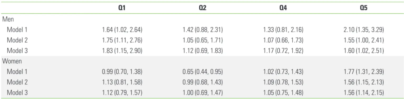
During the follow-up period, 9.4 and 11.0% of men and women developed CKD, respectively. Table 2 shows the effects of SUA levels on CKD development. Higher rates of CKD devel- opment were observed in those with high SUA levels (Q5) than in Q3 among men (adjusted HR, 1.60; 95% CI, 1.02 to 2.51). Furthermore, men participants with low SUA levels (Q1) were also at a significantly higher risk for CKD development
(adjusted HR, 1.83; 95% CI, 1.15 to 2.90). In women, only high SUA levels (Q5), not low levels, showed a high HR (adjusted HR, 1.56; 95% CI, 1.14 to 2.15).
The effects of SUA levels on CKD development were further explored in subgroups stratified by age, BMI, and HTN (Figure 2). The findings were consistent in younger (<60 years), over- weight (BMI ≥23 kg/m2), and hypertensive participants in men. Among the elderly and normal-weight groups in men, SUA levels were non-significantly related with CKD develop- ment. In women, the effects of high SUA levels as a risk factor for CKD development were consistent in elderly and over- weight participants.
Table 2. Risk of chronic kidney disease development according to serum uric acid quintile (Cox regression analysis)1
Q1 Q2 Q4 Q5
Men
Model 1 1.64 (1.02, 2.64) 1.42 (0.88, 2.31) 1.33 (0.81, 2.16) 2.10 (1.35, 3.29)
Model 2 1.75 (1.11, 2.76) 1.05 (0.65, 1.71) 1.07 (0.66, 1.73) 1.55 (1.00, 2.41)
Model 3 1.83 (1.15, 2.90) 1.12 (0.69, 1.83) 1.17 (0.72, 1.92) 1.60 (1.02, 2.51)
Women
Model 1 0.99 (0.70, 1.38) 0.65 (0.44, 0.95) 1.02 (0.73, 1.43) 1.77 (1.31, 2.39)
Model 2 1.13 (0.81, 1.58) 0.99 (0.68, 1.43) 1.09 (0.78, 1.53) 1.56 (1.15, 2.13)
Model 3 1.12 (0.79, 1.57) 1.00 (0.69, 1.47) 1.05 (0.75, 1.48) 1.56 (1.14, 2.15)
Values are presented as hazard ratio (95% confidence interval).
Model 1: not adjusted; Model 2: adjusted for age and estimated glomerular filtration rate; Model 3: model 2+smoking, alcohol, exercise, marriage, education, hypertension, diabetes, body mass index, glucose levels, triacylglycerol levels, total cholesterol, and high-density lipoprotein cholesterol.
1Q3 was used as the reference.
Figure 2. Forest plot for subgroup analysis of chronic kidney disease development according to SUA levels (A: men, B: women).
All models were adjusted for age, gender, estimated glomerular filtration rate, smoking, alcohol, exercise, marriage, education, hypertension (HTN), diabetes mellitus, body mass index (BMI), glucose, total cholesterol, triacylglycerol, and high-density lipo- protein. Q3 was used as the reference.
Age <60 y
Age ≥60 y
BMI <23 kg/m2
BMI ≥23 kg/m2
HTN (-)
HTN (+)
0.20 1.0 2.0 4.0 12.0
Q1Q2 Q4Q5
Q1Q2 Q4Q5
Q1Q2 Q4Q5
Q1Q2 Q4Q5
Q1Q2 Q4Q5
Q1Q2 Q4Q5
A Age <60 y
Age ≥60 y
BMI <23 kg/m2
BMI ≥23 kg/m2
HTN (-)
HTN (+)
0.15 0.50 1.0 2.0 4.0 8.0
Q1Q2 Q4Q5
Q1Q2 Q4Q5
Q1Q2 Q4Q5
Q1Q2 Q4Q5
Q1Q2 Q4Q5
Q1Q2 Q4Q5
B
DISCUSSION
In the current cohort study, we analyzed the effects of SUA levels on CKD development in the Korean rural population.
The risk of CKD development was higher among patients with higher SUA levels in both genders. Moreover, higher CKD inci- dence was found in Q1 than in Q3 in men, but not in women.
Finally, the effects of SUA levels on new-onset CKD were con- sistently observed in younger, overweight, and hypertensive men participants.
In the current study, during an approximately 4-year aver- age follow-up period, 9.4 and 11.0% of men and women de- veloped CKD, respectively. In Korea, the prevalence of CKD in adults of age 30 or higher has been reported to be 4.1% [3].
Moreover it showed the prevalence of CKD to increase dra- matically with age, with rates of 7.9% among those aged 60- 69 and 20.4% among those 70 or older [3]. Furthermore, a study of younger adults (average age, 45.4 years) reported that CKD occurred in 7.6% of participants during a follow-up period of 4 years [23]. Therefore, the rate of CKD development observed in the current study seems appropriate.
SUA levels higher than 6 mg/dL are known to be a risk factor for CVD [24]. Furthermore, several studies have reported that a lower eGFR was related to high SUA levels. In a study of 5808 elderly patients, higher SUA levels were associated with higher odds of rapid renal progression (defined as a decrease in the eGFR of ≥3 mL/min/1.73 m2/y) [25].
There are several potential mechanisms through which high SUA levels affect the progression of kidney failure. Hyperurice- mia is known to increasecyclooxygenase-2 expression and lead to vascular smooth cell proliferation, causing HTN and im- pairing kidney function [26]. Increased SUA levels result in re- nal HTN by oxidative stress [27]. Furthermore, SUA causes re- nal inflammation and fibrosis by inducing the secretion of interleukin-1β [28].
Other studies have reported that low SUA levels caused CKD development. Wang et al. [29] reported that SUA levels <2.0 mg/dL increased CKD incidence in Taiwan, albeit without sta- tistical significance. In addition, SUA levels of <5.0 and <3.6 mg/dL in Japanese men and women, respectively, were re- portedly considered as potential predictors of decreased kid- ney function [19]. These studies suggest that SUA levels be- tween 5 to 6 mg/dL may be reasonable, but do not present definitive conclusions [30]. Our study likewise showed that low SUA levels also contributed to CKD development in men.
In the present study, the cut-off values of SUA levels for the lowest quintile were 4.6 and 3.5 mg/dL in men and women, respectively, similar to the suggested levels of 5 to 6 mg/dL [30]. Our findings suggest that the effects of low SUA levels, particularly in men, might be related to the loss of renal func- tion.
The mechanism underlying CKD development via hypouri- cemia has yet to be completely clarified. Hypouricemia is known to place patients at a high risk of developing acute kid- ney failure [31], especially related to exercise [32]. Among pa- tients who developed acute kidney failure, 24% experienced recurrent kidney injury. Furthermore, despite normal creati- nine clearance, chronic lesions were observed. Systemic re- views have shown that acute renal injury is a risk factor for CKD development, and proposed that SUA levels causing re- current acute renal failure might result in CKD [33]. Oxidative stress has also been proposed as a pathway. As uric acid acts as an antioxidant, it reacts with oxidative species, and is then degraded into end-products such as allantoin [10,34]. Thus, low SUA levels may indicate a low antioxidant capacity, result- ing in vascular inflammation. Furthermore, hypouricemia, caused by increased excretion or diminished reabsorption of filtered uric acid, results in nephrolithiasis [35]. These stones are known to worsen renal function via chronic urine acidity [36].
Low SUA levels, but above the hypouricemia cut-off of 2 mg/dL, have been reported to be associated with various out- comes. Hakoda et al. [5] reported various associations be- tween SUA and cardiovascular mortality in both genders in Ja- pan. Kanda et al. [19] reported similar results to those of our study, showing that both high and low SUA levels led to a de- creased eGFR. Therefore, patients with low SUA levels should be regarded as being at risk for various diseases, such as CVD and CKD.
In the current study, the effect of SUA differed by gender.
The study results, after groups were stratified by age, BMI, and the presence of HTN, differed greatly. In both genders, the ef- fect of SUA was consistent in overweight participants. The fact that obesity is a strong risk factor for CKD, with a study even reporting that patients with metabolically healthy obesity had an increased incidence of CKD, might explain these consistent results in both genders [37]. An explanation for the different effects of SUA by age might relate to differences in the hor- monal profiles of men and women. SUA levels are known to remain low until menopause, because of the lowering effects
254
of endogenous estradiol [38]. Female hormones are known to affect renal uric acid transporter expression by suppressing protein levels, such as that of uric acid reabsorptive transport- ers, uric acid transporter 1, and glucose transporter 9 [39].
Therefore, the effect of SUA levels on CKD incidence might show gender-specific relationships with age. The average SUA levels in men and women were 5.7 and 4.3 mg/dL, respective- ly, in this study. Moreover, men with HTN had the highest C-re- active protein and fasting glucose levels, which might imply the presence of inflammation and insulin resistance, which are typical risk factors of CKD development, as well as outcomes of high SUA levels. Therefore, these factors might have con- tributed to gender-specific differences and differences among subgroups.
This study has some limitations that should be considered.
First, albuminuria, one of the factors used to define CKD, was not evaluated in our cohort. However, an eGFR of <60 mL/
min/1.73 m2 is accepted as the definition of CKD in popula- tion-based research [40]. Furthermore, a study with a similar follow-up period of 4 years showed CKD development defined by eGFR to be 7.6% in a younger population (average age, 45.4 years) [23]. Therefore, the finding that CKD developed in 9.4-11.0% of participants in 47 months in the population ana- lyzed in this study (average age, 60.3 years) seems acceptable.
Second, although patients were followed for 2-4 years, the ex- act time of CKD development is unknown; therefore, the time of follow-up data might have differed from the time of CKD development. Moreover, patients who were not followed up were not analyzed in the current study, and as most of the study patients were elderly individuals, the effects of SUA lev- els on CKD development might have been underestimated.
Lastly, since our follow-up time was relatively short, generaliz- ing the study results to long-term effects might be difficult.
Additional data after the 4-year follow-up would be necessary to determine long-term effects.
However, the present study has several strengths. To our knowledge, this was the first study to report that uric acid, es- pecially at low levels, had an effect on CKD development in Korea. Moreover, the study included large number of subjects from a rural cohort study conducted in 3 different areas, in the northern, western and eastern parts of Korea. Therefore, the results are broadly representative of rural communities throughout the nation. The current study was a large prospec- tive analysis, which is beneficial for assessing causal relation- ships between SUA levels and CKD development.
In this study, the risk of CKD development increased with high SUA levels in both genders and with low SUA levels in men. Future studies are needed to determine the appropriate range of SUA levels to reduce the likelihood of CKD develop- ment in men and women.
ACKNOWLEDGEMENTS
This work was supported by a research program funded by the Korea Centers for Disease Control and Prevention (funding code 2004-E71004-00, 2005-E71013-00, 2006-E71002-00, 2007-E71002-00, 2007-E71013-00, 2008-E71004-00, 2009- E71006-00, 2010-E71003-00, 2011-E71002-00, 2012-E71007- 00, 2013-E71008-00).
CONFLICT OF INTEREST
The authors have no conflicts of interest associated with the material presented in this paper.
ORCID
Kwang Ho Mun https://orcid.org/0000-0001-7489-4738 Bo Youl Choi http://orcid.org/0000-0003-0115-5736 Min-Ho Shin https://orcid.org/0000-0002-2217-5624 Dong Hoon Shin https://orcid.org/0000-0002-3623-7013
REFERENCES
1. Couser WG, Remuzzi G, Mendis S, Tonelli M. The contribution of chronic kidney disease to the global burden of major non- communicable diseases. Kidney Int 2011;80(12):1258-1270.
2. Jin DC, Yun SR, Lee SW, Han SW, Kim W, Park J, et al. Lessons from 30 years’ data of Korean end-stage renal disease registry, 1985-2015. Kidney Res Clin Pract 2015;34(3):132-139.
3. Park JI, Baek H, Jung HH. Prevalence of chronic kidney disease in Korea: the Korean National Health and Nutritional Exami- nation Survey 2011-2013. J Korean Med Sci 2016;31(6):915- 923.
4. Iseki K, Ikemiya Y, Inoue T, Iseki C, Kinjo K, Takishita S. Signifi- cance of hyperuricemia as a risk factor for developing ESRD in a screened cohort. Am J Kidney Dis 2004;44(4):642-650.
5. Hakoda M, Masunari N, Yamada M, Fujiwara S, Suzuki G, Ko- dama K, et al. Serum uric acid concentration as a risk factor for cardiovascular mortality: a longterm cohort study of atomic
bomb survivors. J Rheumatol 2005;32(5):906-912.
6. Oda M, Satta Y, Takenaka O, Takahata N. Loss of urate oxidase activity in hominoids and its evolutionary implications. Mol Biol Evol 2002;19(5):640-653.
7. Kim JW, Kwak SG, Lee H, Kim SK, Choe JY, Park SH. Prevalence and incidence of gout in Korea: data from the national health claims database 2007-2015. Rheumatol Int 2017;37(9):1499- 1506.
8. Chen H, Mosley TH, Alonso A, Huang X. Plasma urate and Par- kinson’s disease in the Atherosclerosis Risk in Communities (ARIC) study. Am J Epidemiol 2009;169(9):1064-1069.
9. Annanmaki T, Pessala-Driver A, Hokkanen L, Murros K. Uric acid associates with cognition in Parkinson’s disease. Parkin- sonism Relat Disord 2008;14(7):576-578.
10. Glantzounis GK, Tsimoyiannis EC, Kappas AM, Galaris DA. Uric acid and oxidative stress. Curr Pharm Des 2005;11(32):4145- 4151.
11. Choi HK, Curhan G. Independent impact of gout on mortality and risk for coronary heart disease. Circulation 2007;116(8):
894-900.
12. Chen JH, Chuang SY, Chen HJ, Yeh WT, Pan WH. Serum uric acid level as an independent risk factor for all-cause, cardiovascu- lar, and ischemic stroke mortality: a Chinese cohort study. Ar- thritis Rheum 2009;61(2):225-232.
13. Nakanishi N, Okamoto M, Yoshida H, Matsuo Y, Suzuki K, Tatara K. Serum uric acid and risk for development of hypertension and impaired fasting glucose or type II diabetes in Japanese male office workers. Eur J Epidemiol 2003;18(6):523-530.
14. Kawashima M, Wada K, Ohta H, Terawaki H, Aizawa Y. Associa- tion between asymptomatic hyperuricemia and new-onset chronic kidney disease in Japanese male workers: a long-term retrospective cohort study. BMC Nephrol 2011;12:31.
15. Madero M, Sarnak MJ, Wang X, Greene T, Beck GJ, Kusek JW, et al. Uric acid and long-term outcomes in CKD. Am J Kidney Dis 2009;53(5):796-803.
16. Sturm G, Kollerits B, Neyer U, Ritz E, Kronenberg F; MMKD Study Group. Uric acid as a risk factor for progression of non-diabet- ic chronic kidney disease? The Mild to Moderate Kidney Dis- ease (MMKD) Study. Exp Gerontol 2008;43(4):347-352.
17. Nashar K, Fried LF. Hyperuricemia and the progression of chron- ic kidney disease: is uric acid a marker or an independent risk factor? Adv Chronic Kidney Dis 2012;19(6):386-391.
18. Goicoechea M, Garcia de Vinuesa S, Verdalles U, Verde E, Ma- cias N, Santos A, et al. Allopurinol and progression of CKD and cardiovascular events: long-term follow-up of a randomized
clinical trial. Am J Kidney Dis 2015;65(4):543-549.
19. Kanda E, Muneyuki T, Kanno Y, Suwa K, Nakajima K. Uric acid level has a U-shaped association with loss of kidney function in healthy people: a prospective cohort study. PLoS One 2015;
10(2):e0118031.
20. Suliman ME, Johnson RJ, García-López E, Qureshi AR, Molinaei H, Carrero JJ, et al. J-shaped mortality relationship for uric acid in CKD. Am J Kidney Dis 2006;48(5):761-771.
21. Lee SM, Lee AL, Winters TJ, Tam E, Jaleel M, Stenvinkel P, et al.
Low serum uric acid level is a risk factor for death in incident hemodialysis patients. Am J Nephrol 2009;29(2):79-85.
22. Levey AS, Stevens LA, Schmid CH, Zhang YL, Castro AF 3rd, Feldman HI, et al. A new equation to estimate glomerular fil- tration rate. Ann Intern Med 2009;150(9):604-612.
23. Toyama T, Furuichi K, Shimizu M, Hara A, Iwata Y, Sakai N, et al.
Relationship between serum uric acid levels and chronic kid- ney disease in a Japanese cohort with normal or mildly re- duced kidney function. PLoS One 2015;10(9):e0137449.
24. Fang J, Alderman MH. Serum uric acid and cardiovascular mor- tality the NHANES I epidemiologic follow-up study, 1971-1992.
National Health and Nutrition Examination Survey. JAMA 2000;
283(18):2404-2410.
25. Chonchol M, Shlipak MG, Katz R, Sarnak MJ, Newman AB, Sis- covick DS, et al. Relationship of uric acid with progression of kidney disease. Am J Kidney Dis 2007;50(2):239-247.
26. Johnson RJ, Segal MS, Srinivas T, Ejaz A, Mu W, Roncal C, et al.
Essential hypertension, progressive renal disease, and uric acid:
a pathogenetic link? J Am Soc Nephrol 2005;16(7):1909-1919.
27. Sánchez-Lozada LG, Soto V, Tapia E, Avila-Casado C, Sautin YY, Nakagawa T, et al. Role of oxidative stress in the renal abnor- malities induced by experimental hyperuricemia. Am J Physiol Renal Physiol 2008;295(4):F1134-F1141.
28. Kim IY, Lee DW, Lee SB, Kwak IS. The role of uric acid in kidney fibrosis: experimental evidences for the causal relationship.
Biomed Res Int 2014;2014:638732.
29. Wang S, Shu Z, Tao Q, Yu C, Zhan S, Li L. Uric acid and incident chronic kidney disease in a large health check-up population in Taiwan. Nephrology (Carlton) 2011;16(8):767-776.
30. Bellomo G, Selvi A. Uric acid: the lower the better? Contrib Nephrol 2018;192:69-76.
31. Ohta T, Sakano T, Ogawa T, Kato J, Awaya Y, Kihara H, et al. Ex- ercise-induced acute renal failure with renal hypouricemia: a case report and a review of the literature. Clin Nephrol 2002;
58(4):313-316.
32. Kikuchi Y, Koga H, Yasutomo Y, Kawabata Y, Shimizu E, Naruse
256
M, et al. Patients with renal hypouricemia with exercise-in- duced acute renal failure and chronic renal dysfunction. Clin Nephrol 2000;53(6):467-472.
33. Coca SG, Singanamala S, Parikh CR. Chronic kidney disease af- ter acute kidney injury: a systematic review and meta-analy- sis. Kidney Int 2012;81(5):442-448.
34. Kand’ár R, Záková P, Muzáková V. Monitoring of antioxidant properties of uric acid in humans for a consideration measur- ing of levels of allantoin in plasma by liquid chromatography.
Clin Chim Acta 2006;365(1-2):249-256.
35. Nishizaki N, Fujinaga S, Hirano D, Kanai H, Kaya H, Ohtomo Y, et al. Hereditary renal hypouricemia: a cause of calcium oxa- late urolithiasis in a young female. Clin Nephrol 2012;77(2):
161-163.
36. Tanaka Y, Hatakeyama S, Tanaka T, Yamamoto H, Narita T, Ha- mano I, et al. The influence of serum uric acid on renal func-
tion in patients with calcium or uric acid stone: a population- based analysis. PLoS One 2017;12(7):e0182136.
37. Chang Y, Ryu S, Choi Y, Zhang Y, Cho J, Kwon MJ, et al. Meta- bolically healthy obesity and development of chronic kidney disease: a cohort study. Ann Intern Med 2016;164(5):305-312.
38. Mumford SL, Dasharathy SS, Pollack AZ, Perkins NJ, Mattison DR, Cole SR, et al. Serum uric acid in relation to endogenous reproductive hormones during the menstrual cycle: findings from the BioCycle study. Hum Reprod 2013;28(7):1853-1862.
39. Takiue Y, Hosoyamada M, Kimura M, Saito H. The effect of fe- male hormones upon urate transport systems in the mouse kidney. Nucleosides Nucleotides Nucleic Acids 2011;30(2):
113-119.
40. Bash LD, Coresh J, Köttgen A, Parekh RS, Fulop T, Wang Y, et al.
Defining incident chronic kidney disease in the research set- ting: the ARIC Study. Am J Epidemiol 2009;170(4):414-424.