• 검색 결과가 없습니다.

Minimally Invasive Cardiac Surgery versus Conventional Median Sternotomy for Atrial Septal Defect Closure

N/A
N/A
Protected

Academic year: 2021

Share "Minimally Invasive Cardiac Surgery versus Conventional Median Sternotomy for Atrial Septal Defect Closure"

Copied!
6
0
0

로드 중.... (전체 텍스트 보기)

전체 글

(1)

ISSN: 2233-601X (Print) ISSN: 2093-6516 (Online)

Received: December 23, 2015, Revised: April 10, 2016, Accepted: April 26, 2016, Published online: December 5, 2016

Corresponding author: Kyung-Hwan Kim, Department of Thoracic and Cardiovascular Surgery, Seoul National University Hospital, Seoul National University College of Medicine, 101 Daehak-ro, Jongno-gu, Seoul 03080, Korea

(Tel) 82-2-2072-3971 (Fax) 82-2-765-7117 (E-mail) [email protected]

© The Korean Society for Thoracic and Cardiovascular Surgery. 2016. All right reserved.

This is an open access article distributed under the terms of the Creative Commons Attribution Non-Commercial License (http://creativecommons.org/

licenses/by-nc/4.0) which permits unrestricted non-commercial use, distribution, and reproduction in any medium, provided the original work is properly cited.

Minimally Invasive Cardiac Surgery versus Conventional Median Sternotomy for Atrial Septal Defect Closure

Joon Chul Jung, M.D., Kyung-Hwan Kim, M.D., Ph.D.

Department of Thoracic and Cardiovascular Surgery, Seoul National University Hospital

Background: Median sternotomy is the standard approach for atrial septal defect (ASD) closure. However, minimally invasive cardiac surgery (MICS) has been introduced at many centers in adult/grown-up congenital heart patients. We retrospectively reviewed the results of right anterolateral thoracotomy compared with con- ventional median sternotomy (CMS) for ASD closure at Seoul National University Hospital. Methods: We ret- rospectively analyzed 60 adult patients who underwent isolated ASD closure from January 2004 to December 2013 (42 in the CMS group, 18 in the MICS group). Preoperative, operative, and postoperative data were collected and compared between the 2 groups. Results: The MICS group was younger (44.6 years vs. 32.4 years, p=0.002) and included more females (66.7% vs. 94.4%, p=0.025) than the CMS group. Operation time (188.4 minutes vs. 286.7 minutes, p<0.001), cardiopulmonary bypass time (72.7 minutes vs. 125.8 minutes, p<0.001), and aortic cross-clamp time (25.5 minutes vs. 45.6 minutes, p<0.001) were significantly longer in the MICS group. However, there were no significant differences in morbidity and mortality between groups.

Only chest tube drainage in the first 24 hours (627.1 mL vs. 306.1 mL, p<0.001) exhibited a significant difference. Conclusion: MICS via right anterolateral thoracotomy is an alternative choice for ASD closure. The results demonstrated similar morbidity and mortality between groups, and favored MICS in chest tube drain- age in the first 24 hours.

Key words: 1. Sternotomy

2. Minimally invasive surgery 3. Congenital heart disease 4. Atrial heart septal defects

Introduction

Atrial septal defect (ASD) is a common congenital heart disease with an incidence of 900 per million in the United States [1], and surgical results of ASD clo- sure are favorable [2]. Most patients are diagnosed in childhood, but some patients are diagnosed as adults. Seoul National University Hospital has a sepa- rate children’s hospital, but ASD patients over 15 years old generally visit and undergo surgery at the

adult cardiac center.

Median full sternotomy is the standard approach for surgical ASD closure in children and adults. The ASD closure procedure is relatively simple compared to other complex congenital and adult heart diseases, and more people are concerned about cosmetic out- comes. Therefore, minimally invasive cardiac surgery (MICS) for ASD closure has been introduced at many centers.

Our center has used right anterolateral thoracotomy

https://doi.org/10.5090/kjtcs.2016.49.6.421

(2)

Fig. 1. (A) Femoral artery and vein cannulations are performed. (B) Right internal jugular vein cannulation and right anterolateral thoracotomy are performed.

for ASD closure in adult patients for the past 10 years.

This study retrospectively reviewed the results of right anterolateral thoracotomy for ASD closure and compared the outcomes with conventional median sternotomy (CMS).

Methods

A total of 125 consecutive patients over 15 years old underwent ASD closure at our institution by a single surgeon from January 2004 to December 2013.

Patients who underwent concomitant procedures, such as the Maze procedure, mitral valve annuloplasty, and tricuspid valve annuloplasty, were excluded. Sixty patients with isolated ASD closure constituted the fi- nal study population. The age of the patients was be- tween 16 and 73 years. CMS was performed in 42 patients, and MICS was performed in 18 patients.

Preoperative, operative, and postoperative data were collected from patients’ medical records and com- pared between the 2 groups. This study was appro- ved by the institutional review board of Seoul National University Hospital (1507-007-683).

In the CMS group, standard median sternotomy was performed, and the usual aortic and bi-caval cannula- tions were used. Cardioplegia was infused in an ante- grade manner via the aortic root cannula, and the as- cending aorta was cross-clamped. The right atrium was opened, and the ASD was closed with glutaral- dehyde-fixed auto-pericardium.

Patients in the MICS group were positioned with their right side up by inserting towels under the right back. A small incision (approximately 2–3 cm) was made in the inguinal region for femoral artery and

vein development. A purse string suture was applied

at the common femoral vein, and a venous cannula

was inserted and the tip positioned at the inferior

vena cava (IVC) level under trans-esophageal echo-

cardiography guidance. Another venous cannula was

inserted via the internal jugular vein and positioned

at the superior vena cava (SVC) level. Internal jugular

vein cannulation was aided by ultrasonography-guided

puncture in the operative field. Two venous cannulae

were connected with a Y-connector. A purse string

suture was applied at the common femoral artery,

and an arterial cannula was inserted (Fig. 1A). A small

anterolateral thoracotomy (approximately 6–8 cm)

was made at the sub-mammary line, and the pleural

cavity was entered through the fourth intercostal

space (Fig. 1B). The pericardium was opened and re-

tracted using a suture that passed through the chest

wall via separate incision. The SVC and IVC were

snared down with umbilical tape and a slider after

cardiopulmonary bypass (CPB) was initiated. Aortic

cross-clamp (ACC) was achieved using a Chitwood

DeBakey Clamp (Scanlan International Inc., St. Paul,

MN, USA) through a separate incision at the second

intercostal space. Cardioplegia was infused in an an-

tegrade manner via the aortic root cannula. The right

atrium was opened, and the ASD was closed using

glutaraldehyde-fixed auto-pericardium. During the en-

tire procedure, CO

2

gas was infused into the chest

wall to reduce the risk of air embolism. No thoraco-

scopy or thoracoscopic devices were used. The right

atrium was closed, the ACC was released, and CPB

weaning and decannulation procedures were per-

formed using standard techniques. Thoracotomy and

femoral wounds were closed with special concern for

(3)

Table 1. Preoperative patient characteristics

Characteristic Conventional median sternotomy (n=42)

Minimally invasive cardiac surgery

(n=18) p-value

Sex (female) 28 (66.7) 17 (94.4) 0.025

Age (yr) 44.6±13.8 32.4±11.3 0.002

Body mass index (kg/m

2

) 21.9±2.5 21.7±3.1 0.787

Ejection fraction (%) 58.4±6.8 60.1±5.4 0.309

Hypertension 8 (19.0) 1 (5.6) 0.255

Diabetes mellitus 1 (2.4) 0 >0.99

Coronary artery disease 0 0

Chronic obstructive pulmonary disease 1 (2.4) 0 >0.99

Congestive heart failure 0 0

Transient ischemic attack 0 0

Stroke 0 0

Peripheral vascular disease 0 0

Renal failure 2 (4.8) 0 >0.99

Arrhythmia 1 (2.4) 0 >0.99

Values are presented as number (%) or mean±standard deviation.

Table 2. Operative details

Variable Conventional median sternotomy

(n=42)

Minimally invasive cardiac surgery

(n=18) p-value

Operation time (min) 188.4±37.8 286.7±30.8 <0.001

Cardiopulmonary bypass time (min) 72.7±21.5 125.8±17.8 <0.001

Aortic cross-clamp time time (min) 25.5±9.8 45.6±9.4 <0.001

Lowest rectal temperature (

o

C) 32.0±1.6 27.8±1.8 <0.001

Lowest nasopharyngeal temperature (

o

C) 30.0±2.1 25.4±2.0 <0.001

Intraoperative transfusion 9 (21.4) 1 (5.6) 0.256

Intraoperatively infused blood products (mL) 128.6±312.4 13.9±59.0 0.114

Values are presented as mean±standard deviation or number (%).

cosmetics.

Categorical variables are described using frequency and percentage. Continuous variables are described as the means and standard deviation. Comparisons of categorical variables between 2 groups were per- formed using the chi-square or the Fisher exact test.

Comparisons of continuous variables were performed using the Student t-test or Mann-Whitney U-test after pretests for normal distribution. Statistical significance was accepted when the p-value was <0.05.

Results

The MICS group was younger (44.6±13.8 years vs.

32.4±11.3 years, p=0.002) and included more females (28 [66.7%] vs. 17 [94.4%], p=0.025) than the CMS croup. However, preoperative ejection fraction and

underlying diseases were not significantly different between the 2 groups (Table 1).

The operation time (188.4±37.8 minutes vs. 286.7±

30.8 minutes, p<0.001), CPB time (72.7±21.5 minutes vs. 125.8±17.8 minutes, p<0.001), and ACC time (25.5±9.8 minutes vs. 45.6±9.4 minutes, p<0.001) were significantly longer in the MICS group. The rectal temperature (32.0

o

C±1.6

o

C vs. 27.8

o

C±1.8

o

C, p<0.001) and nasopharyngeal temperature (30.0

o

C±2.1

o

C vs.

25.4

o

C±2.0

o

C, p<0.001) were significantly lower in the MICS group. There were no significant differences in intraoperative transfusion (Table 2).

There were no significant differences in morbidities

and mortalities between groups. Mechanical ventilation

time (14.6±12.2 hours vs. 12.2±7.1 hours, p=0.566),

intensive care unit length of stay (LOS) (37.5±37.1

hours vs. 27.4±11.7 hours, p=0.675), and hospital LOS

(4)

Table 3. Postoperative results

Variable Conventional median sternotomy

(n=42)

Minimally invasive cardiac surgery

(n=18) p-value

Conversion to sternotomy 0

In-hospital mortality 1 (2.4) 0 >0.99

Reoperation for bleeding 2 (4.8) 0 >0.99

Cardiac arrest 0 0

Neurological complication 0 0

Respiratory failure 0 0

Postoperative intra-aortic balloon pump insertion 0 0

Myocardial infarction 0 0

Atrial fibrillation 8 (19.0) 1 (5.6) 0.255

Renal failure 1 (2.4) 0 >0.99

Wound complication 1 (2.4) 0 >0.99

Complication of femoral cannulation 0

Mechanical ventilation time (hr) 14.6±12.2 12.2±7.1 0.566

Intensive care unit LOS (hr) 37.5±37.1 27.4±11.7 0.675

Hospital LOS (day) 7.3±10.9 5.9±1.9 0.746

Chest tube drainage in first 24 hours (mL) 627.1±742.6 306.1±354.2 <0.001

Blood product requirement 8 (19.0) 4 (22.2) 0.740

Postoperatively infused red blood cell (mL) 196.4±598.6 83.3±257.2 0.471

Postoperatively infused total blood products (mL) 367.9±1,226.5 183.9±559.6 0.889

Residual defect 0 0

Early re-intervention 0 0

Late re-intervention 0 0

Values are presented as number (%) or mean±standard deviation.

LOS, length of stay.

(7.3±10.9 days vs. 5.9±1.9 days, p=0.746) were shorter in the MICS group, but these differences were not statistically significant. Postoperative transfusion was also not significantly different. Only chest tube drainage in the first 24 hours (627.1±742.6 mL vs. 306.1±354.2 mL, p<0.001) was significantly different. There were no residual shunts or early or late re-interventions in the 2 groups (Table 3).

Discussion

Several methods of MICS for ASD closure have been reported, including partial sternotomy, right pa- rasternal mini-incision, right anterolateral thoraco- tomy, right posterolateral thoracotomy, video-assisted mini-thoracotomy, robot-assisted surgery, and total thoracoscopic surgery without robotic assistance [3-11].

MICS exhibits a cosmetic advantage. However, MICS has the disadvantages of a restricted operative field and technical difficulties in peripheral cannulation and aortic cross-clamping. Therefore, MICS should ex-

hibit similar outcomes as conventional median ste- rnotomy. Several studies of MICS for ASD closure have been performed, and most results have revealed comparable outcomes between the 2 groups [12-15].

Right anterolateral thoracotomy has the advantage of providing a direct field of vision to the right at- rium, which may be incised and approached for ASD.

Therefore, the skin incision is minimized and ad- justed to the size required for ASD closure. The in- cision may be entirely hidden in the innerwear line.

Our results revealed that the MICS group experi-

enced significantly longer operation times, CPB times,

and ACC times, primarily because femoral vessel de-

velopment and peripheral cannulation are time-con-

suming procedures. The restricted operative field al-

so results in longer CPB and ACC times. Therefore,

we used lower target body temperatures in the MICS

group for major organ protection during the longer

procedure. There were no significant differences in

postoperative morbidities and mortalities. Re-inter-

vention due to residual defects is an important issue,

(5)

but there were no residual defects or re-interventions in either group. Because ASD closure embraces a very low level of overall risk, we think that the absence of a marked difference paradoxically reflects the stable implementation of ASD closure using MICS.

There was no conversion to full sternotomy in the MICS group, which means there was no failure of the minimally invasive procedure. There were also no fe- moral cannulation site complications, such as lym- phocele, wound infection, or pseudoaneurysm, in the MICS group. These results suggest that there were no additional complications following the peripheral can- nulation procedure.

There was one case of hospital mortality in the CMS group. A 64-year-old woman with chronic renal failure underwent ASD closure and intraoperative he- modialysis catheter insertion via the femoral vein.

Vascular injury and intra-abdominal bleeding occurred during catheter insertion. Hematoma evacuation and bleeding control were performed on postoperative day 5. However, ischemic colitis occurred, and the patient died after a long duration of conservative management.

Preoperative characteristics revealed that the MICS group was younger and included more female patients than the CMS group, which reflects the fact that young females are more concerned about cosmetics.

They especially dislike median sternotomy, which creates scars that are easily visible in the neckline.

Naturally, MICS was performed to satisfy the demand for smaller invisible incision. Finally, patients in the MICS group were satisfied with their small incision and favorable postoperative outcomes.

The limitations of this study are its retrospective nature and small population. To overcome selection bias, propensity score matching was applied. But the matching left only 11 patients in each group, there- fore further comparison could not be made. Because we do not bring patients with excellent outcomes back for follow-up in our outpatient clinic several years after surgery, we cannot collect data on cos- metic satisfaction from all patients. Therefore, ob- jective and subjective parameters, such as a visual analogue pain scale, a satisfaction questionnaire, or an exercise test, are absent in this study. Finally, da- ta related to ASD severity, such as Qp/Qs, pulmonary artery systolic pressure, and ASD size, were not com- pared between groups, because cardiac catheterization was not performed in every patient, and ASD size

was not measured in every patient.

In conclusion, MICS via right anterolateral thor- acotomy is an alternative choice for ASD closure. The results demonstrated similar morbidity and mortality between groups, and favored MICS in chest tube drainage in the first 24 hours.

Conflict of interest

No potential conflicts of interest relevant to this article are reported.

References

1. Hoffman JI, Kaplan S. The incidence of congenital heart disease. J Am Coll Cardiol 2002;39:1890-900.

2. Murphy JG, Gersh BJ, McGoon MD, et al. Long-term outcome after surgical repair of isolated atrial septal defect: fol- low-up at 27 to 32 years. N Engl J Med 1990;323:1645-50.

3. Black MD, Freedom RM. Minimally invasive repair of atrial septal defects. Ann Thorac Surg 1998;65:765-7.

4. Cremer JT, Boning A, Anssar MB, et al. Different appro- aches for minimally invasive closure of atrial septal defects.

Ann Thorac Surg 1999;67:1648-52.

5. Grinda JM, Folliguet TA, Dervanian P, Mace L, Legault B, Neveux JY. Right anterolateral thoracotomy for repair of atrial septal defect. Ann Thorac Surg 1996;62:175-8.

6. Massetti M, Babatasi G, Rossi A, et al. Operation for atrial septal defect through a right anterolateral thoracotomy:

current outcome. Ann Thorac Surg 1996;62:1100-3.

7. Yoshimura N, Yamaguchi M, Oshima Y, Oka S, Ootaki Y, Yoshida M. Repair of atrial septal defect through a right posterolateral thoracotomy: a cosmetic approach for fe- male patients. Ann Thorac Surg 2001;72:2103-5.

8. Min HK, Yang JH, Jun TG, et al. Closure of atrial septal de- fects through a video-assisted mini-thoracotomy. Korean J Thorac Cardiovasc Surg 2008;41:568-72.

9. Bonaros N, Schachner T, Oehlinger A, et al. Robotically as- sisted totally endoscopic atrial septal defect repair: in- sights from operative times, learning curves, and clinical outcome. Ann Thorac Surg 2006;82:687-93.

10. Ma ZS, Dong MF, Yin QY, Feng ZY, Wang LX. Totally thor- acoscopic repair of atrial septal defect without robotic as- sistance: a single-center experience. J Thorac Cardiovasc Surg 2011;141:1380-3.

11. Kim JE, Jung SH, Kim GS, et al. Surgical outcomes of congenital atrial septal defect using da VinciTM Surgical Robot System.

Korean J Thorac Cardiovasc Surg 2013;46:93-7.

12. Ryan WH, Cheirif J, Dewey TM, Prince SL, Mack MJ. Safety and efficacy of minimally invasive atrial septal defect closure. Ann Thorac Surg 2003;75:1532-4.

13. Poyrazoglu HH, Avsar MK, Demir S, Karakaya Z, Guler T,

Tor F. Atrial septal defect closure: comparison of vertical

(6)

axillary minithoracotomy and median sternotomy. Korean J Thorac Cardiovasc Surg 2013;46:340-5.

14. Chu MW, Losenno KL, Fox SA, et al. Clinical outcomes of minimally invasive endoscopic and conventional sternot- omy approaches for atrial septal defect repair. Can J Surg

2014;57:E75-81.

15. Sabate Rotes A, Burkhart HM, Suri RM, et al. Minimally in- vasive video-assisted surgical closure of atrial septal defects:

a safe approach. World J Pediatr Congenit Heart Surg

2014;5:527-33.

수치

Fig. 1. (A) Femoral artery and vein  cannulations are performed. (B) Right internal jugular vein cannulation  and right anterolateral thoracotomy are performed.
Table 1. Preoperative patient characteristics
Table 3. Postoperative results

참조

관련 문서

► See the article “Reversibility of Pulmonary Hypertension Following Surgical Atrial Septal Defect Closure in Children with Down Syndrome” in volume 27 on page 247... In an

The 2-dimensional (2-D) echocardiography in right parasternal long axis view revealed right atrial (RA) dilation (Fig. 3A) and interventricular septal flatten- ing to

Minimal, minimally invasive transforaminal lumbar interbody fusion (MI-TLIF); Open, conventional open surgery... 1-5,15)  반면에, 최소침습적 경추간공 요추 추체간

► See the article “Early to Mid-Term Follow-Up Outcomes of Percutaneous Closure of Atrial Septal Defects Using Recent Generation Devices: a Single-Center Experience” in volume 49

Iatrogenic VSD following concomitant septal myectomy with AVR is not a common complication, and may warrant closure. A po- tential mechanism leading to iatrogenic VSD following

The different deployment method in atrial septal defect (ASD) patients with central defect and anterosuperior (AS) rim defect. With sufficient rims, 87% of procedure was

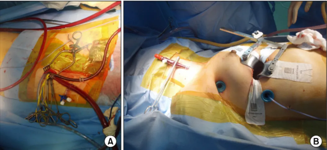
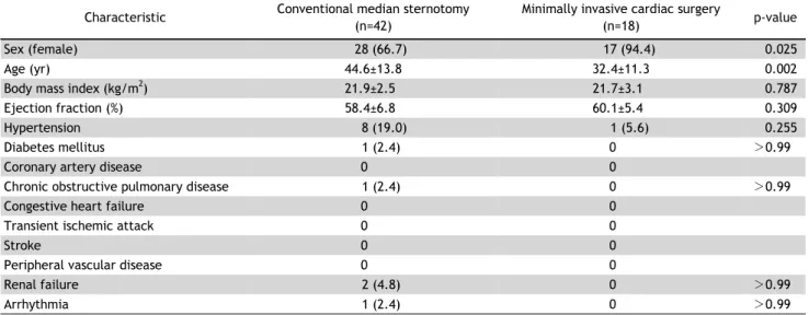
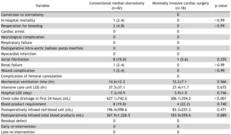
Background: This study aims to evaluate whether or not the method of right vertical axillary minithoracotomy (RVAM) is preferable to and as reliable as conventional

The PVRIs after closure with fenestration were over 3.0 Wood units*m 2 in 11 patients (64.7%), and their pre-closure PVRI, mPAP with oxy- gen, and pre-closure Qp/Qs values