191
Open Access
Morphologic Characteristics and Relating Factors
to the Need of Technical Modification in Transcatheter Closure of Large Atrial Septal Defect (≥25 mm)
Su-Jin Park, MD, Nam Kyun Kim, MD, Jung Ok Kim, MD, Byung Won Yoo, MD, Jae Young Choi, MD and Jun Hee Sul, MD Division of Pediatric Cardiology, Severance Cardiovascular Hospital, Yonsei University Health System, Seoul, Korea
ABSTRACT
Background and Objectives: The rigid coupling between the delivery wire and the right atrial disk has been oc- casionally encountered during transcatheter closure of atrial septal defect (ASD). Therefore the device frequently makes a perpendicular angle, and the leading edge of left atrial disk slips through the defect and prolapses into right atrium (RA) before it is properly placed in the septum. The purpose of this study is to investigate relating fac- tors to the need of technical modification in transcatheter closure of large ASD and to evaluate relevant mor- phologic characteristics of atrial septal rim in this situation. Subjects and Methods: From July, 2003 to May, 2007, 312 patients underwent transcatheter occlusion of ASD with Amplatzer Septal Occluder® (ASO, AGA medical corporation, Golden Valley, MN, USA) at Yonsei Cardiovascular Center and among them 109 patients had large ASD (≥25 mm) and these patients were enrolled in our study. Patients were divided into two groups according to the deploying methods of the device (Group I: standard method, Group II: modified methods). Assessments of the defects and its surrounding rims were made by echocardiography. Results: There were no differences between 2 groups in age, body weight and height except for balloon-stretched diameter (stop-flow technique) and device size. Group II patients with modified methods showed larger balloon-stretched diameter and device size than group I patients with standard method. The mean length of anterosuperior (AS) rim in group II was significantly shorter than group I (p<0.05). As the size of the device used in procedure increased, there was a trend towards increase in the need of modified methods. Conclusion: This study shows that AS rim deficiency and the size of ASD may be the relating factors to the need of technical modification in transcatheter closure of ASD. Therefore, when the initial try with standard method is not successful in large ASD with deficient AS rim, we suggest that changing st- rategy of implantation may save time and efforts and possibly reduce the risk of complications associated with pro- longed procedure. (Korean Circ J 2010;40:191-196)
KEY WORDS:Atrial septal defect; Echocardiography; Amplatzer septal occluder; Congenital heart disease.
Introduction
Transcatheter closure of secundum atrial septal defect (ASD) with Amplatzer Septal Occluder® (ASO, AGA
medical corporation, Golden Valley, MN, USA) has be- come an effective and reliable alternative therapy to op- eration in most patients with ASD.1-6) Compared with surgical repair, transcatheter closure is safer and more phy- siologic, and the treatment results are similar, and as a re- sult, transcatheter closure has surfaced to replace oper- ation in recent years.3-5)
Owing to its design, the ASO can even be used in patients with large ASD associated with deficient rims.
However, the major problem in closing a large ASD is malalignment of the device to the plane of defect. The delivery system of AGA Amplatzer has a rigid coupling between the delivery wire and the right atrial disk, which accounts for the relatively perpendicular orientation of the left atrial disk to the atrial septal plane.7)8) This ch-
Received: October 7, 2009 Revision Received: October 20, 2009 Accepted: October 26, 2009
Correspondence: Jae Young Choi, MD,Division of Pediatric Cardiology, Severance Cardiovascular Hospital, Yonsei University Health System, 250 Seongsan-ro, Seodaemun-gu, Seoul 120-752, Korea
Tel: 82-2-2228-8280, Fax: 82-2-393-3080 E-mail: [email protected]
○ cc This is an Open Access article distributed under the terms of the Creative Commons Attribution Non-Commercial License (http://creativecommons.
org/licenses/by-nc/3.0) which permits unrestricted non-commercial use, distribution, and reproduction in any medium, provided the original work is properly cited.
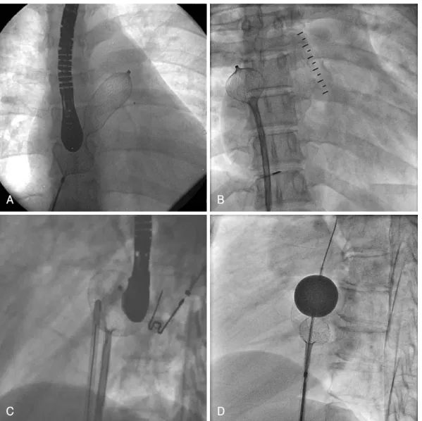
aracter of the device results in the prolapse of the left at- rium (LA) disk to right atrium (RA) during deployment (Fig. 1). There have been many efforts to overcome li- mitations derived from the inherent design of the ASO delivery system and many have succeeded using technical modifications.9-13) The purpose of this study is to inves- tigate the relating factors to the need of technical modi-
fication in transcatheter closure of large ASD (≥25 mm) and to evaluate the relevant morphologic characteristics of atrial septal rim in these patients.
Subjects and Methods
Subjects
A total of 312 patients underwent transcatheter oc- clusion of secundum ASD with ASO at Yonsei Car- diovascular Center from July, 2003 to May, 2007. Among them, 109 (35%) patients (male 34; female 75) had more than 25 mm sized ASD and these patients were en- rolled in the study. The patients were divided by the methods of deployment of ASO into two groups as group I, standard method group, and group II, modified me- thod group. When the initial attempt of standard me- thod failed, modified method was applied.
Echocardiographic and cardiac catheterization parameters
Assessments of the defects were measured by balloon- occlusive diameter (BOD) and its surrounding rims were based on transesophageal or intracardiac echocardiogra-
AAo SVC
PV
IVC
CS
Fig. 1. Diagram showing the relationship between atrial septum and device. The prolapse of left atrial disk during deployment can be observed. SVC: superior vena cava, PV: pulmonary vein, IVC:
inferior vena cava, CS: coronary sinus, AAo: ascending aorta, TV: tricuspid valve.
Fig. 2. Two dimensional echocardiography findings. A: diagram showing the measuring locations of septal rims. B, C and D: measure- ments of the rims with transesophageal echocardiography (TEE). Using the still images obtained by TEE, the rim lengths were meas- ured in the modified 4 chamber view and the short axis view. E and F: measurements of the rims with intracardiac echocardiography (ICE). Using the still images obtained by ICE, the rim lengths were measured in the short axis view and the long axis view. SVC:
superior vena cava, PS: posterosuperior rim, ASD: atrial septal defect, AS: anterosuperior rim, AAo: ascending aorta, PI: posteroinferio rim, AI: anteroinferior rim, CS: coronary sinus, IVC: inferior vena cava, LA: left atrium, LV: left ventricle, RV: right ventricle.
C
E F
D
B A
AAo SVC
PS
ASD AS AI PI
CS IVC
phy, performed at the time of closure. The measure- ment of defect size based on BOD was performed ac- cording to the previous report.14) The analysis of the echocardiographic studies were performed according to the recommendations of the American Society of Echo- cardiogrphy.15) Echocardiographic studies were perform- ed and images were acquired and digitally stored for off- line analysis with each study by experienced physicians.
Anteroinferior (AI) rim was measured as the distance to atrioventricular valve, posterosuperior (PS) rim was measured as the distance to superior vena cava, poste- roinferio (PI) rim was measured as the distance to in- ferior vena cava, and anterosuperior (AS) rim was meas- ured as the distance to aortic posterior wall. The two to- tal septal lengths, anteroposterior septal length (APSL) and superoinferior septal length (SISL) was also meas- ured and calculated. APSL was the total length between PS, ASD, and AI, and SISL was the total length between PI, ASD, and AS. AS rim was measured in the short- axis view, PS and PI rims were measured in the long- axis view or the bicaval view. AI rim was measure in 4- chamber view. In some of the cases, combinations of different views were used to determine the rim length.
Any rim length was considered deficient if its length was less than 5 mm (Fig. 2).
Statistical analysis
The data depicting the parameters of the enrolled sub- jects were presented as mean and standard deviation.
For statistical analysis, Student’s t-test and chi-square test were used. P<0.05 was considered as statistically sig- nificant in all the data.
Results
Characteristics of subjects
The profiles of the enrolled patients are summarized in Table 1. The mean age of the patients in group I was 34.7±15.5 years, 24 were male and 50 were female, the mean weight was 56±13.2 kg, the mean height was 158.8±13.1 cm, and the mean body surface area (BSA) was 1.55±0.24 kg/m2. The mean age of patients in
group II was 32.9±13.6 years, 10 were male and 25 were female, the mean weight was 56.4±12.0 kg, the mean height was 159.2±12.9 cm, and the mean BSA was 1.58±0.23 kg/m2. The mean BOD size in group I and group II were 29.4±4.1 mm and 32.2±3.8 mm, res- pectively. The mean device size in group I was 29.4±
4.0 mm and the mean device size in group II was 32.3
±3.44 mm. The Qp/Qs in group I and II were 2.35±
0.63 and 2.63±0.94, respectively. There were no differ- ences between the 2 groups in age, body weight, sex ra- tio, and height. Group II showed a larger BOD and device size, which was statistically significant (p<0.01).
Qp/Qs ratio in group II was larger than group I, but it wast statistically insignificant. In the 35 patients who underwent modified techniques, 21 patients underwent right upper pulmonary vein (RUPV) technique,9) 9 pa- tients underwent left upper pulmonary vein (LUPV) technique,7) 3 patients underwent dilator assisted tech- nique,16) and 2 patients underwent balloon-assisted te- chnique.10)
Morphologic characteristics of atrial septal rims The morphologic characteristics of the atrial septal rim in the enrolled patients are shown in Table 2. In group I, the mean PS rim length was 13.1±4.9 mm, the mean AI rim length was 15.5±5.3 mm, the mean PI rim length was 11.2±6.1 mm, and the mean AS rim length was 6.4±4.1 mm. In group II, the mean PS rim length was 12.7±4.3 mm, the mean AI rim length was 13.5±4.3 mm, the mean PI rim length was 12.7±4.2
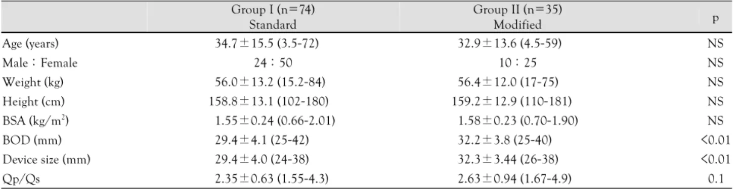
Table 1. Profiles of subjects
Group I (n=74) Standard
Group II (n=35)
Modified p
Age (years) 34.7±15.5 (3.5-72) 32.9±13.6 (4.5-59) NS
Male : Female 24 : 50 10 : 25 NS
Weight (kg) 56.0±13.2 (15.2-84) 56.4±12.0 (17-75) NS
Height (cm) 158.8±13.1 (102-180) 159.2±12.9 (110-181) NS
BSA (kg/m2) 1.55±0.24 (0.66-2.01) 1.58±0.23 (0.70-1.90) NS
BOD (mm) 29.4±4.1 (25-42) 32.2±3.8 (25-40) <0.01
Device size (mm) 29.4±4.0 (24-38) 32.3±3.44 (26-38) <0.01
Qp/Qs 2.35±0.63 (1.55-4.3) 2.63±0.94 (1.67-4.9) <0.10
BSA: body surface area, BOD: balloon occlusive diameter
Table 2. Morphologic characteristics of atrial septal rims Group I (n=74)
Standard
Group II (n=35) Modified p PS rim length(mm) 13.1±4.9 12.7±4.3 NS AI rim length 15.5±5.3 13.5±4.3 NS PI rim length 11.2±6.1 12.7±4.2 NS AS rim length 6.4±4.1 2.5±2.6 <0.01 APSL 51.3±7.6 51.7±8.1 NS SISL 63.1±8.9 66.2±10.2 NS PS: posterosuperior, AI: anteroinferior, PI: posteroinferior, AS:
anterosuperior, APSL: anteroposterior septal length, SISL: supero- inferior septal length
mm, and the mean AS rim length was 2.5±2.6 mm.
The mean length of AS rim in group II was signifi- cantly shorter than group I (p<0.01), but none of the other parameters showed significant difference. The mean APSL in group I and group II were 51.3±7.6 mm and 51.7±8.1 mm, respectively, and the mean SISL in group I and group II were 63.1±8.9 mm and 66.2±
10.2 mm, respectively. Both APSL and SISL did not show any statistical difference between both groups.
In group I with standard method, 48 patients (65%) had central defect and 20 patients (27%) had AS de- fect, shown in Fig. 3A. In group II with modified me- thod, only 6 patients (17%) had central defect, whereas 22 patients (63%) had AS defect, shown in Fig. 3B. In both groups I and II, 54 patients had central defect and 42 patients had AS defect. When the patients with ASD had sufficient rims, 87% of procedure was successfully performed by standard method and 13% needed mo- dified technique, but with AS rim deficiency, modified techniques were needed in 53% of the procedures (Fig.
4). Furthermore, as the size of the device used in the pro- cedure increased, there was a tendency towards increase in the frequency of use of modified methods (Fig. 5).
Discussion
Many factors contribute to the difficulty involved in transcatheter closure of large secundum ASD. Over the course of time, alternative techniques of device deploy- ment have improved, making more ASD amendable us- ing ASO, but they still remain a challenge.
Numerous alternative techniques have been report- ed to facilitate transcatheter closure of large secundum ASDs,11) and our institute utilizes many of the report- ed modified techniques. The LUPV technique (Fig. 6A), which can be used in both children and adults, is en- gagement of LA disk within LUPV, followed by a rapid release of the waist and RA disk.7) The RUPV techni- que (Fig. 6B), recommended, but not limited, to larger patients, is partial deployment of LA disk in RUPV while maintaining contact with the posterior superior septum, followed by quick and successive deployment of the waist and RA disk.9) In the above 2 methods, it is important to place the LA disk exactly in pulmonary vein and the procedure must be performed with gentle movements until the deployment of RA disk, to avoid damage to the atrial wall. Dilator assisted technique, also known as Wahab technique (Fig. 6C), is using a
PS defect AI defect PI defect AS defect Central defect Double defect
PS defect AI defect PI defect AS defect Central defect Double defect 4 (6%) 1 (1%) 1 (1%)
20 (27%)
48 (65%)
Standard method (n=74)
1 (3%) 2 (6%) 4 (11%)
22 (63%) 6 (17%)
Modified method (n=35)
A B
Fig. 3. Anatomical characteristics of defects according to deployment methods. A: in group I with standard method, 48 patients (65%) had central defect and 20 patients (27%) had anterosuperior (AS) defect. B: in group II with modified method, only 6 patients (17%) had cen- tral defect, whereas 22 patients (63%) had AS defect. PS: posterosuperior, AI: anteroinferior, PI: posteroinferior.
100 80 60 40 20 0 (%)
Modified mothod Standard method
Central (n=54) AS (n=42) Fig. 4. The different deployment method in atrial septal defect (ASD) patients with central defect and anterosuperior (AS) rim defect. With sufficient rims, 87% of procedure was successfully performed by standard method and 13% needed modified techni- que, but with AS rim deficiency, modified techniques were need- ed in 53% of the procedure.
25 20 15 10 5 0
Standard (n=74) Modification (n=35)
Device size(mm)
Fig. 5. The different deployment method according to employed device size. As the size of the device used in the procedure in- creased, there was a tendency towards increase in the frequency of use of modified methods.
Case (n)
24 26 28 30 32 34 36 38 100%
0%
92%
8%
78%
22%
78%
22%
45%
55%
50% 50%
45%55% 45%55%
long dilator, usually the delivery sheath being used, to hold the LA disk inside LA, preventing it from pro- lapsing across the defect.16) The balloon-assisted tech- nique (Fig. 6D), first describe by Dalvi et al.10) in 2005, has a concept similar to the Wahab technique, but uses the balloon catheter to support the LA disk from pro- lapsing across the defect. These 4 techniques are the most popular modified techniques used and they were used in our patients who were enrolled in the study. In our institute, the RUPV method is the most frequently applied technique.
Our study revealed that the proportion of AS rim deficiency was higher in patients with modified methods, modified methods were needed more frequently in large- sized ASD. Therefore it demonstrated that AS rim de- ficiency and the size of ASD may be the core relating factors to the need of technical modification in trans- catheter closure of ASD. There have been many studies describing the different modified techniques used in
large ASDs,7)9)10)16) but our study is the first to analyze the factors involved in the decision of whether to em- ploy the modified technique or not. With the predic- tion of a possible need of modified technique, the in- terventionist may be able to prepare, prior to the pro- cedure, which may increase the success rate of the pro- cedure. Also, by preparing, the procedure time may be reduced, which in result may moderate the complica- tions engaged with long procedure time.
Our study was limited to patients of a single institute and only limited interventionists were involved. Fur- ther investigation with a larger pool of patients involv- ing diverse interventionists with different techniques is pertinent in the future.
In conclusion, our study revealed that AS rim defi- ciency and the size of ASD may be the 2 most impor- tant relating factors to the need of technical modifi- cation in transcatheter closure of ASD. Therefore, when the initial attempt with standard method is not suc-
A B
C D
Fig. 6. Modified techniques used in transcatheter closure of large astrial septal defect. A: left upper pulmonary vein technique. B: right upper pulmonary vein technique. C: dilator assisted technique (Wahab technique). D: balloon-assited technique.
cessful, we suggest that changing strategy of implanta- tion may save time and efforts and possibly reduce the risk of complications associated with prolonged procedure.
REFERENCES
1) Rao PS. Catheter closure of atrial septal defects. J Invasive Car- diol 2003;15:398-400.
2) Ebeid MR. Percutaneous catheter closure of secundum atrial sep- tal defects: a review. J Invasive Cardiol 2002;14:25-31.
3) Berger F, Vogel M, Alexi-Meskishvili V, Lange PE. Comparison of results and complications of surgical and Amplatzer device clo- sure of atrial septal defects. J Thoracic Cardiovasc Surg 1999;
118:674-8; discussion 678-80.
4) Chessa M, Carminati M, Butera G, et al. Early and late complica- tions associated with transcatheter occlusion of secundum atrial septal defect. J Am Coll Cardiol 2002;39:1061-5.
5) Du ZD, Hijazi ZM, Kleinman CS, Silverman NH, Larntz K. Com- parison between transcatheter and surgical closure of secundum atrial septal defect in children and adults: results of a multicen- ter nonrandomized trial. J Am Coll Cardiol 2002;39:1836-44.
6) Spies C, Hijazi ZM. Transcatheter closure of secundum atrial sep- tal defects in the elderly. Korean Circ J 2009;39:47-51.
7) Varma C, Benson LN, Silversides C, et al. Outcomes and altern- ative techniques for device closure of the large secundum atrial septal defect. Catheter Cardiovasc Interv 2004;61:131-9.
8) Aeschbacher BC, Chatterjee T, Meier B. Transesophageal echo- cardiography to evaluate success of transcatheter closure of large secundum atrial septal defects in adults using the buttoned de-
vice. Mayo Clin Proc 2000;75:913-20.
9) Berger F, Ewert P, Dhnert I, et al. Interventional occlusion of at- rial septum defects larter than 20 mm in diameter. Z Kardiol 2000;
89:1119-25.
10) Dalvi BV, Pinto RJ, Gupta A. New technique for device closure of large atrial septal defects. Catheter Cardiovasc Interv 2005;64:
102-7.
11) Fu YC, Cao QL, Hijazi ZM. Device closure of large atrial septal defects: technical considerations. J Cardiovasc Med 2007;8:30-3.
12) Kutty S, Asnes JD, Srinath G, Preminger TJ, Prieto LR, Latson LA. Use of a straight, side-hole delivery sheath for improved de- livery of Amplatzer ASD occluder. Catheter Cardiovasc Interv 2007;69:15-20.
13) Staniloae CS, El-Khally Z, Ibrahim R, Dore A, De Guise P, Mer- cier LA. Percutaneous closure of secundum atrial septal defect in adults a single center experience with the Amplatzer septal oc- cluder. J Invasive Cardiol 2003;15:393-7.
14) Zhu W, Cao QL, Rhodes J, Hijazi ZM. Measurement of atrial septal defect size: a comparative study between three-dimensio- nal transesophageal echocardiography and the standard balloon sizing methods. Pediatr Cardiol 2000;21:465-9.
15) Douglas PS, DeCara JM, Devereux RB, et al. Echocardiogra- phic imaging in clinical trials: American Society of Echocardio- graphy Standards for echocardiography core laboratories: en- dorsed by the American College of Cardiology Foundation. J Am Soc Echocardiogr 2009;22:755-65.
16) Wahab HA, Bairam AR, Cao Q, Hijazi ZM. Novel technique to prevent prolapse of the Amplatzer septal occluder through large atrial septal defect. Catheter Cardiovasc Interv 2003;60:543-5.