Outcome of Transcatheter Closure of Oval Shaped Atrial Septal Defect with Amplatzer Septal Occluder
Jinyoung Song,
1Sang Yoon Lee,
2Jae Sook Baek,
2Woo Seub Shim,
2and Eun Young Choi
31Department of Pediatrics, Samsung Medical Center, Sungkyunkwan University School of Medicine, Seoul;
2Department of Pediatrics, Sejong General Hospital, Sejong Heart Institute, Bucheon;
3Department of Pediatrics, Seoul National University Bundang Hosiptal, Seongnam, Korea.
Received: January 30, 2013 Revised: March 15, 2013 Accepted: March 16, 2013
Corresponding author: Dr. Jinyoung Song, Department of Pediatrics,
Samsung Medical Center,
Sungkyunkwan University School of Medicine, 81 Irwon-ro, Gangnam-gu,
Seoul 135-710, Korea.
Tel: 82-2-3410-3538, Fax: 82-2-3410-0043 E-mail: [email protected]
∙ The authors have no financial conflicts of interest.
© Copyright:
Yonsei University College of Medicine 2013 This is an Open Access article distributed under the terms of the Creative Commons Attribution Non- Commercial License (http://creativecommons.org/
licenses/by-nc/3.0) which permits unrestricted non- commercial use, distribution, and reproduction in any medium, provided the original work is properly cited.
Purpose: For the successful completion of transcatheter closure of atrial septal de- fects with the Amplatzer septal occluder, shape of the defects should be considered prior to selecting the device. The purpose of this study is to evaluate the results of a transcatheter closure of oval shaped atrial septal defect. Materials and Meth- ods: From November 2009 until November 2011, cardiac computed tomography was performed on 69 patients who needed a transcatheter closure of atrial septal de- fect. We defined an oval shaped atrial septal defect as the ratio of the shortest diam- eter to the longest diameter ≤0.75 measured using computed tomography. A trans- thoracic echocardiogram was performed one day after and six months after.
Results: The transcatheter closure of atrial septal defect was performed successful- ly in 24 patients in the ovoid group and 45 patients in the circular group. There were no serious complications in both groups and the complete closure rate at 6 months later was 92.3% in the ovoid group and 93.1% in the circular group (p>0.05). The differences between the device size to the longest diameter of the defect and the ratios of the device size to the longest diameter were significantly smaller in the ovoid group (1.8±2.8 vs. 3.7±2.6 and 1.1±0.1 vs. 1.2±0.2). Conclusion: Trans- catheter closure of an oval shaped atrial septal defect was found to be safe with the smaller Amplatzer septal occluder device when compared with circular atrial sep- tal defects.
Key Words: Congenital heart disease, atrial septal defect, transcatheter interven- tion, device closure, cardiac computed tomography
INTRODUCTION
Atrial septal defect (ASD) is one of the most common congenital heart diseases,
with an incidence of approximately 1 per 100 live births, based on a previous re-
port.
1Surgical closure was considered once as the best treatment.
2,3However, de-
vice closure with the Amplatzer septal occluder (ASO) (AGA medical, Golden
Valley, MN, USA) has gradually become a competitive alternative.
4,5For success-
ful device closure of ASD, an accurate size evaluation of the defect, as well as a
detailed morphology description of the defect, is essential. In general, ASD pro-
We decided the device size based on the measured data from various echocardiographic images and images from cardiac computed tomography. Usually, the longest two- di- mensional diameter, among all of the images from trans-tho- racic echocardiogram, cardiac computed tomography and intra-cardiac echocardiogram, was considered for the deci- sion of the device size. From the en face view in the com- puted tomography images, the longest diameter and the shortest diameter were measured (Fig. 1). All diameters of the defect were measured at the end-systolic phase. We typ- ically chose a device that is 0-4 mm larger than the longest diameter of the defect at the first trial, but this depends on the flexibility of the adjacent septal rims. Further, if suc- cessful implantation failed or oversizing was suggested, we changed the size of the device. Before deployment of the device, we pushed the cable and made the device push to- ward the left atrium for one second, and then we confirmed the stability of the device. We referred to this as a modified Minnesota wiggles. We defined the ovoid group with oval shaped septal defect as the ratio of the shortest diameter to the longest diameter (b/a) below 0.75, while the circular group had circular shaped septal defect if the ratio of the shortest diameter to the longest diameter was over 0.75.
6The differences between the longest diameter of the defect and the device size were calculated in the two groups. In addition, the ratios of the device size to the longest diameter of the defect were compared between the two groups. The residual shunt and any complications were checked from a follow-up trans-thoracic echocardiogram on the following day and 6 months later.
Data are expressed as the mean±standard deviation. To compare the results of both groups, a nonparametric Mann- Whitney test and a Fisher’s exact test were performed using SPSS 11.5 (SPSS Inc., Chicago, IL, USA) due to our small vides various shapes including circular, oval and complex
morphology.
6,7Even though good long-term results of a transcatheter closure of ASD with ASO have been report- ed,
8,9it is difficult to find a specific report regarding the trans- catheter closure of ASD, based on various morphologies. We realize that many ASDs are not circular, but oval in devel- oped cardiac tomographic images. When using device clo- sure for non-circular ASD, the size of device should be considered according to the various diameters of the defect.
One diameter of the circular device could be too large for the shortest diameter causing a deformity in residual rims.
The cardiac erosion after successful implantation as one of the most important complications is supposed to occur due to a larger device in the oval defect compared to the circular defect. Therefore, the purpose of the present study is to evaluate the outcome of a transcatheter closure of oval shaped ASD.
MATERIALS AND METHODS
This is a retrospective chart review of patients undergoing transcatheter closure of ASD. The inclusion criteria were all patients with secundum ASD deemed appropriate for trans- catheter closure with ASO. In addition, a cardiac computed tomography was checked before the transcatheter closure of ASD from November 2009 until November 2011 at Se- jong General Hospital in South Korea. Four patients were excluded and among them two patients had 2 or more de- fects, one patient had an extremely complex shape that was overlapped with 2 circular defects, and one patient had apo- lygonal shape upon computed tomography. In total, 69 pa- tients were included and retrospectively reviewed for medi- cal records. For the decision of the transcatheter closure of ASD with ASO, a trans-thoracic echocardiogram was first performed. If the defect was not definite, due to the poor window in the trans-thoracic echocardiogram, trans-esoph- ageal echocardiogram was done in selected patients. The inclusion criteria for the transcatheter closure of ASD with ASO were as follows: 2’ ASD with the evidence of right ven- tricular volume overload, no significant pulmonary arterial hypertension that was irreversible, no significant arrhythmia, no serious problems in other organs and presumptive appro- priate rims for ASO implantation. Cardiac computed tomog- raphy was performed. If necessary, oral or intravenous beta- blockers were used. One operator under the guidance of an
intra-cardiac echocardiogram performed the device closure.
Fig. 1. The reconstructed en face image from cardiac computed tomogra- phy shows the longest diameter (a) and the shortest diameter (b).(Table 1).
In the total population, the differences between the longest diameter of the defect (a) and the device size were 3.0±2.8 (range -7.0 to 11.0) mm. In addition, the ratios of the device size to the longest diameter of the defect were 1.19±0.20 (range 0.84 to 1.93).
A comparison between the two groups indicated that the differences between the longest diameter of the defect and the device size of the ovoid group were significantly small- er than the circular group (1.8±2.8 mm vs. 3.7±2.6 mm, p=0.009). Further, the ratios of the device size to the lon- gest diameter of the defect in the ovoid group were also sig- nificantly smaller than those in the circular group (1.1±0.1 vs. 1.2±0.2, p=0.02) (Table 2).
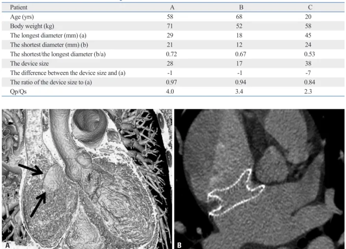
Especially for the three patients in the ovoid group, the differences between the longest diameter of the defect and the device size showed negative numbers, -1, -1 and -7, which meant that the longest diameters of the defect were larger than the waist diameters of the device. In addition, the ratio of the shortest diameter to the longest diameter (b/
a) were 0.72, 0.67 and 0.53, respectively (Table 3). In the last case that showed -7, the longest diameter of the defect was 45 mm and the shortest diameter of the defect was 24 mm (b/a=0.53). For this oval defect, ASO of 38 mm was implanted successfully, and complete closure was confirmed by trans-thoracic echocardiogram immediately and 6 months later even though the immediate cardiac computed tomog- raphy image showed a small gap from the waist of the de- vice to an anterior margin of the defect (Fig. 2). On the oth- er hand, the minimum level of the differences between the longest diameter of the defect and the device size in the cir- cular group was 0 mm.
sample size. p-values <0.05 were considered statistically significant.
The locally appointed ethics committee approved this re- search protocol.
RESULTS
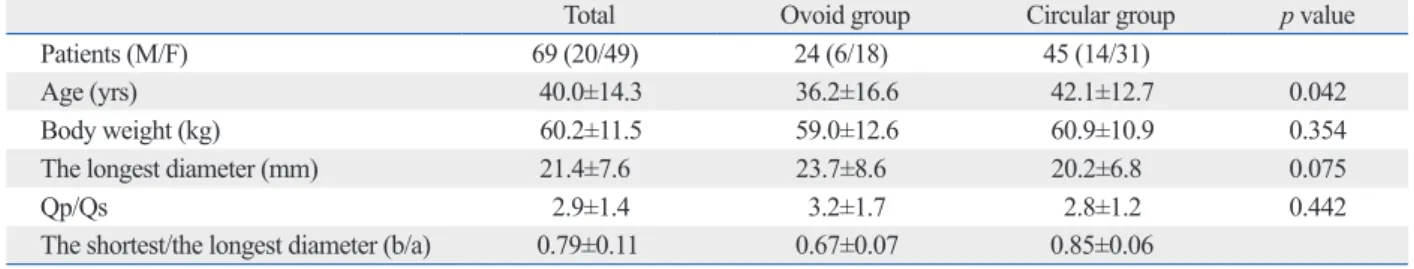
In the computed tomography image of all patients, the lon- gest diameter (a) and the shortest diameter (b) were 21.4±7.6 mm (8.0-45.0 mm) and 16.7±5.6 mm (4.0-31.0 mm), respec- tively. The ratio of the shortest diameter to the longest di- ameter (b/a) was 0.79±0.11 (0.5-1.0). Only three patients had ratios of 1.0, and ten patients had ratios over 0.9. Of the total enrolled patients, 24 patients (34.8%) were assigned in the ovoid group and 45 patients (65.2%) in the circular group (Table 1).
In the ovoid group, data from 6 males and 18 females were reviewed. The mean age was 36.2±16.6 (15-77) years, and the mean body weight was 59.0±12.6 (38-99) kg. From the computed tomography images, the longest diameter (a) was 23.7±8.6 mm, and the ratio (b/a) was 0.67±0.07 (0.5- 0.75). From the catheterization results, the shunt ratio (Qp/
Qs) was 3.2±1.7. In contrast, the circular group consisted of 14 males and 31 females. The mean age was 42.1±12.7 years and the mean body weight was 60.9±10.9 kg. The longest di- ameter (a) and the ratio (b/a) were 20.2±6.8 mm and 0.85±
0.06, respectively. The shunt ratio (Qp/Qs) in the circular group was 2.8±1.2. When we compared the two groups, the body weights, the shunt ratios (Qp/Qs), and the longest di- ameters (a) were not statistically different, but the mean age of the ovoid group was younger than of the circular group
Table 1. Characteristics of the Patients with Oval Shaped Atrial Septal Defect (Ovoid Group) and Circular Atrial Septal Defect (Circular Group) Show No Significant Difference Except Age
Total Ovoid group Circular group p value
Patients (M/F) 69 (20/49) 24 (6/18) 45 (14/31)
Age (yrs) 40.0±14.3 36.2±16.6 42.1±12.7 0.042
Body weight (kg) 60.2±11.5 59.0±12.6 60.9±10.9 0.354
The longest diameter (mm) 21.4±7.6 23.7±8.6 20.2±6.8 0.075
Qp/Qs 2.9±1.4 3.2±1.7 2.8±1.2 0.442
The shortest/the longest diameter (b/a) 0.79±0.11 0.67±0.07 0.85±0.06
Table 2. The Comparison of the Device Size in Ovoid Group with That in Circular Group Shows Smaller Size Used in Ovoid Group
Ovoid group Circular group p value
Size difference (mm) 1.8±2.8 3.7±2.6 0.009
Size ratio 1.1±0.1 1.2±0.2 0.02
Size difference=device size-the longest diameter of defect, size ratio=device size/the longest diameter of defect.
For a successful device closure of ASD, a trans-esopha- geal echocardiogram can assess the size, anatomy, and suit- ability of the lesion for closure. A two-dimensional echo- cardiogram can integrate multiple image planes for the operator to reconstruct a three-dimensional anatomy of ASD, but has limitations of the visualization of an accurate shape During the procedure, no significant complications oc-
curred. The follow-up results showed no embolization, no pericardial effusion, no newly developed arrhythmia, nor mi- tral regurgitation in both groups. Immediate trans-thoracic echocardiogram showed complete closure in 17 of the 24 pa- tients in the ovoid group (70.8%), and follow-up trans-thorac- ic echocardiogram performed 6 months later showed com- plete closure in 12 of the 13 patients in the ovoid group (92.3%). At the same time, the immediate complete closure rate of the circular group was 80.0% (36/45) and at 6 months follow-up was 93.1% (27/29). However, there were no signif- icant differences between the two groups (p>0.05) (Fig. 3).
DISCUSSION
From our study, we could observe that there was a higher incidence of ASD in females than male and completely cir- cular ASD (b/a=1.0) was rare (n=3).
Table 3. The Characteristics of the Oval Shaped Atrial Septal Defects in Three Patients with Negative Number of the Differ- ence between the Device Size and the Longest Diameter of the Defect
Patient A B C
Age (yrs) 58 68 20
Body weight (kg) 71 52 58
The longest diameter (mm) (a) 29 18 45
The shortest diameter (mm) (b) 21 12 24
The shortest/the longest diameter (b/a) 0.72 0.67 0.53
The device size 28 17 38
The difference between the device size and (a) -1 -1 -7
The ratio of the device size to (a) 0.97 0.94 0.84
Qp/Qs 4.0 3.4 2.3
Fig. 2. (A) The tomographic image shows oval shaped atrial septal defect (arrows). (B) There is a small gap between the waist of the device and the anterior margin of the defect on computed tomographic image of patient C immediately after implantation.
Fig. 3. The complete closure rates the next day and 6 months after were not statistically different in both groups (p>0.05).
0 20 40 60 80 100
Ovoid group Control group 71
92
80
93 Next day 6 months later
A B
(%)
vice was much larger than 45 mm and no residual leak was detected by trans-thoracic echocardiogram immediately as well as 6 months later.
It is recommended that oversizing should be avoided due to the risks of mushrooming deformity, impingement on car- diovascular structures, and other serious complications.
15,16Our data showed successful device closure with ASO for oval shaped ASD. In another study, the residual shunt at one day after device closure was found in 8.6% and the re- sidual shunt at three months later was found only in 1.3%
of patients.
4The complete closure rates in our study were a little higher, but we could expect nearly the same results af- terward. There were no differences in the occurrence of complications, immediate, and mid-term complete closure rate. Interestingly, the mean upsizing of the device in the ovoid group was significantly lower than in the circular group, which matched with Zanchetta’s concept.
14Our re- sults showed that the device diameter to be 1.8 mm longer than the longest diameter of defect in ovoid group, whereas a device 3.7 mm longer was used for the circular group.
However, this depends on the ratio (b/a) of the defect.
One limitation of our study is that it is not a randomized control study. This retrospective analysis was based on pa- tients who underwent a transcatheter closure of ASD with ASO, performed by experienced operators. In our study, the evaluation of the defect size and shape was based on cardi- ac computed tomography exclusively so the patients with- out pre-interventional cardiac computed tomography were excluded. Small children were excluded because of the dif- ficulties in cardiac computed tomography; therefore, there could be some selection bias in our study. However, the pre-interventional cardiac computed tomography was ap- plied randomly.
Although not significant, the longest diameters of the de- fects in the ovoid group were bigger than those were in the circular group. This could be another reason for the relative- ly smaller ratio of the device chosen for the closure of de- fects in the ovoid group. The biggest limitation might be that the device size, chosen by these operators, was not based on a constant equation, but on a personal experience. Further, it would be better if we had data on device closure of oval shaped ASD with a balloon sizing method. A formula appli- cable for the proper selection of the device for non-circular ASD is needed. Moreover, our computed tomography sys- tem, which provides low dose radiation, is very beneficial;
however, we are not sure if the computed tomography is su- perior to a three dimensional echocardiogram.
of the defect. However, an accurate dimension related to the shape of the defect is considered important for the suc- cessful transcatheter closure because the ASO is uniform.
There are studies that have described the complex shapes of ASD with a three-dimensional trans-esophageal echocar- diogram.
6,7,9,10However, we did not perform a three-dimen- sional trans-esophageal echocardiogram for our patients’
series. Trans-esophageal echocardiogram requires sedation and can potentially damage the esophagus. Cardiac com- puted tomography can be considered an alternative because Ko, et al.
11reported that cardiac computed tomography is very helpful in the noninvasive evaluation for ASO implan- tation of ASD. Nevertheless, general concerns about cardi- ac computed tomography is the exposure to harmful radia- tion. We did not check the radiation dose for all the patients, but cardiac computed tomography was performed with minimal radiation. We performed cardiac computed tomog- raphy with 0.2-0.6 mSv for small children and 1.7-1.9 mSv for adults. This minimal exposure to radiation helped us evaluate ASD size and morphology by cardiac computed tomography.
When we defined oval shaped ASD as the shortest diam- eter ≤75% of the longest diameter measured from en face image in cardiac computed tomography, oval shaped ASD formed 34.8% of our total patients. Johri, et al.
6reported 42% of oval and 33% of complex ASD shapes in 25 ASDs using a real-time three-dimensional echocardiogram. We realize that many ASDs are not circular in shape.
Regarding the device size, balloon sizing has been con- sidered an integral part of transcatheter closure of ASD with ASO. However, there have been several experiences without balloon sizing.
12,13In one particular case of oval shaped ASD, the effect of a balloon inflation would alter the shape of the defect to conform to the relatively circular shape of the balloon. Moreover, based on Zanchetta’s study,
14the device size in the oval shaped ASD could be smaller than the longest diameter by intra-cardiac echocardiogram measurement: d=√(a×b). Although we did not apply this formula, we had three cases whose differences between the device size and the longest diameter of the defect were be- low zero, and all of them were in the ovoid group. In one patient whose ratio (b/a) was 0.53 and the longest diameter of the defect was 45 mm, we chose an ASO of 38 mm be- cause it was the maximum size of ASO available in our country at that time. This defect was closed successfully.
The waist diameter of the device was smaller than the lon-
gest diameter of the defect, yet the entire length of the de-
al. Three-dimensional transesophageal echocardiography of atrial septal defect: a qualitative and quantitative anatomic study. J Am Soc Echocardiogr 2011;24:600-10.
8. Knepp MD, Rocchini AP, Lloyd TR, Aiyagari RM. Long-term follow up of secundum atrial septal defect closure with the amp- latzer septal occluder. Congenit Heart Dis 2010;5:32-7.
9. Acar P. Three-dimensional echocardiography in transcatheter clo- sure of atrial septal defects. Cardiol Young 2000;10:484-92.
10. Huang X, Shen J, Huang Y, Zheng Z, Fei H, Hou Y, et al. En face view of atrial septal defect by two-dimensional transthoracic echo- cardiography: comparison to real-time three-dimensional trans- esophageal echocardiography. J Am Soc Echocardiogr 2010;23:
714-21.
11. Ko SF, Liang CD, Yip HK, Huang CC, Ng SH, Huang CF, et al.
Amplatzer septal occluder closure of atrial septal defect: evalua- tion of transthoracic echocardiography, cardiac CT, and trans- esophageal echocardiography. AJR Am J Roentgenol 2009;193:
1522-9.
12. Zanchetta M, Onorato E, Rigatelli G, Pedon L, Zennaro M, Car- rozza A, et al. Intracardiac echocardiography-guided transcatheter closure of secundum atrial septal defect: a new efficient device se- lection method. J Am Coll Cardiol 2003;42:1677-82.
13. Gupta SK, Sivasankaran S, Bijulal S, Tharakan JM, Harikrishnan S, Ajit K. Trans-catheter closure of atrial septal defect: Balloon sizing or no Balloon sizing-single centre experience. Ann Pediatr Cardiol 2011;4:28-33.
14. Zanchetta M. On-line intracardiac echocardiography alone for Amplatzer Septal Occluder selection and device deployment in adult patients with atrial septal defect. Int J Cardiol 2004;95:61-8.
15. Amin Z, Hijazi ZM, Bass JL, Cheatham JP, Hellenbrand WE, Kleinman CS. Erosion of Amplatzer septal occluder device after closure of secundum atrial septal defects: review of registry of complications and recommendations to minimize future risk.
Catheter Cardiovasc Interv 2004;63:496-502.
16. Divekar A, Gaamangwe T, Shaikh N, Raabe M, Ducas J. Cardiac perforation after device closure of atrial septal defects with the Amplatzer septal occluder. J Am Coll Cardiol 2005;45:1213-8.
In conclusion, transcatheter closure of ASD with ASO was found safe and effective even for oval shaped ASD.
Oval shaped ASD can be successfully closed with a smaller size of ASO compared to circular ASD.
ACKNOWLEDGEMENTS
We authors thank Dr. Yang Min Kim for providing comput- ed tomography images.
REFERENCES
1. Hoffman JI, Kaplan S. The incidence of congenital heart disease. J Am Coll Cardiol 2002;39:1890-900.
2. Barratt-Boyes BG, Kirklin JW. Cardiac Surgery. New York:
Churchill Livingstone; 1993. p.609-44.
3. Galal MO, Wobst A, Halees Z, Hatle L, Schmaltz AA, Khougeer F, et al. Peri-operative complications following surgical closure of atrial septal defect type II in 232 patients--a baseline study. Eur Heart J 1994;15:1381-4.
4. Masura J, Gavora P, Podnar T. Long-term outcome of transcathe- ter secundum-type atrial septal defect closure using Amplatzer septal occluders. J Am Coll Cardiol 2005;45:505-7.
5. Yew G, Wilson NJ. Transcatheter atrial septal defect closure with the Amplatzer septal occluder: five-year follow-up. Catheter Car- diovasc Interv 2005;64:193-6.
6. Johri AM, Witzke C, Solis J, Palacios IF, Inglessis I, Picard MH, et al. Real-time three-dimensional transesophageal echocardiogra- phy in patients with secundum atrial septal defects: outcomes fol- lowing transcatheter closure. J Am Soc Echocardiogr 2011;24:
431-7.
7. Roberson DA, Cui W, Patel D, Tsang W, Sugeng L, Weinert L, et