Age-related Outcome of Arthroscopic Repair of Isolated Type II Superior Labral Anterior to Posterior Lesions
Jieun Kwon, Yeun Ho Kim, Tae Sung Yeom, Joo Han Oh
Department of Orthopaedic Surgery, Seoul National University Bundang Hospital, Seoul National University College of Medicine, Seongnam, Korea
Background: Repair of superior labral anterior to posterior (SLAP) lesion in patients older than 40 years is controversial. The purpose of this study was to evaluate clinical outcomes of arthroscopic repair of SLAP lesions between younger and older patient groups.
Methods: We reviewed 50 patients with isolated type II SLAP lesions who underwent arthroscopic repair. Patients were divided into 2 groups: group 1 included 20 patients aged <40 years, and group 2 included 30 patients aged ≥40 years. Functional outcome at the final follow-up was assessed using a visual analog scale for pain and satisfaction, American Shoulder and Elbow Surgeons form, Constant score, University of California at Los Angeles score, and periodic change in range of motion (ROM). Anatomical outcome was evaluated using computed tomography (CT) arthrography at least 1 year after surgery.
Results: No significant differences in functional scores or postoperative ROM were observed between the 2 groups. In group 2, later recovery of ROM (forward flexion, p=0.025; internal rotation, p=0.034) and lower satisfaction score (p=0.06) were observed for atrau- matic patients (n=16) compared to patients with traumatic injury (n=14). Fifteen patients in group 1 (15/17, 88%) and 21 patients in group 2 (21/26, 81%) demonstrated a healed labrum on postoperative CT arthrography, and this difference was not significant.
Conclusions: The results of this study suggest that arthroscopic repair of type II SLAP lesions can yield good functional and anatomical outcomes regardless of age, if patient selection is adequate. However, the delay in ROM recovery and lower satisfaction, particularly in older patients without traumatic injury, should be considered.
(Clin Shoulder Elbow 2015;18(1):36-42)
Key Words: Superior labrum anterior and posterior; Age; Arthroscopy Clinics in Shoulder and Elbow Vol. 18, No. 1, March, 2015
http://dx.doi.org/10.5397/cise.2015.18.1.36
Received October 24, 2014. Revised February 7, 2015. Accepted February 8, 2015.
Correspondence to: Joo Han Oh
Department of Orthopaedic Surgery, Seoul National University Bundang Hospital, 82 Gumi-ro 173beon-gil, Bundang-gu, Seongnam 463-707, Korea
Tel: +82-31-787-7197, Fax: +82-31-787-4056, E-mail: [email protected] Financial support: None. Conflict of interests: None.
Introduction
Lesions of the superior glenoid labrum–biceps complex were first described by Andrews et al.,1) while superior labral anterior to posterior (SLAP) lesions were subsequently classified ac- cording to four types by Snyder et al.2) Type II SLAP lesions are unstable lesions of the superior labrum–biceps anchor complex that are detached from the superior glenoid rim, and develop as a result of traction or compression injury or repetitive overhead activity.3) The majority of clinical outcome studies regarding type II SLAP lesions have been conducted with patients younger than 40 years of age.4-6) As a result, the repair of SLAP lesion in patients older than 40 years of age remains controversial. Some
authors have reported that surgical repair of type II SLAP lesion in older patients might lead to persistent postoperative pain and joint stiffness; thus biceps tenotomy or tenodesis has been advocated instead, particularly when associated with rotator cuff disorders.7,8) Neri et al.9) reported that although there was a significant difference in change of postoperative range of mo- tion (ROM) according to age, there was no significant difference in final American Shoulder and Elbow Surgeons (ASES) score between younger and older patients. Schrøder et al.10) reported that good results were achieved with surgical repair of isolated SLAP lesion regardless of age. Alpert et al.11) also reported that there were no differences in visual analog scale (VAS) score for pain, ASES score, Simple Shoulder Test score, Short Form-12
score, and postoperative ROM between 2 age-based cohorts (<40 years and ≥40 years).10) Some of the above mentioned studies included patients with rotator cuff tears or concomitant procedures such as subacromial decompression, distal clavicle resection, or capsular release.11) Based on a current literature review, no study regarding postoperative anatomical healing us- ing imaging studies after repair of an isolated type II SLAP lesion has been reported. The purposes of this study were to investigate clinical characteristics of isolated type II SLAP lesions according to patient age, and to compare functional and anatomical outcomes of arthroscopic repair of such lesions between younger and older patient groups.
Methods
Patient Selection
This study was approved by the Institutional Review Board of the Seoul National University College of Medicine. We retro- spectively reviewed 308 patients who underwent arthroscopic repair of type II SLAP lesion between January 2004 and January 2010; 240 patients who underwent concomitant procedures such as repair of rotator cuff tear, capsulolabral reconstruction for shoulder instability, capsular release for adhesive capsulitis, repair of labral tear, or subacromial decompression and acromioplasty for impingement syndrome, or who had glenohumeral arthritis were excluded. Finally, out of 68 patients (7%) who met the criteria for isolated type II SLAP lesion, 50 (73%) were followed- up for at least 2 years postoperatively. All patients had positive results of a physical examination specific for SLAP lesion.12) They also underwent preoperative computed tomography (CT) arthrography to detect leakage of contrast media through the biceps anchor with detachment from the bony glenoid. Of the patients who had a SLAP lesion on CT arthrography, those with failed conservative treatment for a minimum of 6 months elect- ed to proceed with surgery.
The study group included 45 male and 5 female patients with a mean age of 42 years (range, 21 to 66 years). Patients were divided into 2 groups according to age: group 1 included 20 patients aged under 40 years, and group 2 included 30 patients aged over 40 years.
Group 1 comprised 19 men and 1 woman with a mean age of 33 years (range, 21 to 39 years). The dominant arm was in- volved in 14 patients (70%). Fifteen patients (75%) had memory of a distinct injury such as falling on an outstretched hand, trac- tion, direct compression, or forceful abduction and external rotation of the shoulder. Ten patients (50%) participated in over- head sports.
Group 2 comprised 26 men and 4 women with a mean age of 47 years (range, 40 to 66 years). The dominant arm was involved in 22 patients (71%). Fourteen patients (47%) had memory of traumatic injury. Only 4 patients (13%) participated
in overhead sports.
Surgical Technique
All surgical procedures were performed by one senior sur- geon. The patient was placed in the lateral decubitus position under general anesthesia, and traction of the involved arm was applied with 30o abduction and 10o flexion. An approximately 4.5 kg weight was applied according to the constitution of the patient. The superior labrum complex was palpated with a probe to determine the type of SLAP lesion. When the superior labrum was elevated more than 5 mm with a cartilaginous crack, hemorrhagic spots or inflammatory granulation tissues beneath the detached superior labrum were observed, or the pathologic
‘peel back’ phenomenon during abduction and external rotation occurred, the lesion was confirmed as a type II SLAP lesion.3) The glenohumeral joint was also systematically evaluated to determine the presence of any other lesion, especially glenohu- meral arthritis, articular-side tear of the rotator cuff tendon, long head of the biceps tear, or other labral lesion.
Repair of the superior labrum was performed using a trans- rotator cuff portal (TRCP).13) After subchondral bone of the supraglenoid tubercle was exposed using a high-speed burr, 2 posterior holes were drilled at the 10- or 11-o’clock position and the base of the biceps anchor around the 12- or 1-o’clock position. A suture hook loaded with No. 2 Polydioxanone (PDS;
Ethicon, Somerville, NJ, USA) was introduced through the TRCP and pierced the posterosuperior labrum at the base of the biceps tendon. Then, a strand of the PDS was retrieved through the anterior portal. An open utility loop of bioabsorbable knotless suture anchors (Bioknotless anchor system; Mitek, Norwood, MA, USA) was retrieved from the TRCP through the anterior portal using the shuttle relay technique with PDS. The anchor was introduced from the TRCP with proper tension of the utility loop and balance loop (No. 2 PDS), and inserted into the drilled hole by capturing one strand of the closed anchor loop. After the repair was completed, we checked for firm reattachment of the labrum to the glenoid with a probe, and released the posterosu- perior capsule for early recovery of ROM.
Postoperative Rehabilitation
The same rehabilitation protocol was applied to all patients.
For 4 weeks, patients were immobilized in a brace (Acro Assist 50A1; Ottobock, Duderstadt, Germany) with neutral rotation and 30o abduction, but the brace was removed intermittently to begin passive shoulder motion and scapulothoracic exercises in the supine position from the first day after surgery. After weaning from the brace, active motion exercises were allowed, and gain- ing full ROM was encouraged until 2 months after surgery. Then, muscle-strengthening exercises with a resistance band (Thera- band; The Hygienic Co., Akron, OH, USA) were started, and sports including overhead activity were gradually allowed from 5
to 6 months after surgery.
Outcome Assessment
Functional outcome was evaluated using a VAS for pain and satisfaction, ASES form, Constant score, and University of Cali- fornia at Los Angeles (UCLA) shoulder rating scale at 1 year post- operatively and then annually. Shoulder ROM was measured at 3 months, 6 months, and 1 year after surgery, and then annually.
Physical examinations were performed by 2 trained shoulder surgeons using a standard universal goniometer. Both examiners measured forward flexion, internal rotation and external rotation and the average values of these measurements were calculated.
Anatomical outcome was estimated using CT arthrography at least 1 year postoperatively (17 patients in group 1 [85.0%], 26 in group 2 [86.7%]). Failure of labral healing to the glenoid was determined by leakage of contrast media through the biceps anchor with detachment from the bony glenoid (Fig. 1). How- ever, dye filling at the anterosuperior quadrant of the labrum, especially below the 1-o’clock position, was not considered a failure.14) All radiologic interpretations were done by a single musculoskeletal radiologist, while preoperative and various follow-up functional outcome measurements were evaluated by a single clinical examiner who was blinded to the current study.
Statistical Analysis
All statistical analyses were performed using SPSS version 12.0 software (SPSS Inc., Chicago, IL, USA). The independent t- test, paired t-test, chi-square test, Fisher’s exact test, and Mann- Whitney U-test were used, and a p-value of <0.05 was consid-
ered statistically significant.
Results
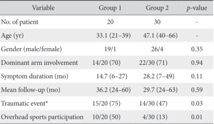
For demographic and clinical data (Table 1), there were no significant differences in sex, arm dominance, or symptom dura- tion between the 2 groups. However, in group 1 traumatic event (p=0.03) and participation in overhead sports (p=0.01) were more frequently associated with development of initial symp- toms. Traumatic event was defined as an episode of injury (such as falling on an outstretched hand, traction, direct compression,
Fig. 1. The failure of labral healing was determined by leakage of contrast media through the biceps anchor with detachment (arrow) from the bony glenoid.
Table 1. Demographics and Clinical Characteristics of Patients
Variable Group 1 Group 2 p-value
No. of patient 20 30 -
Age (yr) 33.1 (21–39) 47.1 (40–66) -
Gender (male/female) 19/1 26/4 0.35
Dominant arm involvement 14/20 (70) 22/30 (71) 0.94 Symptom duration (mo) 14.7 (6–27) 28.2 (7–49) 0.11 Mean follow-up (mo) 36.2 (24–60) 29.7 (24–63) 0.59
Traumatic event* 15/20 (75) 14/30 (47) 0.03
Overhead sports participation 10/20 (50) 4/30 (13) 0.01 Values are presented as number only, median (range), or number (%).
Group 1: aged <40 years, Group 2: aged ≥40 years.
*Patients’ memory regarding distinct shoulder injury such as fall on out- stretched hand, traction, direct compression, and forceful abduction and external rotation of shoulder.
Table 2. Functional Outcomes of Patients
Variable Group 1 Group 2 p-value
Pain VAS
Preoperative 5.9 ± 1.5 5.4 ± 1.8 0.28
Postoperative 1.2 ± 2.2 0.9 ± 1.8 0.69
ASES
Preoperative 58.1 ± 12.7 59.0 ± 17.5 0.85 Postoperative 91.4 ± 13.1 90.8 ± 12.9 0.89 Constant
Preoperative 55.1 ± 6.9 50.1 ± 10.1 0.11 Postoperative 65.2 ± 7.3 66.4 ± 8.1 0.63 UCLA
Preoperative 22.6 ± 5.1 21.8 ± 4.1 0.54 Postoperative 32.8 ± 4.2 33.5 ± 1.9 0.45
Satisfaction VAS 7.9 ± 2.5 8.9 ± 1.2 0.09
Values are presented as mean ± standard deviation.
Group 1: aged <40 years, Group 2: aged ≥40 years, VAS: visual analog scale, ASES: American Shoulder and Elbow Society, UCLA: University of California at Los Angeles.
or forceful abduction and external rotation of the shoulder), as opposed to an atraumatic mechanism, occurring with insidious onset and/or associated with repetitive overuse activity.
After a mean follow-up period of 32.8 months (range, 24 to 63 months), both groups showed significant improvements in functional scores from preoperative to postoperative (all p<0.05). However, no significant differences in postoperative VAS score for pain, ASES form, Constant score, or UCLA score were observed between the 2 groups (Table 2). For postopera- tive change in ROM (forward flexion, external rotation, internal rotation), there were no significant differences between the 2 groups at the final follow-up (all p>0.05, Table 3). However, in
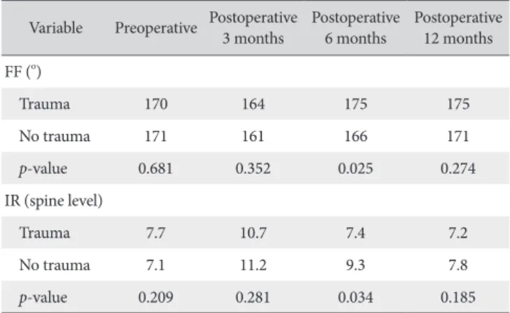
group 2, regarding periodic change in ROM, forward flexion (p=0.025) and internal rotation (p=0.034) at 6 months postop- eratively were significantly lower in atraumatic patients than in patients with traumatic injury (Table 4), and their VAS score for satisfaction was lower (p=0.06, Fig. 2).
For anatomical healing of the superior labrum on CT arthrog- raphy (Table 5), 15 patients in group 1 (15/17, 88%) and 21 pa- tients in group 2 (21/26, 81%) demonstrated a healed labrum;
this difference was not significant (p=0.42). Of patients who had an unhealed labrum (7/43, 16%), 2 patients reported dissatisfac- tion with satisfaction VAS scores below 5 points. One of these patients underwent revision surgery and achieved a satisfaction
Table 3. Postoperative Change in Range of Motion for Patients (Mean Values)
Variable Group 1 Group 2 p-value
FF (o) +1.0 +1.8 0.79
ER arm at side (o) +3.3 +1.1 0.56
ER 90o abduction (o) +14.8 +18.4 0.58
IR (spine level) +1.25 +0.2 0.11
Group 1: aged <40 years, Group 2: aged ≥40 years, FF: forward flexion, ER:
external rotation, IR: internal rotation.
Table 4. Mean Value of Periodic Range of Motion according to Traumatic Injury in Group 2
Variable Preoperative Postoperative 3 months Postoperative
6 months Postoperative 12 months FF (o)
Trauma 170 164 175 175
No trauma 171 161 166 171
p-value 0.681 0.352 0.025 0.274
IR (spine level)
Trauma 7.7 10.7 7.4 7.2
No trauma 7.1 11.2 9.3 7.8
p-value 0.209 0.281 0.034 0.185
Group 2: aged ≥40 years, FF: forward flexion, IR: internal rotation.
Table 5. Anatomical Healing of Superior Labrum Using Computed Tomogra- phy Arthrography
Variable Patients with
healed labrum Patients with
unhealed labrum p-value
Group 1 15 (88) 2 (12)
Group 2 21 (81) 5 (19)
Total 36 7 0.42
Values are presented as number (%).
Group 1: aged <40 years, Group 2: aged ≥40 years.
Trauma 9.5
9.0
8.5
8.0
7.5
No trauma 7.0
p=0.06
Visualanaloguescale
Fig. 2. Satisfaction visual analogue scale in group 2 (2 included 30 patients aged ≥40 years) was shown.
Table 6. Characteristics of Patients Who Had Unhealed Labrum
Patient no. Age (yr) Pain VAS Satisfaction VAS Time to revision (mo) Treatment
1 34 8 4 15 Arthroscopic debridement
2 38 0 9 - Conservative management
3 40 0 10 - Conservative management
4 42 0 8 - Conservative management
5 43 5 5 - Refuse revision, conservative management
6 45 3 10 - Conservative management
7 47 2 9 - Conservative management
VAS: visual analog scale.
VAS score of 9 points at 12 months after revision surgery. The other patient refused revision surgery and underwent conserva- tive management including steroid injection, medication, and physical therapy. At the final follow-up, 36 months after the index surgery, the patient achieved a satisfaction VAS score of 8 points. Five patients who had excellent satisfaction VAS scores (range, 8 to 10 points) underwent conservative management (Table 6).
Discussion
Initial studies14-16) have demonstrated only early improvement after arthroscopic debridement of detached SLAP lesion, with worsening outcomes over time. Although controversy exists with regard to whether surgical treatment is better than conservative treatment for type II SLAP lesions, various fixation techniques to stabilize SLAP lesions have been developed, including transos- seous sutures17) and arthroscopic repair using suture anchors,18) staples,19) screws,7) or bioabsorbable tacks.20) According to a recent systematic review, outcomes of SLAP lesion repair were excellent for patients not involved in throwing or overhead ac- tivity, but were less predictable for those who did participate in overhead activity.21) However, most clinical outcome studies re- garding type II SLAP lesions have assessed patients younger than 40 years of age, and there is a lack of acceptable knowledge regarding the clinical characteristics and appropriate treatment for middle-aged patients with such lesions.
We suggest that the pathogenic mechanism was different between the younger and older age groups and that type II SLAP lesions in older patients were less related to traumatic in- jury or participation in overhead sports compared with lesions in younger patients. Typically, traction or compression injury and repetitive overhead activity are common causes of type II SLAP lesions in young patients,3) whereas such lesions in older patients are highly associated with glenohumeral osteoarthritis and rotator cuff disorders (>40 years).4) Burkhart and Morgan22) proposed the peel back mechanism mainly in younger athletes who participate in overhead sports. Differences in the patho- genic mechanism of SLAP lesions between older and younger patients could affect clinical outcome after arthroscopic labral repair. Katz et al.,23) who analyzed a subset of patients with poor outcomes after SLAP lesion repair, suggested that a history of trauma at initial symptom onset may have been a risk factor for poor outcome. On the other hand, Brockmeier et al.24) reported that patient-reported satisfaction was significantly higher for pa- tients with a traumatic etiology, although final outcome scores and objective measurements did not differ significantly regard- less of traumatic injury.
According to the current literature, surgical repair of type II SLAP lesions in patients aged >40 years is still controversial.
Some authors advocate nonoperative treatment for patients with
SLAP lesion. Due to age-related degeneration of the labrum, surgical repair may lead to postoperative pain and stiffness.23) Khetia et al.25) reported that patients aged >40 years rarely had labral lesions as a cause of pain, and at least some patients were better managed with biceps tenodesis. In a recent study, Boi- leau et al.8) advocated the use of biceps tenotomy or tenodesis for patients with SLAP lesion, particularly when associated with rotator cuff tear. On the contrary, Neri et al.9) reported good or excellent results in properly indicated patients who underwent isolated type II SLAP lesion repair regardless of age, and that os- teoarthritis of the glenohumeral joint was associated with lower functional score and inability to return to pre-injury levels of activity. Alpert et al.11) also concluded that arthroscopic repair of isolated type II SLAP lesions could yield good to excellent results in patients both older and younger than 40 years, and that there were no significant differences in functional scores, satisfaction level, or willingness to undergo the same surgical procedure again between the 2 groups. In a minimum 5-year follow- up study by Schrøder et al.,10) good long-term outcomes were achieved after repair of isolated SLAP lesions regardless of age or sex. Similar to these studies,9-11) in the current study arthroscopic repair of isolated type II SLAP lesion yielded good functional outcome regardless of age, because the appropriate surgical indications, procedures, and postoperative rehabilitation were followed. Considering that postoperative stiffness is known to be a common complication after surgical repair of type II SLAP lesions, the current results of postoperative ROM, especially external rotation at 90˚ abduction, were satisfactory in both groups at the final follow-up, and the change in ROM did not differ significantly between the 2 age groups. We believe that adequate patient selection excluding those with concomitant lesions such as glenohumeral arthritis, and rotator cuff tear, pro- duced good results However, in the older age group, atraumatic patients showed slower recovery of ROM (forward flexion and internal rotation at 6 months postoperatively) than patients with traumatic injury. Therefore, the pathogenic mechanism of SLAP lesions is an important factor that can affect surgical outcome, particularly in older patients.
Anatomical outcomes using CT arthrography confirmed that there was no significant difference in labral healing between the 2 groups. However, there were 7 patients with an unhealed labrum, 5 of whom were more than 40 years old. Thus, we believe that there is a need for analysis of anatomical outcome according to age using a larger sample size so that we can bet- ter advise potential failure of SLAP lesion repair to patients older than 40 years of age.
There are several limitations in the current study. First, due to the paucity of surgical candidates for isolated type II SLAP lesion, we analyzed only a small number of patients using a retrospec- tive study design. Therefore, this study is compelled to have inherent errors in data collection with relatively weak statistical
power. However, only patients with isolated type II SLAP lesions and without concomitant procedures such as rotator cuff repair, capsulolabral reconstruction, anterior capsular release, sub- acromial decompression, and acromioplasty were analyzed in order to remove any confounding variables that could influence the outcomes, and this homogeneity of the study population would support the results. Second, although the mean follow-up period was 32.8 months with a minimum of 24 months, there still might be the need for a long-term follow-up cohort study.
Finally, repair of an isolated SLAP lesion in a 66-year-old patient would not be recommended, although other possible colesions were absent. As described, the surgical strategy for isolated type II SLAP lesion is currently controversial, thus a prospective ran- domized comparative study should be conducted to determine which procedure is beneficial in each age group.
Conclusion
The results of this study suggest that arthroscopic repair of type II SLAP lesions can yield good functional and anatomical outcomes regardless of age, if patient selection is adequate.
However, the delay in ROM recovery and lower satisfaction, particularly in older patients without traumatic injury, should be considered.
References
1. Andrews JR, Carson WG Jr, McLeod WD. Glenoid labrum tears related to the long head of the biceps. Am J Sports Med.
1985;13(5):337-41.
2. Snyder SJ, Karzel RP, Del Pizzo W, Ferkel RD, Friedman MJ.
SLAP lesions of the shoulder. Arthroscopy. 1990;6(4):274-9.
3. Nam EK, Snyder SJ. The diagnosis and treatment of superior labrum, anterior and posterior (SLAP) lesions. Am J Sports Med. 2003;31(5):798-810.
4. Kim TK, Queale WS, Cosgarea AJ, McFarland EG. Clinical fea- tures of the different types of SLAP lesions: an analysis of one hundred and thirty-nine cases. J Bone Joint Surg Am. 2003;
85(1):66-71.
5. Park JH, Lee YS, Wang JH, Noh HK, Kim JG. Outcome of the isolated SLAP lesions and analysis of the results according to the injury mechanisms. Knee Surg Sports Traumatol Arthrosc.
2008;16(5):511-5.
6. Park S, Glousman RE. Outcomes of revision arthroscopic type II superior labral anterior posterior repairs. Am J Sports Med.
2011;39(6):1290-4.
7. Rhee YG, Lee DH, Lim CT. Unstable isolated SLAP lesion:
clinical presentation and outcome of arthroscopic fixation. Ar- throscopy. 2005;21(9):1099.
8. Boileau P, Parratte S, Chuinard C, Roussanne Y, Shia D, Bick- nell R. Arthroscopic treatment of isolated type II SLAP lesions:
biceps tenodesis as an alternative to reinsertion. Am J Sports Med. 2009;37(5):929-36.
9. Neri BR, Vollmer EA, Kvitne RS. Isolated type II superior labral anterior posterior lesions: age-related outcome of arthroscopic fixation. Am J Sports Med. 2009;37(5):937-42.
10. Schrøder CP, Skare O, Gjengedal E, Uppheim G, Reikerås O, Brox JI. Long-term results after SLAP repair: a 5-year follow-up study of 107 patients with comparison of patients aged over and under 40 years. Arthroscopy. 2012;28(11):1601-7.
11. Alpert JM, Wuerz TH, O’Donnell TF, Carroll KM, Brucker NN, Gill TJ. The effect of age on the outcomes of arthroscopic re- pair of type II superior labral anterior and posterior lesions. Am J Sports Med. 2010;38(11):2299-303.
12. Oh JH, Kim JY, Kim WS, Gong HS, Lee JH. The evaluation of various physical examinations for the diagnosis of type II su- perior labrum anterior and posterior lesion. Am J Sports Med.
2008;36(2):353-9.
13. Oh JH, Kim SH, Lee HK, Jo KH, Bae KJ. Trans-rotator cuff por- tal is safe for arthroscopic superior labral anterior and posterior lesion repair: clinical and radiological analysis of 58 SLAP le- sions. Am J Sports Med. 2008;36(10):1913-21.
14. Altchek DW, Warren RF, Wickiewicz TL, Ortiz G. Arthroscopic labral debridement. A three-year follow-up study. Am J Sports Med. 1992;20(6):702-6.
15. Cordasco FA, Steinmann S, Flatow EL, Bigliani LU. Arthroscop- ic treatment of glenoid labral tears. Am J Sports Med. 1993;
21(3):425-30.
16. Payne LZ, Jokl P. The results of arthroscopic debridement of glenoid labral tears based on tear location. Arthroscopy.
1993;9(5):560-5.
17. Field LD, Savoie FH 3rd. Arthroscopic suture repair of superior labral detachment lesions of the shoulder. Am J Sports Med.
1993;21(6):783-90.
18. Kim SH, Ha KI, Kim SH, Choi HJ. Results of arthroscopic treat- ment of superior labral lesions. J Bone Joint Surg Am. 2002;
84(6):981-5.
19. Yoneda M, Hirooka A, Saito S, Yamamoto T, Ochi T, Shino K.
Arthroscopic stapling for detached superior glenoid labrum. J Bone Joint Surg Br. 1991;73(5):746-50.
20. Samani JE, Marston SB, Buss DD. Arthroscopic stabilization of type II SLAP lesions using an absorbable tack. Arthroscopy.
2001;17(1):19-24.
21. Gorantla K, Gill C, Wright RW. The outcome of type II SLAP repair: a systematic review. Arthroscopy. 2010;26(4):537-45.
22. Burkhart SS, Morgan CD. The peel-back mechanism: its role in producing and extending posterior type II SLAP le- sions and its effect on SLAP repair rehabilitation. Arthroscopy.
1998;14(6):637-40.
23. Katz LM, Hsu S, Miller SL, et al. Poor outcomes after SLAP repair: descriptive analysis and prognosis. Arthroscopy. 2009;
25(8):849-55.
24. Brockmeier SF, Voos JE, Williams RJ 3rd, Altchek DW, Cordas- co FA, Allen AA; Hospital for Special Surgery Sports Medicine and Shoulder Service. Outcomes after arthroscopic repair of type-II SLAP lesions. J Bone Joint Surg Am. 2009;91(7):1595-
603.
25. Khetia EA, Curtis A, Miller SL. Factors of failure in SLAP repair (SS-51). Arthroscopy. 2007;23(6):e26.