Degeneration Exists along the Entire Length of the Supraspinatus Tendon in Patients with a Rotator Cuff Tear
Chris Hyunchul Jo , Mee Soo Chang1
Departments of Orthopedic Surgery and 1Pathology, SMG-SNU Boramae Medical Center, Seoul, Korea
Background: The purposes of the study were to examine rotator cuff tendon degeneration with respect to harvesting location, to deter- mine a rationale for debridement of the torn end, and thus, to determine adequate debridement extent.
Methods: Twenty-four patients with a full-thickness rotator cuff tear were included in the study. Tendon specimens were harvested dur- ing arthroscopic rotator cuff repair from three locations; from torn ends after minimal regularization of fraying (native end group, NE group), from torn ends after complete freshening of the frayed end (freshened end group, FE group), and from the macroscopically intact portion just distal to the musculotendinous junction (musculotendinous junction group, MTJ group). Control samples were harvested from patients admitted for surgery for proximal humerus fracture. Harvested samples were evaluated using a semi-quantitative grading scale.
Results: Mean total degeneration scores in the NE group (13.3 ± 3.21), the FE group (12.5 ± 2.30), and in the MTJ group (10.8 ± 3.10) were significantly higher than those in the normal control group (5.0 ± 2.87; all p<0.001). Mean total degeneration score in the NE group was significantly higher than that in the MTJ group (p=0.012), but was not from that of the FE group. Mean total degeneration score in the FE group was not significantly different from that of the MTJ group.
Conclusions: Tendon degeneration exists throughout the entire tendon to the macroscopically intact portion of full-thickness rotator cuff tear. Therefore, aggressive debridement to grossly normal appearing, bleeding tendon is unnecessary for enhancing healing after repair.
(Clin Shoulder Elbow 2015;18(2):61-67)
Key Words: Rotator cuff tendon; Rotator cuff tear; Degeneration; Histology; Rotator cuff repair
Copyright © 2015 Korean Shoulder and Elbow Society. All Rights Reserved. pISSN 2383-8337
Clinics in Shoulder and Elbow Vol. 18, No. 2, June, 2015 http://dx.doi.org/10.5397/cise.2015.18.2.61
Received September 16, 2014. Revised November 2, 2014. Accepted December 7, 2014.
Correspondence to: Chris Hyunchul Jo
Department of Orthopedic Surgery, SMG-SNU Boramae Medical Center, 20 Boramae-ro 5-gil, Dongjak-gu, Seoul 156-707, Korea Tel: +82-2-870-2315, Fax: +82-2-870-3864, E-mail: [email protected]
Financial support: This research was supported by the Bio & Medical Technology Development Program (No. 2011-0019773) through the National Research Foundation of Korea (NRF) funded by the Korean Ministry of Science, ICT and Future Planning. Conflict of interests: None.
Introduction
Rotator cuff disease is one of the most common sources of shoulder pain seen by physicians.1) The prevalence of symp- tomatic rotator cuff disease increases with age and the disease is present in 2.8% of those older than 30 years and in 21% of those older than 70.1,2) In the United States, rotator cuff disease is responsible for more than 4.5 million physician visits annually, and over 300,000 rotator cuff repairs are performed annually at a cost exceeding US $3 billion.3,4) Thus, rotator cuff disease poses high socioeconomic costs and burdens.
Several theories have been proposed to explain the etiol- ogy of rotator cuff tear. These theories could be categorized as
extrinsic or intrinsic.5) Extrinsic theories include impingement, overuse, and multifactorial, whereas intrinsic theories include hypoperfusion, degeneration, degeneration-microtrauma, apop- tosis, and extracellular matrix modifications. Nonetheless, the actual etiologies of rotator cuff tears remain to be elucidated.
A number of histological studies have emphasized the impor- tance of degeneration. Kannus and Józsa6) demonstrated char- acteristic histological changes in tendons that rupture spontane- ously. In this study, degenerative changes were evident in 865 of 891 cases (97%) and included features of hypoxic degenerative tendinopathy, mucoid degeneration, tendolipomatosis, or cal- cifying tendinopathy, which were either present in isolation or combination. Nirschl7) described changes in rotator cuff tendons,
such as, disorganization and fragmentation of collagen architec- ture and infiltration of fibroblasts and vascular tissue. Hashimoto et al.8) described different patterns of degenerative change in partial and full thickness rotator cuff tears. Longo et al.9) reported histological characteristics with respect to tear size, and con- cluded that small-sized rotator cuff tears have greatest healing potential. However, few studies have investigated the extent of degeneration along the whole length of rotator cuff tendons.
The purposes of the present study were to investigate degen- eration of the supraspinatus tendon histologically in patients with a full-thickness rotator cuff tear with respect to harvest location, to determine a rationale for torn end debridement, and thus, to determine an adequate extent of debridement. Our hypothesis was that degeneration scores would be dependent on harvesting location.
Methods
Study Design and Patients
This study was approved by our institutional review board, and all patients provided informed consent. The inclusion crite- ria were a full-thickness rotator cuff tear treated by arthroscopic surgery and the availability of tissue samples of rotator cuff ten- don harvested at time of surgery. The exclusion criteria applied were inflammatory arthritis (including rheumatoid arthritis), a his- tory of acute trauma or infection, a subacromial injection within the previous 3 months, systemic conditions associated with chronic pain, isolated subscapularis tear, rotator cuff arthropathy, calcific tendinitis, and retear. The normal control group included patients admitted for surgery of a proximal humerus fracture with no history of shoulder injury or disease.
Tendon Harvest and Histological Assessments
Rotator cuff tendons were harvested during arthroscopic ro- tator cuff repair using a basket forceps. Specimens of 3×3 mm were taken from three locations, that is, from torn ends after minimal regularization of fraying (native end group, NE group), from torn ends after complete freshening of the frayed end (freshened end group, FE group), and from the macroscopically intact portion just distal to the musculotendinous junction (mus- culotendinous junction group, MTJ group). In the control group, specimens were harvested from the middle portion of the supra- spinatus tendon after opening the rotator cuff interval for biceps tenotomy using a basket forceps.
Harvested tendon samples were fixed with 5 ml of 10% for- malin in plastic pathology containers, dehydrated, embedded in paraffin, sectioned at 4 μm, and stained with H&E. Three sections were prepared per tendon, and one of these three was randomly selected and examined under a light microscope.
Whole areas of sections were examined and the most severely degenerated area with the worst degeneration score was select-
ed for analysis.10) The examination was performed by a fellow- ship trained orthopedic surgeon and a pathologist, and results were assessed for inter-observer reliability by having the ortho- pedic surgeon re-assess slides one week after first examinations.
Each slide was evaluated using the semi-quantitative grading scale originally devised by Aström and Movin and modified by Maffulli (requoted from reference).10-12) The parameters included in the scale were fiber structure, fiber arrangement, rounding of nuclei, regional variations in cellularity, increased vascular- ity, decreased collagen stainability and hyalinization. A 4-point scoring system was used, where 0 indicated a normal appear- ance, 1 slightly abnormal, 2 moderately abnormal, and 3 mark- edly abnormal. The following scheme was used: fiber structure (0=linear, no interruption, 3=short with early truncation); fiber arrangement (0=well ordered and regular, 3=no pattern identi- fied); appearance of nuclei (0=flat, 3=rounded); regional varia- tions in cellularity (0=uniform; 3=marked regional variations);
vascularity (0=absent, 3=high); collagen stainability (0=vivid, 3=pale); and hyalinization (0=absent, 3=high).10) Total tendon degeneration scores for a given slide could vary from 0 (normal) to 21 (severely degenerated).
Statistical Analysis
Scale values of histological parameters and total degenera- tion scores were compared using the t-test or analysis of vari-
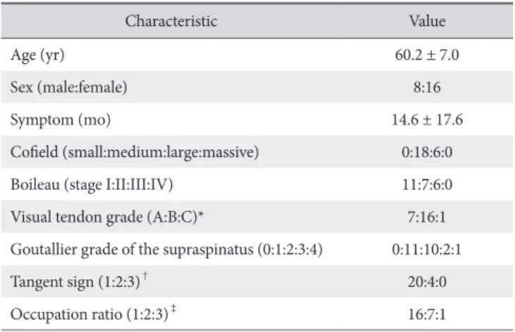
Table 1. Baseline Characteristics of Patients
Characteristic Value
Age (yr) 60.2 ± 7.0
Sex (male:female) 8:16
Symptom (mo) 14.6 ± 17.6
Cofield (small:medium:large:massive) 0:18:6:0
Boileau (stage I:II:III:IV) 11:7:6:0
Visual tendon grade (A:B:C)* 7:16:1
Goutallier grade of the supraspinatus (0:1:2:3:4) 0:11:10:2:1
Tangent sign (1:2:3)† 20:4:0
Occupation ratio (1:2:3)‡ 16:7:1
Values are presented as mean ± standard deviation or number only.
*Tendon grade assesses rotator cuff quality using three gross tendon criteria;
(1) fraying over half of the tendon thickness; (2) delamination of the supra- spinatus tendon, and (3) thinning of less than half of the normal thickness.
A, none of these criteria were met; B, fraying or delamination was identified;
and C, both fraying and delamination or thinning were present (regardless of other criteria). †The tangent sign assesses muscle atrophy of the supraspina- tus. Grade 1 (negative) means that the superior border of the supraspinatus was superior to the line tangential to the coracoid and scapular spine; grade 2 (borderline) means that the superior border was located about the tangential line; grade 3 (positive) means that the superior border was inferior to the tangential line. ‡Occupation ratio means the ratio of the cross-sectional area of the supraspinatus to the fossa. Grade 1, 0.6 to 1; grade 2, 0.4 to 0.6; and grade 3, <0.4.
ance (ANOVA) with Bonferroni tests for multiple comparisons.
To validate the tendon histopathology grading system used, we assessed its intra- and inter-observer reliabilities using kappa sta- tistics. Kappa values were interpreted according to convention:
excellent when κ was between 0.81 and 1.0; high between 0.61 and 0.80; moderate between 0.41 and 0.60; fair between 0.21 and 0.40; and poor 0.20 or less. The analysis was performed us- ing SPSS ver. 13.0 (SPSS Inc, Chicago, IL, USA), and significance was accepted for p-values of <0.05 throughout.
Results
Patients
Twenty-four patients with a full-thickness rotator cuff tear were included in the study (Table 1). Mean patient age was 60.2 ± 7.0 years, and there were 8 males and 16 females. The majority of the patients had a medium-sized tear of Goutallier grade 1 or 2 and a Tangent sign of 1. The normal control group contained nine patients of average age 62.8 ± 17.7 years. At surgery, no patient in the control group showed any evidence of rotator cuff tear in bursal or articular sides.
Histologic Assessments
1) Intra- and inter-observer reliabilities
Intra- and inter-observer reliability testing showed high to excel- lent reliability, except for the assessment of hyalinization (Table 2).
2) General morphology
The microscopic appearances of tendons from patients dif- fered from those of controls (Fig. 1). In specimens from controls,
Table 2. Intra- and Inter-observer Reliabilities
Parameter Intra-observer Inter-observer
Fiber structure 0.836 0.860
Fiber arrangement 0.913 0.854
Rounding of the nuclei 0.612 0.712
Variations in cellularity 0.731 0.759
Increased vascularity 0.742 0.773
Decreased stainability 0.884 0.808
Hyalinization 0.538 0.412
Total 0.796 0.746
Fig. 1. Histology of a torn rotator cuff tendon from a normal control (A), the native end (B), the freshened end (C), and from near the musculotendinous junc- tion (D). (A) Fiber structure, 0; fiber arrangement, 0; rounding of the nuclei, 1; regional variation in cellularity, 0; increased vascularity, 0; decreased collagen stainability, 0; hyalinization, 0; total degeneration score 1. (B) Fiber structure, 3; fiber arrangement, 3; rounding of the nuclei, 2; regional variations in cellularity, 2; increased vascularity, 3; decreased collagen stainability, 2; hyalinization, 2; total degeneration score 17. (C) Fiber structure, 3; fiber arrangement, 3; rounding of the nuclei, 2; regional variations in cellularity, 3; increased vascularity, 0; decreased collagen stainability, 2; hyalinization, 2; total degeneration score 15. (D) Fiber structure, 2; fiber arrangement, 2; rounding of the nuclei, 2; regional variations in cellularity, 2; increased vascularity, 1; decreased collagen stainability, 2; hyalin- ization, 2; total score 13 (A–D: H&E, ×200).
A B
D C
Table 3. Parameters and the Total Degeneration Scores of Tendons Harvested from Normal Controls, Native Ends, Freshened Ends, and Musculotendinous Junctions
Parameter Control Native end Freshened end Musculotendinous junction p-value*
Fiber structure 1.4 ± 0.53 2.0 ± 0.72 2.3 ± 0.74 2.0 ± 0.62 0.298
Fiber arrangement 1.2 ± 0.83 2.3 ± 0.44 2.3 ± 0.64 2.1 ± 0.50 0.262
Rounding of the nuclei 0.9 ± 0.60 2.0 ± 0.72 1.8 ± 0.56 1.3 ± 0.64 0.002
Variations in cellularity 0.6 ± 0.73 2.0 ± 0.83 1.9 ± 0.78 1.7 ± 0.87 0.353
Increased vascularity 0.4 ± 0.73 1.6 ± 0.78 1.8 ± 0.74 1.6 ± 0.88 0.434
Decreased stainability 0.2 ± 0.67 1.6 ± 0.88 0.9 ± 0.78 0.7 ± 0.69 0.708
Hyalinization 0.2 ± 0.44 1.8 ± 1.37 1.5 ± 1.22 1.4 ± 0.88 0.001
Total 5.0 ± 2.87 13.3 ± 3.21 12.5 ± 2.30 10.8 ± 3.10 0.012
Values are presented as mean ± standard deviation.
*p-values for comparisons between the native end, freshened end, and musculotendinous junction groups were calculated using analysis of variance with Bonfer- roni’s post hoc adjustment.
Fig. 2. Comparisons between the control group and the native end (A), freshened end (B), and musculotendinous junction (MT junction) (C) groups. Each pa- rameter in these 3 groups had a significantly greater degeneration score than in the control group, except for nuclear rounding and hyalinization in the MT junc- tion group. (D) Fiber structures, fiber arrangements, regional variations in cellularity, increased vascularity, and decreased collagen stainability were no different in these three groups, whereas nuclear rounding and hyalinization were significantly different (p=0.002 and 0.001, respectively).
Control Freshened end Native end
Control
Fiber structur
e Fiber arrangement
s Nuclei
Cellularity Vascularity StainabilityHyalinizatio n 3
2
1
Degenerationscore
0
A
Nuclei
Cellularity Vascularity StainabilityHyalinizatio n 3
2
1
Degenerationscore
0
B
MT junction Control
Nuclei
Cellularity Vascularity StainabilityHyalinizatio n 3
2
1
Degenerationscore
0
C
Nuclei
Cellularity Vascularity StainabilityHyalinizatio n 3
2
1
Degenerationscore
0
D Native end Freshened end MT junction
p=0.080
p=0.079
p=0.002
p=0.001 Fiber
structur e
Fiber arrangement
s
Fiber structur
e Fiber arrangement
s
Fiber structur
e Fiber arrangement
s
collagen fibers were arranged close and parallel to each other with slight waviness (Fig. 1A).13) Nuclei were flattened and spin- dle-shaped, sometimes arranged in rows between the collagen fibers, and distributed evenly. Vascular bundles were inconspicu- ous and usually ran parallel to and alongside collagen fibers.
Colors of collagen fibers were deep pink-red after H&E staining.
Hyalinization was rarely found.
Specimens from the NE, FE, and MTJ groups showed char- acteristics of degeneration, including loss of fine fiber structure and parallel arrangement, fewer and rounded nuclei, increased cellularity and vascularity, reduced and paler H&E staining, and some evidence of hyalinization (Fig. 1A–C).
3) Native ends
The degeneration scores of the 7 parameters were signifi- cantly higher in the NE group than in the control group (all p<0.01 except for fiber structure p=0.044; Table 3, Fig. 2A).
Mean total degeneration score in the NE group (13.3 ± 3.21) was significantly higher than that of the control group (5.0 ± 2.87, p<0.001; Fig. 3A).
4) Freshened ends
Degeneration scores of all 7 parameters were significantly higher in the FE group than in the control group (all p<0.01 ex- cept for hyalinization p=0.024; Table 3, Fig. 2B). Mean total de- generation score in the FE group (12.5 ± 2.30) was significantly higher than that of the control group (5.0 ± 2.87, p<0.001; Fig.
3B).
5) Musculotendinous junctions
Mean degeneration scores of fiber structure, fiber arrange- ment, regional variations in cellularity, increased vascularity, and decreased collagen stainability were significantly higher in the MTJ group than in the control group, whereas nuclear round- ing and hyalinization scores were not (p=0.080 and 0.078, re- spectively; Table 3, Fig. 2C). Mean total degeneration score was significantly higher in the MTJ group (10.8 ± 3.10) than in the control group (5.0 ± 2.87, p<0.001; Fig. 3C).
6) Native ends, freshened ends, and musculotendinous junctions
Degeneration scores of fiber structure, fiber arrangement,
Fig. 3. Total degeneration scores in the native end (A), freshened end (B), and musculotendinous junction (MT junction) (C) groups as compared with the con- trol group. All three groups showed significantly greater total degeneration scores than the control group. (D) Total degeneration scores in the native end group were no different to those of the freshened end group, but differed significantly from those of the MT junction group (p=0.012). Mean total degeneration scores of the freshened and MT junction groups were non-significantly different (p=0.120).
Control 21
18 15 12 9 6 3
Native end
Totaldegenerationscore
0
A
Control 21
18 15 12 9 6 3
Freshened end
Totaldegenerationscore
0
B
Control 21
18 15 12 9 6 3
MT junction
Totaldegenerationscore
0
C
Native end 21
18 15 12 9 6 3
Freshened end
Totaldegenerationscore
0
D
MT junction
p<0.001 p=0.005
p=0.003 p=1.000 p=0.012 p=0.120
5.0
13.3
5.0
12.5
5.0
10.8
13.3 12.5
10.8
regional variations in cellularity, increased vascularity, and de- creased collagen stainability were not significantly different in the 3 groups (all p>0.05; Table 3, Fig. 2D). However, scores for nuclear rounding and hyalinization were significantly different between the 3 groups (p=0.002 and 0.001, respectively). Fur- thermore, mean total degeneration scores of the 3 groups dif- fered significantly (p=0.012; Fig. 3D). Mean total degeneration score in the NE group was significantly greater than in the MTJ group (p=0.012), but was not significantly different from that of the FE group. The mean total degeneration score of the FE group was not significantly different from that of the MTJ group.
Discussion
This study shows that degeneration of the supraspinatus ten- don in patients with a full-thickness rotator cuff tear is greater than that in patients without a rotator cuff tear, and that degen- eration is present not only at tear ends, but also in macroscopi- cally intact portions of musculotendinous junctions. Further- more, degrees of degeneration were not significantly different between native ends before debridement and freshened ends after debridement (p=1.000) or between freshened ends and macroscopically intact portions just distal to musculotendinous junctions (p=0.120). However, degrees of degeneration were significantly different between native ends and musculotendi- nous junctions (p=0.012). These results suggest that aggressive debridement of frayed torn ends to macroscopically intact or bleeding tendon is unnecessary, because healthy tendon free of degeneration cannot be achieved in patients with a full-thickness rotator cuff tear. In addition, these results suggest that surgeons need a strategy to reverse or regenerate torn, degenerated rota- tor cuff tendons and achieve secure fixation of torn tendon ends at their anatomical locations.
One of strengths of this study is that it includes investigations of degeneration at three different locations, including a macro- scopically intact location, in torn rotator cuff tendons. Whereas many histological studies have addressed rotator cuff degenera- tion and tears,8,9,14-17) few studies have included specimens from a macroscopically intact portions.13,18,19) Furthermore, it is worth mentioning that all of these studies demonstrated degeneration in macroscopically intact tendon. In addition, some authors have reported apoptosis, an important cause of rotator cuff ten- dinopathy,20) in macroscopically intact tendons. In particular, Lee et al.21) found no differences between apoptotic cell profiles in biopsy material from torn edges and a 1-cm-deep region proxi- mal to the margins of torn supraspinatus tendons. The results of the present study concur with these findings, and also show that degrees of degeneration in grossly intact tendons do not differ significantly from those at freshened ends.
With respect to the extent of debridement of frayed torn ends, previous recommendations could be classified as; tradi-
tional, radical, and minimal. Surgeons have been customarily taught that they should resect the torn, frayed tendon end to bleeding, healthy looking tissue,22) and attach it to a cancellous trough to enhance healing.23,24) For rotator cuff repair, Neer25) also recommended careful resection of the torn edge to increase the likelihood of tendon healing. However, these traditional pro- cedures are not supported by solid scientific evidence, and some authors have suggested quite different treatment options.13,21,26-29)
Goutallier et al.15) in a histological study, recommended radical debridement. In this previous study, histological lesions in ten- dinous stumps were found to correspond roughly to tendinous macroscopic lesions requiring resection. However, as this radical debridement would cause a large amount of tension, they also recommended use of special procedures called musculo-tendi- noplasties. On the other hand, Uhthoff et al.26) reported there is no scientific basis for unnecessary shortening of the tendon, be- cause the tendon itself does not contribute to healing, and rec- ommended that simple regularization of frayed ends is sufficient.
In a histological study, Longo et al.13) also suggested that it is not necessary to excessively freshen torn tendon to bleeding tissue because the macroscopically intact supraspinatus tendon is also degenerated and because the causes of healing failure are not limited to the ends of torn tendons. Recently, Fabiś et al.19) re- ported that the expressions of caspases 9, 8 and 3, Bax, and tu- mor necrosis factor-alpha significantly diminished from the distal to the proximal parts of the torn edges of supraspinatus tendons, and suggested that resection 4 to 7 mm from torn edges may enhance the healing process by achieving a reasonable compro- mise between apoptotic and inflammatory processes and the mechanical aspects of rotator cuff reconstruction. The results of this study also confirmed that levels of degeneration in grossly intact locations of rotator cuff tendons were similar to those ob- served at more lateral torn ends. Taken these results together, we suggest that freshening of the torn end should be limited to the removal of most lateral fraying.
The limitations of this study include the small numbers of pa- tients included, the lack of en bloc biopsy specimens, the use of a semi-quantitative grading system, the use of only H&E staining, no evaluation of myxoid, lipoid, or calcific degeneration, and potential rotator cuff problems in the control group.
Conclusion
The findings of this study demonstrate that tendon degen- eration in patients with a full-thickness rotator cuff tear occurs throughout the entire rotator cuff tendon to macroscopically in- tact portions. Minimal freshening of lateral frayed ends, resulted in degrees of degeneration in freshened ends that were not significantly different from those observed in macroscopically intact portions, which suggests aggressive debridement to grossly normal looking, bleeding tendon is unnecessary.
References
1. Hermans J, Luime JJ, Meuffels DE, Reijman M, Simel DL, Bierma-Zeinstra SM. Does this patient with shoulder pain have rotator cuff disease?: The Rational Clinical Examination systematic review. JAMA. 2013;310(8):837-47.
2. Chard MD, Hazleman R, Hazleman BL, King RH, Reiss BB.
Shoulder disorders in the elderly: a community survey. Arthritis Rheum. 1991;34(6):766-9.
3. Oh LS, Wolf BR, Hall MP, Levy BA, Marx RG. Indications for rotator cuff repair: a systematic review. Clin Orthop Relat Res.
2007;455:52-63.
4. Colvin AC, Egorova N, Harrison AK, Moskowitz A, Flatow EL.
National trends in rotator cuff repair. J Bone Joint Surg Am.
2012;94(3):227-33.
5. Via AG, De Cupis M, Spoliti M, Oliva F. Clinical and biologi- cal aspects of rotator cuff tears. Muscles Ligaments Tendons J.
2013;3(2):70-9.
6. Kannus P, Józsa L. Histopathological changes preceding sponta- neous rupture of a tendon. A controlled study of 891 patients.
J Bone Joint Surg Am. 1991;73(10):1507-25.
7. Nirschl RP. Rotator cuff tendinitis: basic concepts of pathoetiol- ogy. Instr Course Lect. 1989;38:439-45.
8. Hashimoto T, Nobuhara K, Hamada T. Pathologic evidence of degeneration as a primary cause of rotator cuff tear. Clin Or- thop Relat Res. 2003;(415):111-20.
9. Longo UG, Franceschi F, Ruzzini L, et al. Light microscopic histology of supraspinatus tendon ruptures. Knee Surg Sports Traumatol Arthrosc. 2007;15(11):1390-4.
10. McCarrel TM, Minas T, Fortier LA. Optimization of leukocyte concentration in platelet-rich plasma for the treatment of ten- dinopathy. J Bone Joint Surg Am. 2012;94(19):e143.
11. Aström M, Rausing A. Chronic Achilles tendinopathy. A survey of surgical and histopathologic findings. Clin Orthop Relat Res.
1995;(316):151-64.
12. Movin T, Gad A, Reinholt FP, Rolf C. Tendon pathology in long-standing achillodynia. Biopsy findings in 40 patients. Acta Orthop Scand. 1997;68(2):170-5.
13. Longo UG, Franceschi F, Ruzzini L, et al. Histopathology of the supraspinatus tendon in rotator cuff tears. Am J Sports Med.
2008;36(3):533-8.
14. Matthews T, Brinsden M, Athanasou N, Rees J, Carr A. Predic- tion of rotator cuff repair failure by histological analysis. Shoul- der Elbow. 2009;1(1):10-4.
15. Goutallier D, Postel JM, Van Driessche S, Voisin MC. Histo- logical lesions of supraspinatus tendons in full thickness tears of the rotator cuff. Rev Chir Orthop Reparatrice Appar Mot.
2005;91(2):109-13.
16. Fukuda H, Hamada K, Yamanaka K. Pathology and pathogen- esis of bursal-side rotator cuff tears viewed from en bloc histo- logic sections. Clin Orthop Relat Res. 1990;(254):75-80.
17. Sano H, Ishii H, Trudel G, Uhthoff HK. Histologic evidence of degeneration at the insertion of 3 rotator cuff tendons: a com- parative study with human cadaveric shoulders. J Shoulder Elbow Surg. 1999;8(6):574-9.
18. Castagna A, Cesari E, Gigante A, Conti M, Garofalo R. Metal- loproteases and their inhibitors are altered in both torn and intact rotator cuff tendons. Musculoskelet Surg. 2013;97 Suppl:39-47.
19. Fabiś J, Szemraj J, Strek M, Fabiś A, Dutkiewicz Z, Zwier- zchowski TJ. Is resection of the tendon edge necessary to en- hance the healing process? An evaluation of the homeostasis of apoptotic and inflammatory processes in the distal 1 cm of a torn supraspinatus tendon: part I. Shoulder Elbow Surg. 2014;
23(12):1772-8.
20. Yuan J, Murrell GA, Wei AQ, Wang MX. Apoptosis in rotator cuff tendonopathy. J Orthop Res. 2002;20(6):1372-9.
21. Lee HJ, Kim YS, Ok JH, Song HJ. Apoptosis occurs throughout the diseased rotator cuff. Am J Sports Med. 2013;41(10):2249- 55.
22. DePalma AF. Ruptures of the musculotendinous cuff. In: De- Palma AF, ed. Surgery of the shoulder. Philadelphia: Lippin- cott; 1950. 112-37.
23. Jones JR, Smibert JG, McCullough CJ, Price AB, Hutton WC.
Tendon implantation into bone: an experimental study. J Hand Surg Br. 1987;12(3):306-12.
24. McLaughlin H. Lesions of the musculotendinous cuff of the shoulder. The exposure and treatment of tears with retraction.
Bone Ioint Surg Am. 1944;24:31.
25. Neer CS. Shoulder reconstruction. Philadelphia: WB Saun- ders; 1990.
26. Uhthoff HK, Trudel G, Himori K. Relevance of pathology and basic research to the surgeon treating rotator cuff disease. J Orthop Sci. 2003;8(3):449-56.
27. St Pierre P, Olson EJ, Elliott JJ, O’Hair KC, McKinney LA, Ryan J. Tendon-healing to cortical bone compared with healing to a cancellous trough. A biomechanical and histological evaluation in goats. J Bone Joint Surg Am. 1995;77(12):1858-66.
28. Shaieb MD, Singer DI, Grimes J, Namiki H. Evaluation of ten- don-to-bone reattachment: a rabbit model. Am J Orthop (Belle Mead NJ). 2000;29(7):537-42.
29. Soda Y, Sumen Y, Murakami Y, Ikuta Y, Ochi M. Attachment of autogenous tendon graft to cortical bone is better than to cancellous bone: a mechanical and histological study of MCL reconstruction in rabbits. Acta Orthop Scand. 2003;74(3):322-6.