Arthroscopic Decompression of an Inferior Paralabral Cyst of the Shoulder in an Elderly Patient: A Case Report
Ju-Oh Kim, Ki-Yong An , Hwang-Se Bong, Kyu-Jung Lee, Woong-Bae Min
Department of Orthopedic Surgery, Gwangju Veterans Hospital, Gwangju, Korea
The widespread use of diagnostic radiography, especially using magnetic resonance imaging, has helped to increase the diagnosis of paralabral cysts in patients with chronic shoulder pain. These paralabral cysts are frequent in the anterior, the superior, and the posterior compartment of the shoulder joint but are rare in the inferior compartment. Paralabral cysts in the shoulder appear particularly in men in their third and fourth decades but rarely in elderly patients. We report a case of an inferior paralabral cyst in an elderly patient whom we treated through arthroscopic decompression.
(Clin Shoulder Elbow 2015;18(4):266-268)
Key Words: Shoulder; Cyst; Arthroscopy; Cystoreduction surgical procedures
Clinics in Shoulder and Elbow
CiSE
Copyright © 2015 Korean Shoulder and Elbow Society. All Rights Reserved.
This is an Open Access article distributed under the terms of the Creative Commons Attribution Non-Commercial License (http://creativecommons.org/licenses/by-nc/4.0) which permits unrestricted non-commercial use, distribution, and reproduction in any medium, provided the original work is properly cited.
pISSN 2383-8337 eISSN 2288-8721
CASe RepORt
Clinics in Shoulder and elbow Vol. 18, No. 4, December, 2015 http://dx.doi.org/10.5397/cise.2015.18.4.266
Received October 4, 2015. Revised November 27, 2015. Accepted December 10, 2015.
The findings of this paper has been presented at the 2015 Autumn Conference of the Korean Orthopedic Society.
Correspondence to: Ki-Yong An
Department of Orthopedic Surgery, Gwangju Veterans Hospital, 99 Cheomdanwolbong-ro, Gwangsan-gu, Gwangju 62284, Korea Tel: +82-62-602-6162, Fax: +82-62-602-6164, E-mail: [email protected]
Financial support: None. Conflict of interests: None.
The increased use of diagnostic magnetic resonance imaging (MRI) in patients with chronic shoulder pain has correlated with the increase in the diagnosis of paralabral cysts. Paralabral cysts are commonly associated with labral or capsule tears through which synovial fluid seeps out into the extra-articular space;
the labral or the capsule tear essentially becomes a ‘one-way- valve’.1,2) Paralabral cysts are present in 2% to 4% of the popula- tion, most of whom are 30- to 40-year-old men. They are rarely present in the elderly population. Generally, the cysts are located in the posterior, the superior, or the anterior compartment of the shoulder and are rarely located in the inferior compartment.3) The symptoms of pain and dysfunction can vary according to the position of the cysts. In this study, we present a case of a paralabral cyst in the inferior compartment of the shoulder in a 79-year-old man whom we treated using arthroscopic decom- pression.
Case Report
The 79-year-old man admitted to our hospital presented with a chief complaint of left shoulder pain that began and deteriorat-
ed without particular trauma from a month before. Specifically, he complained of pain around the posterior compartment of the shoulder, and without particular reason he complained that the pain exacerbated at night. At the time of admittance, the patient scored a visual analogue scale for pain of 6 points. Through physical examination, we found that the range of motion of the left shoulder showed a forward elevation of 160o, an external rotation of 40o, an internal rotation of 40o, and an abduction of 120o; the patient displayed slight restriction in motion. We did not observe other physical or neurological abnormalities that indicated instability. Visually, we could not find evidence for atrophy of the muscles, and muscle strength was within the nor- mal range. Because symptoms prevailed with drug therapy, we carried out plain radiography and MRI. At the preoperative MRI, we found and diagnosed a ganglion cyst, which was fused to the articular surface, with a dimension of 2.5×1.5×2 cm at the inferior labrum, and an inferior labral tear (Fig. 1). Despite con- servative treatment of the paralabral cyst through drug therapy, rest, and modified activity for 3 months, the symptoms did not improve; thus, we decided to treat the patient surgically.
Under general anesthesia, we placed the patient in the right
Arthroscopic Decompression of an Inferior paralabral Cyst of the Shoulder in an elderly patient Ju-Oh Kim, et al.
www.cisejournal.org
267
lateral decubitus position. Arthroscopically, we found that the labral tear was situated in the anteroinferior compartment and was directed in the 5 and 6 o’clock direction (Fig. 2A). Using a probe, we expanded the articular-cyst junction and compressed the axilla to drain the mucinous liquid from the inferior labrum (Fig. 2B). We performed an arthroscopic debridement of the inferior labral tear using a Shaver and an ArthroCare. We did not perform a decompression of the subacromial space. As for the rehabilitation of the patient, he was applied an arm sling to immobilize the arm for three postoperative days after which we permitted active motion. A month postoperatively, we car- ried out a follow-up MRI. We found that the cyst disappeared completely without indications of recurrence and that pain was resolved (Fig. 3). At the 6 months follow-up, an ultrasonography confirmed that there were no signs of recurrence and that pain and night pain of the left shoulder which were the chief com- plaints of the patients had improved to a visual analogue scale score of 1 point.
Fig. 3. Postoperative proton density-weighted coronal oblique image reveals complete resolution of the cyst.
A B
Fig. 2. (A) The arthroscopic photograph in left shoulder shows an anteroinferior labral tear. (B) Cyst decompression and debride- ment of the anteroinferior labral tear were performed.
A B
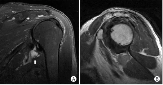
Fig. 1. (A) Preoperative magnetic resonance imaging of the left shoulder with T2-weighted fat-suppressed coronal oblique view demon- strates a 25×15 mm-sized multi-septated cyst (arrow) at the inferior aspect of the glenoid neck. (B) Proton density-weighted sagittal oblique view shows the inferior labral tear.
268
www.cisejournal.orgClinics in Shoulder and elbow Vol. 18, No. 4, December, 2015
Discussion
The prevalence of paralabral cysts of the shoulder is 2% to 4%
in the general population. Paralabral cysts are located in the fol- lowing compartments of the labrum in descending order of oc- currence: the posterior labral compartment in 57% of paralabral cysts; the anterior compartment in 21%; the superior compart- ment in 14%; and the inferior compartment in 8%.3) Westerhe- ide et al.1) and Westerheide and Karzel2) suggested that the prin- cipal cause of paralabral shoulder cysts is a labral tear, through which a unidirectional out-flux of synovial fluid occurs–the un- derlying pathophysiological mechanism of paralabral cysts. With widespread use of diagnostic radiography, especially the use of MRI, the diagnosis of paralabral cysts has increased in patients who present with shoulder pain. The use of radiography has in- creased in part because a diagnosis based on clinical symptoms, medical history, and physical examination, the findings of which are ambiguous, are difficult.4) Ultrasound and computed tomog- raphy (CT) are also useful diagnostic tools. Ganglion shoulder cysts are relatively rare causes of shoulder pain, and, in those with recurrent, chronic shoulder pain, which reflects a refractory cyst, the symptoms should be differentiated from those of de- generative arthritis, rheumatoid arthritis, osteomyelitis, or malig- nant or benign tumors. The pathophysiology of the symptoms of paralabral shoulder cysts may derive from the labral lesion itself or from cyst-induced compression of the supracapsular nerve.4-6)
Conservative treatment usually precedes surgical treatment.
Physiotherapy or exercise may relieve symptoms to an extent, but it is difficult to anticipate spontaneous resolution of cysts.
Past studies have detected through CT, ultrasonography, and MRI a high rate of refractory cyst after simple aspiration.7) If with conservative treatment symptoms persist upon clinical examina- tion or abnormalities exist in the results of electromyographic or nerve conduction test, the patient should opt for surgical treatment. Arthroscopy, which is usually the method of choice for surgery, is advantageous in that it can be used to concomi- tantly treat other lesions of the joint. Youm et al.8) reported that arthroscopic repair of the labral tear without cyst decompression showed good clinical outcomes. Whereas Jeong et al.9) reported that arthroscopic repair of the labral tear with decompression of the inferior labral cyst also showed good clinical outcomes. In accordance with these findings, Abboud et al.10) found through 9 arthroscopic decompression of cysts that a significant difference in clinical outcomes was not seen between those who received decompression alone and those who received it with concomi- tant labral repair, showing that good clinical outcomes can be achieved without recurrence with arthroscopic decompression alone. In this report, we found that in an elderly patient with a labral tear and a paralabral cyst of the inferior compartment,
combined with a degenerative joint, an arthroscopic debride- ment and a cyst decompression without repair of the labral tear showed good clinical outcomes without occurrence of a refrac- tory cyst.
Paralabral cysts of the inferior shoulder are relatively rare and generally occur in young, active men. If, conversely, they occur idiopathically in elderly patients, we found that arthroscopic de- compression and debridement is one of the possible treatment methods that we can perform. Further, when treating these patients orthopedic surgeons should remember that a one-way- valve effect may lead to recurrence of the cysts.
References
1. Westerheide KJ, Dopirak RM, Karzel RP, Snyder SJ. Supra- scapular nerve palsy secondary to spinoglenoid cysts: results of arthroscopic treatment. Arthroscopy. 2006;22(7):721-7.
2. Westerheide KJ, Karzel RP. Ganglion cysts of the shoulder:
technique of arthroscopic decompression and fixation of as- sociated type II superior labral anterior to posterior lesions.
Orthop Clin North Am. 2003;34(4):521-8.
3. Tung GA, Entzian D, Stern JB, Green A. MR imaging and MR arthrography of paraglenoid labral cysts. AJR Am J Roentgenol.
2000;174(6):1707-15.
4. Iannotti JP, Ramsey ML. Arthroscopic decompression of a gan- glion cyst causing suprascapular nerve compression. Arthros- copy. 1996;12(6):739-45.
5. Lichtenberg S, Magosch P, Habermeyer P. Compression of the suprascapular nerve by a ganglion cyst of the spinoglenoid notch: the arthroscopic solution. Knee Surg Sports Traumatol Arthrosc. 2004;12(1):72-9.
6. Kessler MA, Stoffel K, Oswald A, Stutz G, Gaechter A. The SLAP lesion as a reason for glenolabral cysts: a report of five cases and review of the literature. Arch Orthop Trauma Surg.
2007;127(4):287-92.
7. Piatt BE, Hawkins RJ, Fritz RC, Ho CP, Wolf E, Schickendantz M. Clinical evaluation and treatment of spinoglenoid notch ganglion cysts. J Shoulder Elbow Surg. 2002;11(6):600-4.
8. Youm T, Matthews PV, El Attrache NS. Treatment of patients with spinoglenoid cysts associated with superior labral tears without cyst aspiration, debridement, or excision. Arthroscopy.
2006;22(5):548-52.
9. Jeong JJ, Panchal K, Park SE, et al. Outcome after arthroscopic decompression of inferior labral cysts combined with labral repair. Arthroscopy. 2015;31(6):1060-8.
10. Abboud JA, Silverberg D, Glaser DL, Ramsey ML, Williams GR. Arthroscopy effectively treats ganglion cysts of the shoul- der. Clin Orthop Relat Res. 2006;444:129-33.