Biomechanical Test for Repair Technique of Full-thickness Rotator Cuff Tear
Chae-Ouk Lim, Kyoung-Jin Park
Department of Orthopaedic Surgery, Chungbuk National University College of Medicine, Cheongju, Korea
The arthroscopic rotator cuff repair is now considered a mainstream technique with highly satisfactory clinical results. However, concerns remain regarding healing failures for large and massive tears and high revision rate. In recent decades, various repair strategies and con- struct configurations have been developed for rotator cuff repair with the understanding that many factors contribute to the structural integrity of the repaired construct. The focus of biomechanical test in arthroscopic repair has been on increasing fixation strength and restoration of the footprint contact characteristics to provide early rehabilitation and improve healing. These include repaired rotator cuff tendon-footprint motion, increased tendon-footprint contact area and pressure, and tissue quality of tendon and bone. Recent studies have shown that a transosseous tunnel technique provides improved contact area and pressure between rotator cuff tendon and inser- tion footprint, and the technique of using double rows of suture anchors to recreate the native footprint attachment has been recently described. The transosseous equivalent suture bridge technique has the highest contact pressure and fixation force. In this review, the biomechanical tests about repair techniques of rotator cuff tear will be reviewed and discussed.
(Clin Shoulder Elbow 2016;19(1):51-58)
Key Words: Biomechanical test; Rotator cuff repair; Full-thickness rotator cuff tear; Transosseous equivalent repair
Copyright © 2016 Korean Shoulder and Elbow Society. All Rights Reserved. pISSN 2383-8337
Clinics in Shoulder and Elbow Vol. 19, No. 1, March, 2016 http://dx.doi.org/10.5397/cise.2016.19.1.51
Received September 27, 2015. Revised December 22, 2015. Accepted January 14, 2016.
Correspondence to: Kyoung-Jin Park
Department of Orthopaedic Surgery, Chungbuk National University College of Medicine, 1 Chungdae-ro, Seowon-gu, Cheongju 28644, Korea Tel: +82-43-269-6077, Fax: +82-43-274-8719, E-mail: [email protected]
Financial support: None. Conflict of interests: None.
Introduction
Rotator cuff tears can be asymptomatic, however often they result in pain and functional disability. Arthroscopic rotator cuff repair is now considered a mainstream technique with highly satisfactory clinical results.1) Although many advantages of ar- throscopic techniques exist, concern remains regarding healing failures for large and massive tears.2) Many factors have been blamed for the failure of rotator cuff repair, including blood sup- ply, tendon or bone quality, and fatty infiltration of rotator cuff musculature.3-5) Recent research has focused on improving the strength and durability of rotator cuff repairs to decrease this high rate of failure. Many factors contribute to an optimal repair.
Initial fixation strength is an essential consideration in optimizing rotator cuff repair and therefore, numerous biomechanical stud- ies have focused on elucidating the strongest devices, knots, and repair configurations for rotator cuff repair.6-8) To restore the foot-
print area, numerous arthroscopic techniques have been stud- ied. Recently, the transosseous-equivalent (TOE) technique has become popular with sutures from the medial row of anchors coming across the rotator cuff and being secured laterally in the footprint. However, there is still considerable debate regarding the optimal biomechanical arthroscopic repair construct.
There was little article to review for biomechanical study of rotator cuff repair. Therefore, in this review, the biomechanical concepts behind rotator cuff repair techniques will be reviewed and discussed. This review focused on recent biomechanical testing for full thickness rotator cuff repair using conventional single row repair, double row repair and TOE technique.
What Is Biomechanical Testing for Rotator Cuff Tear?
Tensile testing is generally used to quantify the biome chanical
characteristics and structural integrity of repaired constructs (Fig. 1). The tensile testing includes both cyclic loading and load- ing to failure. For each cycle of the cyclic loading, two important biomechanical parameters are determined.9) The first parameter is the linear stiffness of the construct, defined as the slope of the linear portion of the load-elongation curve with units of N/mm.
The second parameter is the hysteresis of the construct, de- fined as the differences in area under the loading and unloading curves on the load-elongation curve. This parameter represents energy dissipated in the construct during each cycle of loading and unloading. This energy can be dissipated in many ways, including suture anchor-bone slippage, knot slippage, and tissue fiber alignment. From the load to failure tests, four important biomechanical parameters are determined. The first is the lin- ear stiffness of the construct, which is determined in the same fashion as described for cyclic loading. The next parameter is yield load and deformation, which is the load and deformation at which the load/elongation curve deviates from linearity; that is, when the stiffness begins to decrease. Prior to reaching yield
load and deformation, the construct is in the elastic load and de- formation range where all deformation is recoverable; however once surpassing the yield load permanent plastic deformation occurs. The next parameter is the ultimate load and deforma- tion, which represents the maximum load and deformation sustained by the construct prior to failure. Lastly, the energy ab- sorbed by the construct can be calculated at both yield load and ultimate load and deformation by calculating the area under the load-elongation curve. The final biomechanical parameter com- monly used is gap formation which is unique to reconstructed tendon bone complex. The gap formation was defined as the space created at the lateral edge of the tendon at the repair site.
The rotator cuff repair constructs were tested using a materials testing machine, a custom shoulder fixture device. When the specimen was mounted securely, nonreflective black paint was used to make markers on the specimens to be used for the video digitizing system. A video digitizing system that involved video recording of the markers, computer digitization of the markers, creation of centroids representing the center of the markers, and the calculation of distances with software program (Fig. 2).
The Mechanical Properties of Repair Technique & Bone Attachment
Historically, anchors with different suture configurations have been used to improve the biomechanical integrity of rotator cuff repairs in open procedures. Simple, mattress, and other suture configurations such as the Mason-Allen stitch have been biome- chanically compared throughout the literature.10) A Mason-Allen suture has been shown to have a higher tensile load and ulti- mate tensile strength when compared with simple and mattress suture configurations. Gerber et al.11) studied current techniques of rotator-cuff repair to assess their mechanical properties and consider potential improvements. To determine current practice by expert shoulder surgeons, they interviewed ten members Fig. 1. Typical materials testing load-elongation curve for cyclic loading (cycle 1)
and load to failure with relevant parameters.
Cycle 1 Load
The load-elongation curve
Ultimate load
Yield load
Linear stiffness
Energy absorbed to failure Hysteresis
Elongation
Fig. 2. (A) Th e rotator cuff repair constructs were tested using a materials testing machine, a custom shoulder fi xture device. (B) Nonre- fl ective black paint was used to make markers on the specimens to be used for the video digitizing system.
A B
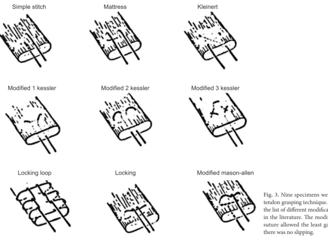
of the European Shoulder and Elbow Society and 20 active members of the American Shoulder and Elbow Surgeons who reported on the methods they used for tendon grasping of their preferred suture material. And then, this study tested the suture materials most frequently used, measuring elongation under load and ultimate tensile strength of knotted threads. Non-absorbable braided polyester and absorbable polyglactin and polyglycolic acid sutures best combined ultimate tensile strength and stiff- ness. In this study, they compared the mechanical properties of nine different techniques of tendon grasping, using 159 normal infraspinatus tendons from sheep (Fig. 3). There was no slipping within the tendon of the modified Mason-Allen technique, the locking suture or the locking loop suture. By contrast, the simple stitches and the mattress sutures slipped out at moderate loads of 184 to 270 N. The modified Mason-Allen suture allowed the least gap formation; however the differences between that and the first modification of the Kessler and the Kleinert suture were not statistically significant.
Structural failure at the repair site is the most common com- plication of rotator cuff repair. A single-row repair does not ade- quately reproduce the anatomic insertion and may not optimize fixation strength. A double-row repair provides a more anatomic repair by creating a larger area of contact of the repaired tendon with the tuberosity. Waltrip et al.12) hypothesized that double- row fixation provides a biomechanically stronger construct than
single- row fixation. Each matched pair underwent a double- row ‘anatomic’ repair on one side and sequential single row repair on the opposite side. Rotator cuff repair with a double- row technique provides greater initial fixation strength than that achieved with a single-row repair. The two layers of bony fixa- tion achieved with the anatomic repair were the key to the in- creased strength of fixation. Since the anatomic repair has more points of fixation to the tuberosity, it should have a greater area of contact than single-row repairs. Consequently, a double-row repair should lead to a larger insertion and improved biologic healing.
Suture Anchor vs. Transosseous Suture Fixation
The optimal method of reconstruction remains highly con- troversial. There is no consensus as to which one of the 2 most commonly used procedures (suture anchor or transosseous su- ture fixation) provides enhanced tendon-to-bone healing and, ultimately, a stronger repair (Fig. 4). Gerber et al.11) indepen- dently reported that suture anchor repairs were comparable or weaker than traditional transosseous reconstructions, whereas other investigators found that suture anchors outperformed the transosseous suture repair.13,14) Apreleva et al.15) tested to compare the area of the intact supraspinatus insertion with the
Fig. 3. Nine specimens were tested for each tendon grasping technique. Th is fi gure shows the list of diff erent modifi cation of single row in the literature. Th e modifi ed Mason-Allen suture allowed the least gap formation and there was no slipping.
Simple stitch
Modified 1 kessler
Locking loop
Mattress
Modified 2 kessler
Locking
Kleinert
Modified 3 kessler
Modified mason-allen
repair-site areas of the reattached supraspinatus tendon after 2 transosseous suture and 2 suture-anchor repairs of a simulated supraspinatus tear. A supraspinatus tear was created and 4 repair techniques were evaluated: transosseous simple suture (TOS), transosseous mattress suture, suture-anchor simple suture, suture-anchor mattress suture. The 3-dimensional outlines of the reconstructed supraspinatus insertion were digitized after each repair. None of the tested repairs restored the area of the original supraspinatus insertion. The larger repair-site area of the TOS repair suggests that this technique provides better potential for healing and, ultimately, greater strength of repair. Among the transosseous repairs, the simple suture technique appeared to be superior to the mattress suture technique. Because TOS re- pair had 2 fixation points, one of which is located more distally on the humeral head, it allowed the free edge of the tendon to be laid down on the bone underneath the suture more laterally, therefore, expanding the repair site area.
In the case of suture-anchor repair, which is widely used dur- ing arthroscopic surgery, the anchors were placed at the medial border of the original insertion, closer to the articular surface.
The result was a smaller surface contact area between the ten- don and bone. This suggests that if suture anchors are used in a repair, a more lateral placement of the anchors may be better because it may increase the repair-site area. The stress con- centration constitutes one of the biomechanical factors of the material failure, Sano et al.16) attempted to compare the stress distribution after a cuff repair using the suture anchor fixation or the transosseous suture fixation. Single-row fixation, double-row fixation, and transosseous suture fixation were simulated. The suture anchor models revealed that the stress extended from the site of the suture anchor to the bursal surface of the tendon, whereas no significant stress concentration was observed in the transosseous model. The torn cuff tendons show diffuse histolog- ic degeneration, which weakens their biomechanical strength. In the healing process after repair, such poor quality tendon might cause a failure, especially under the high stress concentration.17) The highest stress concentration in the transosseous model was
observed at the site of attachment with the suture thread inside the bony trough, which might indicate that the weakest link in this repair technique is the suture thread itself. The suture anchor repair technique showed higher stress concentration inside the tendon than transosseous suture fixation. Maximizing the con- tact area between tendon and tuberosity at the rotator cuff foot- print enhances the biological healing process, in turn improving the mechanical strength and function of the repaired tendon.18) The study of Park et al.19) was to compare the contact pressures, and the area distribution of these pressures, over a repaired rotator cuff footprint resulting from commonly used techniques for rotator cuff repair. They hypothesize that the transosseous repair techniques create a larger footprint of contact, over which a greater contact pressure is present, when compared with the suture anchor techniques. The result have shown that the tran- sosseous repair technique provides a more pressure-producing contact area for potential healing and more mean pressure, or focal fixation, over a defined tuberosity footprint when com- pared to either of the suture anchor techniques. The transosse- ous suture provides greater compressive force over a larger area, when compared to suture anchor repair techniques. In contrast, the sutures for the suture anchor technique predominantly pro- vide circumferential tension around the tendon while providing relatively little compression between the tendon and bone. Park et al.,20) who quantified the amount of motion between the re- paired tendon and bone in a transosseous repair versus a single- row construct, found that the transosseous suture repair allowed less motion in internal and external rotation compared to the single-row construct. This is important, as the ideal repair con- struct would minimize motion in the immediate postoperative setting in order to allow healing of the tendon to bone.
Single-Row Repair vs. Double-Row Repair
Transosseous techniques with open procedures were origi- nally shown to restore a greater percentage of the supraspinatus tendon insertion.15) With the current trend away from open Fig. 4. (A) Isolated suture anchor repair schematic diagram. (B) Isolated transosseous tunnel repair schematic diagram.
A B
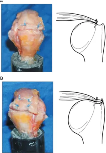
repairs, new techniques have been devised to improve the res- toration of the footprint arthroscopically. It has been suggested that this could be accomplished with the use of a double row suture anchor fixation technique. Initial reports demonstrated that standard arthroscopic repairs using a single row of anchors did not adequately restore the footprint contact area.21) The technique of using double rows of suture anchors to re-create the native footprint attachment has been recently described.22) This double-row technique has been shown to closely re-create the repair site of the footprint insertion and would theoretically improve the ability of the tendon to heal to bone. Double-row repair was created to increase the footprint contact area and dis- tribute the stress over multiple fixation points. Lo and Burkhart21) then described their arthroscopic technique for a double-row repair. The medial sutures are passed through the medial aspect of the tendon in a mattress fashion and tied down. The lateral sutures are then passed through the tendon in a simple suture formation and tied down, providing medial row and lateral row fixation to increase the contact area of the repair (Fig. 5).
In addition, the majority of the studies23) showed improved
biomechanical characteristics of a double-row repair compared with a single-row repair. A study by Domb et al.,24) who com- pared high-tension double-row and medialized single-row con- structs, found that the high-tension double-row repair fared better than the medialized single row construct. There was significantly decreased displace ment at first cycle, stiffness in the final cycle, and ultimate load to failure. The authors concluded that when possible, a retracted tear should be repaired with a double-row con struct. A study by Ma et al.25) tested a standard double-row repair with three different single-row repairs: the Mason- Allen stitch, massive cuff stitch, and two simple sutures. The double- row construct was as good as or better than all single-row repair constructs in all parameters tested. Finite element models were also used by Sano et al.16) to characterize stress concentration at the repair site in a single-row and double-row. In the double- row construct, there was more stress in the medial row of anchors than the lateral row. This is advantageous for chronic rotator cuff tears with a degenerated tendon where the tendon cannot tolerate large amounts of strain. The study of Nelson et al.26) is to determine the surface area of the repair interface of a double-row technique and a single-row modified Mason-Allen configuration as well as to biomechanically compare the strength of fixation of the 2 repairs. Double-row suture anchor fixation restores a greater percentage of the anatomic footprint when compared with a single-row Mason-Allen technique. For smaller tears, a single-row modified Mason-Allen suture technique may provide sufficient strength; however for large amenable tears, a double row can provide both strength and increased surface area for healing. In cyclic testing Milano et al.27) found that a double-row repair construct significantly outperformed a single- row repair construct. The double-row repair group required 600 cycles to reach 5 mm of elongation (failure), whereas the single- row repair group repaired under tension failed at a mean of 23 cycles. They concluded that a double row technique should be considered for large, unstable tears, especially if the single-row repair would be under tension. Tuoheti et al.22) reported the double-row repair technique to have a repair contact area 60%
greater than that of the single-row repair technique. They also showed that double-row repair restored 42% more area when compared with a traditional transosseous rotator cuff repair. Wall et al.28) reviewed the literature of all biomechanical studies com- paring double-row vs single-row repair techniques. Fifteen stud- ies were identified and reviewed. Nine studies showed a statisti- cally significant advantage to a double-row repair with regards to biomechanical strength, failure, and gap formation. Three studies produced results that did not show any statistical advan- tage. Five studies that directly compared footprint reconstruction all demonstrated that the double-row repair was superior to a single row repair in restoring anatomy. The current literature re- veals that the biomechanical properties of a double-row rotator cuff repair are superior to a single-row repair.
Fig. 5. (A) Th e construct of single-row repair technique & schematic diagram.
(B) Th e construct of double-row repair technique & schematic diagram.
B A
The New Repair Technique:
Transosseous Equivalent Repair
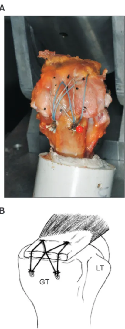
The arthroscopic TOE suture bridge (TOE-SB) technique was developed to maximize tendon-to-bone compression by bridging the medial suture limbs to lateral suture anchors (Fig.
6), which results in compression of the tendon onto the rotator cuff footprint. TOE-SB repair provides significantly more contact area and pressure over a repaired rotator cuff footprint than a double-row suture anchor repair.29) By placing the lateral row of anchors orthogonal from the rotator cuff-loading vector, a com- pression vector over the tendon is created to increase pressure at the footprint, greatly increasing the contact pressure along the repaired tendon in the TOE repair compared to the double- row repair. Behrens et al.30) compared the initial in vitro tensile fixation strength of a TOE-SB rotator cuff repair construct with a traditional transosseous (TO) suture construct. There were no statistically significant differences in gap formation at the repair
sites under low or high load conditions between TOE-SB and TO techniques. The arthroscopic TOE-SB technique is compara- ble in initial fixation strength to the traditional TO simple suture repair technique. In a study by Bisson et al.31) comparing the bio- mechanical performance of transosseous–suture anchor and su- ture bridge rotator cuff repairs, the suture bridge repair showed similar biomechanical performance during cyclic and load-to- failure testing as a transosseous–suture anchor repair, which has historically been performed in open or mini-open fashion. Park et al.20) hypothesized that a TOE repair would demonstrate im- proved tensile strength and gap formation between the tendon and tuberosity when compared with a double-row technique.
The TOE rotator cuff repair technique improves ultimate failure loads when compared with a double-row technique. Stiffness and gap formation are similar for both techniques. And the TOE rotator cuff repair technique can improve pressurized contact area and mean pressure between the tendon and footprint when compared with a double-row technique. A TOE repair helps restore footprint dimensions and provides a stronger re- pair than the double-row technique, which may help optimize healing biology. A study by Mazzocca et al.32) tested a single- row, double-row, TOE, and suture-chain TOE. The suturechain TOE uses FiberChain (Arthrex, Naples, FL, USA) to connect the medial and lateral rows; however unlike the TOE, the anterior and posterior anchors are not connected. This study emphasizes the importance of a strong, stable rotator cuff repair in the im- mediate postoperative period, and it shows that a TOE provides the strongest initial construct and potential for healing. Busfield et al.33) investigated the importance of the medial row knots in a TOE configuration. In both groups with supraspinatus tears, a TOE configuration was used, however in one, the medial row was fixed with a knotless suture anchor while a standard anchor with knots was used in the other group. Biomechanical testing showed greater gap formation during cyclic loading and yield load in the knotless group as well as a decreased ultimate load.
Therefore, the authors concluded that a medial row of knots provides a biomechanically stronger construct compared with knotless fixation.
Conclusion
Recent biomechanical test has focused on improving the strength and durability of rotator cuff repairs to decrease this high rate of failure. It has been proposed that the goals of rotator cuff repair are as follows: (1) an initially strong construct with, (2) minimal gap formation, and (3) footprint stability that will allow the tendon to heal to bone during the rehabilitation period. Bio- mechanical test has contributed to the best results of rotator cuff repair and it will contribute more in the future.
Fig. 6. Th e construct of the transosseous-equivalent suture bridge technique (A) and the schematic diagram (B).
GT: greater tubercle, LT: lessor tubercle.
A
GT
LT B
References
1. Bennett WF. Arthroscopic repair of full-thickness supraspinatus tears (small-to-medium): a prospective study with 2- to 4-year follow-up. Arthroscopy. 2003;19(3):249-56.
2. Galatz LM, Ball CM, Teefey SA, Middleton WD, Yamaguchi K.
The outcome and repair integrity of completely arthroscopi- cally repaired large and massive rotator cuff tears. J Bone Joint Surg Am. 2004;86(2):219-24.
3. Goutallier D, Postel JM, Bernageau J, Lavau L, Voisin MC. Fatty muscle degeneration in cuff ruptures. Pre- and postoperative evaluation by CT scan. Clin Orthop Relat Res. 1994;(304):78- 83.
4. Barber FA, Feder SM, Burkhart SS, Ahrens J. The relationship of suture anchor failure and bone density to proximal humerus location: a cadaveric study. Arthroscopy. 1997;13(3):340-5.
5. Liu SH, Baker CL. Arthroscopically assisted rotator cuff repair:
correlation of functional results with integrity of the cuff. Ar- throscopy. 1994;10(1):54-60.
6. Caldwell GL, Warner JP, Miller MD, Boardman D, Towers J, Debski R. Strength of fixation with transosseous sutures in rota- tor cuff repair. J Bone Joint Surg Am. 1997;79(7):1064-8.
7. Craft DV, Moseley JB, Cawley PW, Noble PC. Fixation strength of rotator cuff repairs with suture anchors and the transosseous suture technique. J Shoulder Elbow Surg. 1996;5(1):32-40.
8. Demirhan M, Atalar AC, Kilicoglu O. Primary fixation strength of rotator cuff repair techniques: a comparative study. Arthros- copy. 2003;19(6):572-6.
9. Lee TQ. Current biomechanical concepts for rotator cuff re- pair. Clin Orthop Surg. 2013;5(2):89-97.
10. Schneeberger AG, von Roll A, Kalberer F, Jacob HA, Gerber C.
Mechanical strength of arthroscopic rotator cuff repair tech- niques: an in vitro study. J Bone Joint Surg Am. 2002;84(12):
2152-60.
11. Gerber C, Schneeberger AG, Beck M, Schlegel U. Mechani- cal strength of repairs of the rotator cuff. J Bone Joint Surg Br.
1994;76(3):371-80.
12. Waltrip RL, Zheng N, Dugas JR, Andrews JR. Rotator cuff re- pair. A biomechanical comparison of three techniques. Am J Sports Med. 2003;31(4):493-7.
13. Reed SC, Glossop N, Ogilvie-Harris DJ. Full-thickness rotator cuff tears. A biomechanical comparison of suture versus bone anchor techniques. Am J Sports Med. 1996;24(1):46-8.
14. Burkhart SS, Diaz Pagàn JL, Wirth MA, Athanasiou KA. Cyclic loading of anchor-based rotator cuff repairs: confirmation of the tension overload phenomenon and comparison of su- ture anchor fixation with transosseous fixation. Arthroscopy.
1997;13(6):720-4.
15. Apreleva M, Ozbaydar M, Fitzgibbons PG, Warner JJ. Rotator cuff tears: the effect of the reconstruction method on three- dimensional repair site area. Arthroscopy. 2002;18(5):519-26.
16. Sano H, Yamashita T, Wakabayashi I, Itoi E. Stress distribu- tion in the supraspinatus tendon after tendon repair: suture anchors versus transosseous suture fixation. Am J Sports Med.
2007;35(4):542-6.
17. Gerber C, Schneeberger AG, Perren SM, Nyffeler RW. Experi- mental rotator cuff repair. A preliminary study. J Bone Joint Surg Am. 1999;81(9):1281-90.
18. Ahmad CS, Stewart AM, Izquierdo R, Bigliani LU. Tendon- bone interface motion in transosseous suture and suture anchor rotator cuff repair techniques. Am J Sports Med.
2005;33(11):1667-71.
19. Park MC, Cadet ER, Levine WN, Bigliani LU, Ahmad CS. Ten- don-to-bone pressure distributions at a repaired rotator cuff footprint using transosseous suture and suture anchor fixation techniques. Am J Sports Med. 2005;33(8):1154-9.
20. Park MC, Elattrache NS, Ahmad CS, Tibone JE. “Transosse- ous-equivalent” rotator cuff repair technique. Arthroscopy.
2006;22(12):1360.
21. Lo IK, Burkhart SS. Double-row arthroscopic rotator cuff re- pair: re-establishing the footprint of the rotator cuff. Arthros- copy. 2003;19(9):1035-42.
22. Tuoheti Y, Itoi E, Yamamoto N, et al. Contact area, contact pressure, and pressure patterns of the tendon-bone interface after rotator cuff repair. Am J Sports Med. 2005;33(12):1869-74.
23. Kim DH, Elattrache NS, Tibone JE, et al. Biomechanical comparison of a single-row versus double-row suture an- chor technique for rotator cuff repair. Am J Sports Med.
2006;34(3):407-14.
24. Domb BG, Glousman RE, Brooks A, Hansen M, Lee TQ, ElAttrache NS. High-tension double-row footprint repair com- pared with reduced-tension single-row repair for massive rota- tor cuff tears. J Bone Joint Surg Am. 2008;90 Suppl 4:35-9.
25. Ma CB, Comerford L, Wilson J, Puttlitz CM. Biomechanical evaluation of arthroscopic rotator cuff repairs: double-row compared with single-row fixation. J Bone Joint Surg Am.
2006;88(2):403-10.
26. Nelson CO, Sileo MJ, Grossman MG, Serra-Hsu F. Single-row modified mason-allen versus double-row arthroscopic rotator cuff repair: a biomechanical and surface area comparison. Ar- throscopy. 2008;24(8):941-8.
27. Milano G, Grasso A, Zarelli D, Deriu L, Cillo M, Fabbriciani C.
Comparison between single-row and double-row rotator cuff repair: a biomechanical study. Knee Surg Sports Traumatol Ar- throsc. 2008;16(1):75-80.
28. Wall LB, Keener JD, Brophy RH. Double-row vs single-row rotator cuff repair: a review of the biomechanical evidence. J Shoulder Elbow Surg. 2009;18(6):933-41.
29. Park MC, Idjadi JA, Elattrache NS, Tibone JE, McGarry MH, Lee TQ. The effect of dynamic external rotation comparing 2 footprint-restoring rotator cuff repair techniques. Am J Sports Med. 2008;36(5):893-900.
30. Behrens SB, Bruce B, Zonno AJ, Paller D, Green A. Initial fixa- tion strength of transosseous-equivalent suture bridge rotator cuff repair is comparable with transosseous repair. Am J Sports Med. 2012;40(1):133-40.
31. Bisson LJ, Manohar LM. A biomechanical comparison of tran- sosseous-suture anchor and suture bridge rotator cuff repairs in cadavers. Am J Sports Med. 2009;37(10):1991-5.
32. Mazzocca AD, Bollier MJ, Ciminiello AM, et al. Biomechani- cal evaluation of arthroscopic rotator cuff repairs over time.
Arthroscopy. 2010;26(5):592-9.
33. Busfield BT, Glousman RE, McGarry MH, Tibone JE, Lee TQ. A biomechanical comparison of 2 technical variations of double- row rotator cuff fixation: the importance of medial row knots.
Am J Sports Med. 2008;36(5):901-6.