Hyun Il Lee1, Ho Young Ryu, Sang-Jun Shim1, Jae Chul Yoo
Department of Orthopaedic Surgery, Samsung Medical Center, Sungkyunkwan University School of Medicine, Seoul, 1Department of Orthopedic Surgery, Gangneung Asan Hospital, University of Ulsan Collage of Medicine, Gangneung, Korea
Background: The purpose of this study was to evaluate the postoperative magnetic resonance imaging (MRI) results of minimal-tying (one medial-row tie among 4 medial-row sutures) on the medial-row in double–row suture-bridge configuration (2×2 anchor with 4×4 suture stands).
Methods: From 2011 March to 2012 July, 79 patients underwent arthroscopic rotator cuff repair using 2×2 anchor double-row configu- ration. The mean age was 61.3 years (range, 31–81 years). Two double-loaded suture anchors were used for medial-row. Four medial- row stitches were made with only one medial-row knot-tying (the most anterior suture). Lateral-row was secured using the conventional suture-bridge anchor technique; all 4 strands were used for each anchor. Repair integrity was evaluated with MRI at mean 6.2 months postoperatively. Retear and the pattern of retear, change of fatty infiltration, and muscle atrophy of supraspinatus were evaluated using pre- and postoperative MRI.
Results: Repaired tendon integrity was 38 for type I, 30 for type II, 6 for type III, 4 for type IV, and 1 for type V, according to Sugaya clas- sification. Considering type IV/V as retear, the rate was 6.3% (5 out of 79 patients). Medial cuff failure was observed in 4 patients. Fatty atrophy of supraspinatus was significantly improved postoperatively according to Goutallier grading (p=0.01). The level of muscle atro- phy of supraspinatus was not changed significantly after surgery.
Conclusions: Minimal tying technique with suture configuration of four-by-four strand double-row suture-bridge yielded a lower retear rate (6.3%) in medium to large rotator cuff tears.
(Clin Shoulder Elbow 2015;18(4):197-205)
Key Words: Rotator cuff; Tendon; Suture anchor; Shoulder; Arthroscopy
Received September 26, 2014. Revised December 9, 2014. Accepted December 13, 2014.
Correspondence to: Jae Chul Yoo
Department of Orthopaedic Surgery, Samsung Medical Center, Sungkyunkwan University School of Medicine, 81 Irwon-ro, Gangnam-gu, Seoul 06351, Korea
Tel: +82-2-3410-3509, Fax: +82-2-3410-0061, E-mail: [email protected] Financial support: None. Conflict of interests: None.
Introduction
The purpose of rotator cuff repair is the restoration of the normal anatomy by repairing the cuff tendon onto the footprint as accurately as possible. Double-row technique, which utilizes multiple suture anchors in two separate rows, was developed to increase the contact area of tendon to bone. Recently intro- duced suture bridge technique has the added advantage not only of increasing surface contact and mean pressure between bone and tendon but also resistance to shear and rotational force.1,2) This is the arthroscopic version of open transosseous
repair, initially introduced as medial-row single-mattress suture- bridge repair technique.3) In the original technique, the medial row was tied by horizontal mattress suture to grasp the medial rotator cuff and resultant suture strand after tying was used for suture bridging. With advance in arthroscopic technique, various repair methods have been introduced to provide better repair configuration and clinical success.2,4-7) Most studies focused on the technical aspect to enhance biomechanical strength at time zero.2,3,7) Medial row configuration is of particular interest since the biomechanical strength is known to be affected by the mode of medial cuff grasping.2,8) For example, double-mattress suture
with 4 medial knot-tyings was shown to be twice as strong as single mattress-suture with 2 medial knot-tyings at time zero.2) Currently, the majority of techniques adopted the knot-tying methods in medial row to strongly grasp the medial tendinous portion, two or more knot-tying is usually performed.2-6) A re- cent systematic review also underscored that the knot-tying of the medial row is superior to non-tying methods in the biome- chanical aspect.7) However, there is an increasing concern that strangulation of the medial tendinous portion can be elicited by multiple medial knot-tying, resulting in subsequent deteriora- tion of tendon viability.4,5) Strangulation was speculated as one of the causes of medial cuff failure, which is a more frequently- encountered condition in double row technique.4-6) In fact, an unexpectedly high retear rate was reported with current medial knot-tying technique, in spite of enhanced time zero biome- chanical strength.4,6,9)
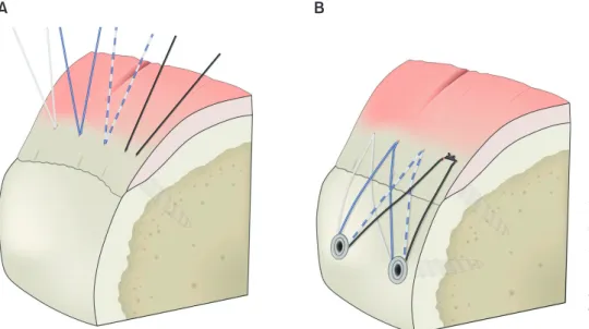
To avoid excessive strangulation in medial row, we modified the medial knot-tying method to minimal-tying technique. In the modified technique, just one knot was made in the most anterior region of the supraspinatus (the most tendinous portion of the supraspinatus; Fig. 1, 2) to minimize the number of knot- tying in medial row. After minimal-tying, we applied conven- tional suture bridge repair using 4 suture limbs for each knotless suture anchor (total 2 knotless suture anchors), finally making for 4×4 repair configuration.
The purpose of this study was to report on the repair integrity of modified suture bridge technique for patients with medium and large size full-thickness supra-infraspinatus tendon tears. We evaluate the postoperative magnetic resonance imaging (MRI) to investigate retear rate and mode of failure. We hypothesized that the minimal tying technique would result in equivalent or lower retear rate compared with previously published results af- ter conventional suture-bridge technique.
Methods
Permission was obtained from the Institutional Review Board of Samsung Medical Center before conduct of this retrospective review (IRB File No.2013-06-009-001). Inclusion criterion was patients with medium or large size rotator cuff tear (usually 2 to 4 cm anteroposterior width of supra/infraspinatus tendon tear) covered with minimal-tying double row suture bridge technique (specifically 4×4 configuration with two medial suture anchors and two lateral knotless anchors). Medium and large size tears were specially selected since these sizes are the best candidate for the current technique. Tear size was categorized according Fig. 2. The most anterior region of supraspinatus (blue arrowed line) represents the most tout tendinous portion, which is fit to optimal fixation by single me- dial knot tying. Red curved line indicates larger muscular portion of posterior portion of supraspinatus.
Fig. 1. A schematic drawing of the minimal medial-row tie with suture-bridge technique.
(A) Total four-threads (8 suture limbs) from two suture anchors penetrated through the medial cuff. (B) Lateral-row was secured us- ing the conventional suture-bridge anchor technique; all 4 suture limbs from each thread were used for each lateral-row knotless anchor.
The most anterior suture was tied among 4 strands.
A B
to the previously reported classification; small (<1 cm), medium (1–3 cm), large (3–5 cm), and massive (>5 cm).10) Tear pat- tern was not considered important if we can repair type I or II.
Type I was complete repair with coverage of the lateral end of the greater tuberosity footprint and type II was complete repair to the medial one half or less of the footprint.11) A total of 339 shoulders were treated between March 2011 and July 2012.
Among these, 98 shoulders met with inclusion criterion. Exclu- sion criteria were as follows: 1) patients who did not undergo postoperative MRI at 6 months after surgery (16 patients); 2) Re- vision surgery (2 patients); and 3) associated avulsion fracture of greater tuberosity (1 patient); other concomitant pathology such as biceps lesion, acromion-clavicular joint arthritis, frozen shoul- der, and subscapularis tear or related procedures were not rea- sons for exclusion (Table 1). The remaining 79 shoulders in 79 patients were finally included. Forty-seven women and 32 men were affected. Fifty-four shoulders were on the right side. Domi- nant arms were involved in 56 shoulders (69%). The average age of the patients was 61.3 years (range, 31–81 years). The average duration of shoulder pain before surgery was 33.4 months (range, 3–360 months). A complete physical examination including ac- tive shoulder range of motion was performed preoperatively.
Surgical Technique
The surgical procedures were performed by a single surgeon (J.C.Y.) with the patient under general anesthesia in the lateral decubitus position. After evaluation of range of motion (ROM), gentle manipulation was performed in cases with motion limita- tion. The glenohumeral joint was then examined. A tenodesis or tenotomy of the long head of the biceps was performed if nec-
placement were determined with a spinal needle, two suture anchors were inserted for the medial row (Fig. 3A). We used two suture anchors loaded with two No. 2 synthetic suture materials in the medial row from the following manufacturers (Genesys CrossFT OC 4.5 mm [ConMed Linvatec, Largo, FL, USA], Healix BR 4.5 mm [Dupey Mitek, Norwood, MA, USA], and Twinfix Ultra HA 5.5 mm [Smith & Nephew, Andover, MA, USA]). The anterior anchor was just posterior to the biceps tendon on the humeral head before entering into the groove. The posterior anchor was roughly 1 cm from the anterior anchor. Both limbs from each of the 2 sutures were passed through the tendon us- ing the Spectrum suture shuttling device (ConMed Linvatec) or the Scorpion Needle (Arthrex, Naples, FL, USA) at 3 to 4 mm lateral from the musculotendinous junction (Fig. 3B). The first stitch was passed at the most anterior tendon of supraspinatus;
the second stitch was usually on the mid-portion of supraspina- tus (Fig. 1). The third stitch was on the most anterior portion of the infraspinatus tendinous portion. The fourth stitch was 5 to 7 mm posterior to the third stitch. Each stitch included two limbs of suture strand; therefore, finally, a total of 8 suture limbs would pierce the medial tendon (Fig. 1). After all sutures were passed, the most anterior one was tied by horizontal mattress suture.
Lateral-row was secured using the conventional suture-bridge anchor technique; all 4 suture limbs from each stitch were used for each anchor (Fig. 3C, D). The lateral fixation points for knot- less suture anchors (Poplock 3.5 mm; ConMed Linvatec) were placed 1 cm distal and lateral to the lateral edge of footprint in- sertion.
Postoperative Rehabilitation
A 30o abduction brace was kept for 5 weeks. No passive movement was allowed during that period. Passive ROM was enforced as 5 weeks of immobilization, which was continued for 6 weeks. After roughly 3 months, active strengthening exercise was permitted.
Radiographic Evaluation
Final follow-up rate of postoperative MRI was 83.1% (79 pa- tients out of 95 patients). MRI was performed using a 3.0 T MR imager (Gyroscan Intera Achieva; Philips Medical Systems, Best, Netherlands). Patients underwent postoperative MRI at average 6.2 months (range, 6.0–7.4 months) after the operation. Tendon integrity and retear rate after repair were evaluated by analyzing the oblique coronal and sagittal T2 image according to classifica-
Adhesive capsulitis 13 (16.5)
Cartilage lesion 5 (6.3)
Acromio-clavicular arthritis 15 (19.0) Associated procedures
Biceps tenotomy or tenodesis 58 (73.4) Subscapularis debridement or repair 54 (68.4)
Subacromial decompression 52 (65.8)
Coraco-acromial ligament release 70 (88.6)
Pancapsular release 10 (12.7)
Mumford procedure 1 (1.3)
tion of Sugaya.12) Briefly, type I indicates a repaired cuff with suf- ficient thickness; type II indicates sufficient thickness with a par- tial high-intensity; type III indicates insufficient thickness; type IV indicates a minor discontinuity in more than one slice; and type V indicates the presence of major discontinuity. Sugaya classifi- cation IV and V are regarded as full-thickness retear. Sugaya clas- sification was measured by two independent observers (observer 1 and 2) at two time points separated by 1 month. We resolved discrepancies between the two observers with repeated review and discussion. Retear was further documented as medial cuff failure or lateral cuff (footprint) failure to speculate mode of failure (type I and type II retear suggested by Cho et al.5)). Fatty degeneration of supraspinatus was measured using Goutallier grade.13) This grading system classified the fatty degeneration ac- cording to 5 stages; stage 0: normal muscle, stage 1: muscle with some fatty streaks is shown, stage 2: more fatty infiltration, but still more muscle than fat, stage 3: the amount between fat and muscle is similar, and stage 4: more fat is presented than muscle.
Muscle atrophy of supraspinatus was evaluated on the oblique sagittal T2-weighted images medial to the level of the coracoids process suggested by Warner et al.14) This is the 4-stage grading as normal, mild, moderate, or severe. Normal was given a score
of 0; mild, 1; moderate, 2; and severe, 3.
Statistical Analysis
Fatty infiltration and muscle atrophy before and after the op- eration was compared using Wilcoxon signed rank test. Cohen’s unweighted kappa was calculated for inter-observer and intra- observer reliability of Sugaya classification. Strength of agree- ment was poor when kappa value <0.20, fair when 0.21 to 0.4, moderate when 0.41 to 0.60, good when 0.61 to 0.8, and very good when 0.81 to 1.0.15) All statistical analyses were performed using IBM SPSS Statistics ver. 19.0 (IBM Co., Armonk, NY, USA) with α level set at 0.05.
Results
There were 65 patients with medium tear and 14 patients with large tears. Mean preoperative pain visual analogue scale score was 4.8 ± 2.2. Average American Shoulder and Elbow Surgeons and Constant-Murley shoulder score was 46.6 and 51.0, respectively. The mean preoperative active ROM of for- ward elevation was 146o (range, 60o–170o). Mean external rota- tion at side was 42o (range, 0o–70o) and median internal rotation
A B
C D
Fig. 3. (A) Two medial row suture anchors, which are double-loaded with No. 2 sutures, are placed in the medial aspect of the foot- print. (B) A stitch for the medial cuff is made via a curved suture hook and shuttled relay. (C) After the most anterior knot-tying (arrow), lateral fixation was performed using a knotless suture anchor with 4 suture limbs from each thread. (D) The final results after the same step repeated for the posterior side of the lateral row.
observed in 1 patient (Fig. 4). Detailed information of patients with retear is shown in Table 3.
Fatty infiltration of supraspinatus according to Goutallier grad- ing was improved in 24 patients, worsened in 6 patients, and tied in 49 patients. This postoperative improvement was statisti-
worsened in 17 patients, and tied in 41 patients. Average preop- erative grade was 0.9 and 1.2 in patients with an intact cuff and with a retear, respectively, and these grades were not changed after surgery.
The inter-observer reliability (kappa value) of Sugaya classifi- cation was average 0.320 (fair agreement, average of the value of 4 combinations [0.349, 0.382, 0.296, and 0.256]; Table 4).
Intra-observer reliability of observer 1 was 0.517 (moderate agreement) and that of observer 2 was 0.764 (good agreement).
Discussion
To minimize strangulation in the medial row, we modified the repair technique to minimal tying method in the medial row.
Our question is to know whether our modified technique is bet- ter or at least comparable to the pre-existing standard method in the radiological aspect (in terms of tendon healing assessed by
A B C
Fig. 4. Magnetic resonance imaging of the shoulder: coronal T2-weighted image demonstrating (A) an intact healed rotator cuff after repair (B) retear at the medial portion of the cuff (medial cuff failure) (C) retear at the lateral portion of the cuff (lateral cuff failure).
Table 2. Repair Integrity after Modified Minimal Tying Suture Bridge Rotator Cuff Repair
Type* Number (%)
I 38 (48.1)
II 30 (38.0)
III 6 (7.6)
IV 4 (5.1)
V 1 (1.3)
*According to Sugaya classification.
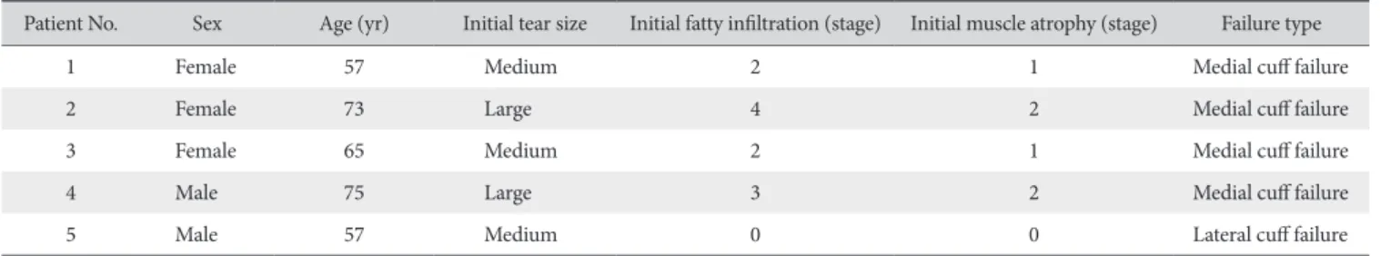
Table 3. Detailed Information of Retear Patients
Patient No. Sex Age (yr) Initial tear size Initial fatty infiltration (stage) Initial muscle atrophy (stage) Failure type
1 Female 57 Medium 2 1 Medial cuff failure
2 Female 73 Large 4 2 Medial cuff failure
3 Female 65 Medium 2 1 Medial cuff failure
4 Male 75 Large 3 2 Medial cuff failure
5 Male 57 Medium 0 0 Lateral cuff failure
postoperative MRI). The results showed a retear rate of 6.3% af- ter mean 6.2-month follow-up. We hypothesized that the mul- tiple knot-tying in the medial row would result in strangulation of the tendinous portion; thus, minimal tying would alleviate excessive strangulation and result in better healing in vivo. For a better biological environment, tying was minimized to the most anterior point. The most anterior portion was selected because the tendinous portion in the anterior supraspinatus is the most
prominent and strong to repair. Anterior third of the supraspina- tus is histologically distinct from other portions of the rotator cuff and has a higher ultimate failure load than the middle and pos- terior parts.16,17) Although the mechanical strength at time zero might be weaker than multiple knot-tying methods, minimal tying methods showed relatively low retear rate, which at least proved that all medial row ties were not necessary.
Significant retear rate (25%–90%) was reported with early arthroscopic rotator cuff repair, either by single row or double row.18) Recent suture bridge technique had generally reduced the retear rate compared with more conventional tech- nique.4,6,8,9,19,20) Many authors reported the retear rate from 8%
to 33% (Table 5).4,6,8,9,19-22) Most techniques utilized 2 to 4 medial knot-tying in the medial row and obtained an approximately 20% retear rate, which is still high.23-25) In the current series of 79 patients, only 6.3% of patients experienced retear on 6.2-month follow-up MRI. This finding suggested that our methods are at Table 4. Inter-observer Reliability of Sugaya Classification (Kappa Value)
Observer 2
First measurement Second measurement Observer 1
First measurement 0.349 0.382
Second measurement 0.296 0.256
Fig. 5. Assessment of fatty infiltration (A) and muscle atrophy (B) at two time points (preoperative vs. postoperative).
A
Preoperative Postoperative
No.ofpatients
Grade 0 50
45 40 35 30 25 20 15 10 5 0
Fatty infiltration
Grade 1 Grade 2 Grade 3 Grade 4
No.ofpatients
None 60
50
40
30
20
10
0
Muscle atrophy
Mild Moderate Severe
4 7
21 37
44
21
7 12
5 0
19 25
49
40
10
1 4 10
Preoperative Postoperative
B
Goutallier grading Table 5. Reported Failure Rates after Suture Bridge Repair
Study (year) No. of case Tear size Timing of
evaluation (mo) Technique No. of knot tying
in medial row* Failure rate (%)
Frank et al. (2008)21) 25 ND 15 Conventional SB 2 12
Voigt et al. (2010)9) 51 Isolated supraspinatus tear 12 Conventional SB 2 29
Park et al. (2010)20) 50 Medium to large† 12 Conventional SB NS‡ 8
Cho et al. (2011)4) 123 Small to massive 9 Conventional SB 2§ 33
Choi et al. (2012)19) 34 Medium to large† 28 Double pulley SB 3 18
Rhee et al. (2012)8) 51 ND 7 Conventional SB 2 19
Gerhardt et al. (2012)6) 20 Medium to large 23 Double mattress SB 4 25
Kim et al. (2013)22) 26 Medium to large 28 Conventional SB 2 20
ND: not documented, SB: suture bridge.
*This number of knot tying in medial row could be applicable when two suture anchors were used. †The result of medium and large size was specifically extracted from the original article. ‡Medial knots were tied, but the exact number was not specified in the original article. §The number of knots was speculated with the figure, since the exact number was not specified in the original article.
Although the relationship between the number of medial knot-tying and the retear rate is not clear, concern about strangu- lation has already been hypothesized in many literature studies after observation of medial cuff failure.4-6) There was also another attempt to reduce strangulation of medial tendon by techni- cal modification. Mason-Allen configuration was used instead of knot-tying in medial row by Rhee et al.,8) and the retear rate was significantly lower (5.9% from 51 shoulders) compared with knot-tying suture bridge technique (18.6% from 59 shoulders).
Taken together with the current study, it seems that the minimal medial row tying technique could enhance the healing of rotator cuff tendon.
Knot-tying method has been known to be stronger than non- tying methods in the biomechanical aspect, especially in time zero.2,7) Systematic review indicated that the time zero strength of knot-tying was superior to non-tying method. However, biomechanical or structural advantages do not always translate into a superior clinical outcome.26) Despite weaker time zero strength, the current results showed a relatively low retear rate, suggesting the importance of the in vivo healing process. The additional advantage of the current technique along with the reduction of strangulation is technical simplicity. For example, Mason-Allen modification is a more time-consuming procedure.
Our technique is relatively simple and straightforward with re- duced operation time.
From this analysis, we also wanted to know where the weak point of the repair is in minimal tying method. Retear may result from suture cutting through the tendon, eyelet breakage, anchor loosening, suture material breakage or surgical knot loosening.2) Because of the introduction of stronger suture anchor and suture materials, the weakest point of repair has shifted to suture-ten- don interface. The failure of suture-tendon interface could occur in medial musculotendinous junction or footprint in double row repair.27) The incidence of lateral cuff failure (footprint detach- ments) was reduced with introduction of the suture bridge technique. Previous studies reported that retear in conventional suture bridge repair had occurred at musculotendinous junction in 46%, 66.7%, and 74.1% of cases, respectively.5,9,28) Because we suspected that medial cuff failure originated in part from medial knot-tying, we expected that the main mode of failure in the modified technique would be other than medial cuff tears.
However, despite minimal knot-tying, the dominant failure mode was still medial cuff failure (80% in the current study). Pos- sible explanation would be that even though we did a minimum
muscle atrophy of supraspinatus, suggesting a technical failure or bad compliance of patients during rehabilitation as a reason of failure.
Fatty infiltration and muscular atrophy of supraspinatus or in- fraspinatus was known as one of the factors determining the fate of repair integrity and functional outcome.13) We also observed that the average grade of Goutallier fatty infiltration and muscu- lar atrophy was slightly higher in the retear group, preoperatively (2.2 vs. 1.9 and 1.2 vs. 0.9, respectively). However, because the number of retear patients was small (just five patients) in the current study, we could not attempt a detailed factor analysis to lead to conclusive findings. The reversibility of supraspinatus muscle atrophy after surgery is also a matter of debate. Both atrophy and fatty infiltration were known to have progressed over the course of the study after the operation.29) On the con- trary, there were also reports that showed minimal progression in healed repairs or even improvement in muscle atrophy23) and fatty infiltration.13) Our results showed that fatty infiltration was minimally improved after repair; however, muscle atrophy was unchanged even if repair was successful. A recent study reported that muscle atrophy measured with occupation ratio was revers- ible.30) They showed that 42.4% of patients had improved and 17.3% had worsened at minimum 1-year follow-up, while in the current study, 26.6% and 21.5% of patients showed improve- ment and worsening, respectively. They concluded that muscle atrophy could be improved in cases of successful rotator cuff repair. In the current study population, most patients had nor- mal or minimal muscular atrophy in the preoperative situation, which might be one of the reasons that little change was shown after surgery.
There are several limitations in this study. First, this is a pre- liminary review to assess rotator cuff repair site integrity after case series and thus the inherent weakness of level IV study ex- ists. The use of historical data for controls is another weakness.
Clinical results such as pain, strength, and function should be evaluated and compared with other techniques in subsequent study. Direct comparison between knot-tying and minimal ty- ing methods could provide more direct answers on the primary question. Second, the follow-up period for MRI might be too short to observe the overall retear rate. However, it was suffi- cient time to evaluate initial healing of repaired tendon. Multiple evaluations with longer follow-up might be more informative, although the cost is a problem. Serial evaluation with ultrasonog- raphy might be a good alternative.20) One recent study reported
that all retears occurred prior to 6 months after surgery in serial ultrasound follow-up up to 24 months, with a 41% retear rate.9) Another author also reported a high frequency of retear within the first 6 months (75.0% [6 of 8]), justifying the evaluation at 6 months.19)
Conclusion
Minimal tying technique with suture configuration of four-by- four strand double-row suture-bridge yielded a lower retear rate (6.3%) in medium to large rotator cuff tears.
Acknowledgements
The author would like to thank Multimedia department of Samsung Medical Center for providing us with medical illustra- tion.
References
1. Burkhart SS, Cole BJ. Bridging self-reinforcing double-row rota- tor cuff repair: we really are doing better. Arthroscopy. 2010;
26(5):677-80.
2. Pauly S, Kieser B, Schill A, Gerhardt C, Scheibel M. Biome- chanical comparison of 4 double-row suture-bridging rotator cuff repair techniques using different medial-row configura- tions. Arthroscopy. 2010;26(10):1281-8.
3. Park MC, Elattrache NS, Ahmad CS, Tibone JE. “Transosseous- equivalent” rotator cuff repair technique. Arthroscopy. 2006;
22(12):1360.e1-5.
4. Cho NS, Lee BG, Rhee YG. Arthroscopic rotator cuff repair using a suture bridge technique: is the repair integrity actually maintained? Am J Sports Med. 2011;39(10):2108-16.
5. Cho NS, Yi JW, Lee BG, Rhee YG. Retear patterns after ar- throscopic rotator cuff repair: single-row versus suture bridge technique. Am J Sports Med. 2010;38(4):664-71.
6. Gerhardt C, Hug K, Pauly S, Marnitz T, Scheibel M. Arthro- scopic single-row modified mason-allen repair versus double- row suture bridge reconstruction for supraspinatus tendon tears: a matched-pair analysis. Am J Sports Med. 2012;40(12):
2777-85.
7. Mall NA, Lee AS, Chahal J, et al. Transosseous-equivalent rotator cuff repair: a systematic review on the biomechani- cal importance of tying the medial row. Arthroscopy. 2013;
29(2):377-86.
8. Rhee YG, Cho NS, Parke CS. Arthroscopic rotator cuff re- pair using modified Mason-Allen medial row stitch: knotless versus knot-tying suture bridge technique. Am J Sports Med.
2012;40(11):2440-7.
9. Voigt C, Bosse C, Vosshenrich R, Schulz AP, Lill H. Arthroscop- ic supraspinatus tendon repair with suture-bridging technique:
functional outcome and magnetic resonance imaging. Am J Sports Med. 2010;38(5):983-91.
10. DeOrio JK, Cofield RH. Results of a second attempt at surgi- cal repair of a failed initial rotator-cuff repair. J Bone Joint Surg Am. 1984;66(4):563-7.
11. Yoo JC, Ahn JH, Yang JH, Koh KH, Choi SH, Yoon YC. Correla- tion of arthroscopic repairability of large to massive rotator cuff tears with preoperative magnetic resonance imaging scans.
Arthroscopy. 2009;25(6):573-82.
12. Sugaya H, Maeda K, Matsuki K, Moriishi J. Functional and structural outcome after arthroscopic full-thickness rotator cuff repair: single-row versus dual-row fixation. Arthroscopy. 2005;
21(11):1307-16.
13. Goutallier D, Postel JM, Gleyze P, Leguilloux P, Van Driessche S.
Influence of cuff muscle fatty degeneration on anatomic and functional outcomes after simple suture of full-thickness tears.
J Shoulder Elbow Surg. 2003;12(6):550-4.
14. Warner JJ, Higgins L, Parsons IM 4th, Dowdy P. Diagnosis and treatment of anterosuperior rotator cuff tears. J Shoulder Elbow Surg. 2001;10(1):37-46.
15. Landis JR, Koch GG. The measurement of observer agreement for categorical data. Biometrics. 1977;33(1):159-74.
16. Itoi E, Berglund LJ, Grabowski JJ, et al. Tensile properties of the supraspinatus tendon. J Orthop Res. 1995;13(4):578-84.
17. Lake SP, Miller KS, Elliott DM, Soslowsky LJ. Effect of fiber distribution and realignment on the nonlinear and inho- mogeneous mechanical properties of human supraspinatus tendon under longitudinal tensile loading. J Orthop Res.
2009;27(12):1596-602.
18. Cummins CA, Murrell GA. Mode of failure for rotator cuff re- pair with suture anchors identified at revision surgery. J Shoul- der Elbow Surg. 2003;12(2):128-33.
19. Choi CH, Kim SK, Cho MR, et al. Functional outcomes and structural integrity after double-pulley suture bridge rotator cuff repair using serial ultrasonographic examination. J Shoul- der Elbow Surg. 2012;21(12):1753-63.
20. Park JY, Siti HT, Keum JS, Moon SG, Oh KS. Does an ar- throscopic suture bridge technique maintain repair integrity?:
a serial evaluation by ultrasonography. Clin Orthop Relat Res.
2010;468(6):1578-87.
21. Frank JB, ElAttrache NS, Dines JS, Blackburn A, Crues J, Ti- bone JE. Repair site integrity after arthroscopic transosseous- equivalent suture-bridge rotator cuff repair. Am J Sports Med.
2008;36(8):1496-503.
22. Kim KC, Shin HD, Cha SM, Lee WY. Comparison of repair integrity and functional outcomes for 3 arthroscopic suture bridge rotator cuff repair techniques. Am J Sports Med. 2013;
41(2):271-7.
23. Gerber C, Fuchs B, Hodler J. The results of repair of massive tears of the rotator cuff. J Bone Joint Surg Am. 2000;82(4):505- 15.
Joint Surg Am. 2010;92(3):732-42.
27. Trantalis JN, Boorman RS, Pletsch K, Lo IK. Medial rotator cuff failure after arthroscopic double-row rotator cuff repair. Ar-
supraspinatus muscle atrophy truly irreversible after surgical re- pair of rotator cuff tears? Clin Orthop Surg. 2013;5(1):55-65.