Introduction
Ischemic neuropathy describes any injury of a pe- ripheral nerve caused by either vessel occlusion or reduced oxygen content in the blood supply.1 This condition may result from Extracorporeal Membrane Oxygenation (ECMO) as proximal major arteries get
occluded. Among several types of ECMO, Veno-Arterial (VA) type is frequently used to support patients with circulatory failure. VA ECMO functions by draining blood from major veins and returning it to the arterial system, commonly using femoral artery as the cannula- tion site because of its easy access.2 Although ECMO is a crucial intervention to support patients with critical illness, the number of complications tend to increase along with the number of procedures carried out.2 In previous studies, ischemic monomelic neuropathies (IMN) have been reported as a complication from the use of large size cannulas ranging from 20∼52%.3,4 However, reported cases of multiple mononeuropa-
ISSN 1229-6066 https://doi.org/10.18214/jkaem.2018.20.1.42 J Korean Assoc EMG Electrodiagn Med 20(1):42-47, 2018
J Korean Assoc
Electrodiagn MedEMG
Copyright © by Korean Association of EMG Electrodiagnostic Medicine
This is an Open Ac cess article distributed under the terms of the Creative Commons Attribution Non-Commercial License (http://creativecommons.org/licenses/by-nc/4.0) which permits unrestricted non-commercial use, distribution, and reproduction in any medium, provided the original work is properly cited.
Received January 5, 2018
Revised (1st) April 3, 2018, (2nd) May 25, 2018 Accepted May 29, 2018
Corresponding Author: Hee-Kyu Kwon
Department of Physical Medicine & Rehabilitation, Korea University Anam Hospital, Korea University College of Medicine, 73 Inchon-ro, Seongbuk- gu, Seoul 02841, Korea
Tel: 82-2-920-6471, Fax: 82-2-929-9951, E-mail: [email protected]
체외막산소공급의 합병증으로 인한 허혈성신경병증: 증례보고
맹원준, 이아리, 문현임, 권희규
고려대학교 의과대학 고려대학교안암병원 재활의학과교실
Ischemic Neuropathy Possibly as a Complication of Extracorporeal Membrane Oxygenation Management: A Case Report
Won Jun Meng, Ahry Lee, Hyun Im Moon, Hee-Kyu Kwon
Department of Physical Medicine & Rehabilitation, Korea University Anam Hospital, Korea University College of Medicine, Seoul, Korea
Ischemic neuropathy can result from either vessel occlusion or reduced oxygen content in the blood. We report a case of distal ischemic mononeuropathies involving multiple limbs that have developed after the use of Veno-Arterial Extracorporeal Membrane Oxygenation (VA ECMO). A 37-year old female with suspected acute myocarditis and postpartum cardiomyopathy has developed weakness and tingling sense in all the distal extremities after VA ECMO.
Electrodiagnostic study was performed 4 weeks after the onset of the symptoms and the findings suggested multiple mononeuropathies in all four limbs, especially in the distal components. Ischemic neuropathy in multiple limbs can develop as a consequence of generalized limb ischemia, although it is frequently overlooked.
Key Words: ischemic neuropathy, limb ischemia, extracorporeal membrane oxygenation
thies after ECMO are quite rare. In this study, we report a case of distal ischemic mononeuropathies involving multiple limbs that have occurred following VA ECMO.
Case Report
1) Clinical history
A 37-year old female was transferred from a local hospital for ECMO with suspected diagnosis of acute myocarditis and postpartum cardiomyopathy with estimated left ventricular ejection fraction of 11%
in January 2016. The patient initially complained of dyspnea, which had started about two to three days before her visit to the local hospital. She had given birth 2 months before the onset of the symptoms and had no known history of underlying diseases.
Veno-arterial (VA) ECMO was applied in our hospital via cannulations on the right femoral vein and left femoral artery. Then, the patient was referred to us for electrodiagnostic examination 4 weeks after the onset.
2) Physical examination
The patient complained of tingling sense in both lower limbs, weakness of her left foot, and decreased grasp power in the right hand. The strengths of her extremities were as follows: hip extensors and knee flexors grade 5/5 bilaterally; ankle dorsiflexors (right/left), grade 5/3; great toe extensors (right/left), grade 3/2; ankle plantar flexors, grade 3, bilaterally;
elbow flexors and extensors, grade 5, bilaterally;
finger abductors (right/left), grade 3/4. Overall, more proximal muscles were normal in strength, while distal muscles tended to be weaker. Light touch sensation was reduced in the right 1st to 4th fingers and palm.
3) Electrodiagnostic examination (1) Methods
The entire nerve conduction studies (NCS) were completed with percutaneous supramaximal stimula- tions using a hand-held stimulator with two metal elec- trodes placed 3.0 cm apart. Motor NCS were performed
with the standardized technique, recording from 1cm tin disc electrodes over the motor points (appropriate muscle belly). Sensory NCS were performed antidromi- cally, with a bar-type electrode where the reference electrode was approximately 4 cm apart from the ac- tive electrode. The needle electrode examination was performed with a standard monopolar needle elec- trode.
(2) Nerve conduction studies
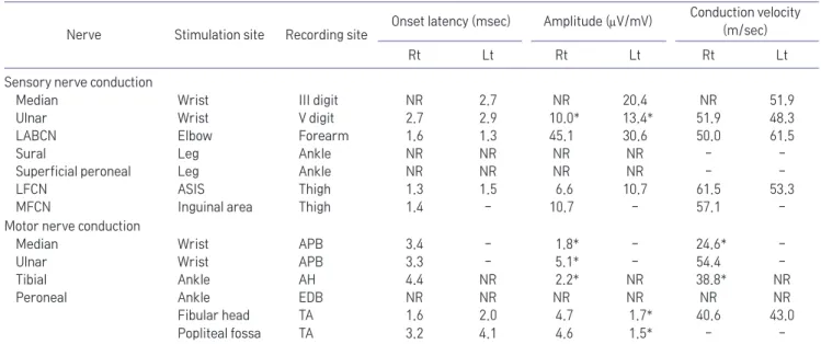
The initial motor and sensory NCS performed are shown in Table 1. Bilateral peroneal and left tibial motor as well as bilateral sural and superficial sensory responses were unobtainable. Likewise, right median and bilateral ulnar sensory responses were unobtainable or very low in amplitude. However, sensory conduction studies of the proximal limbs, such as medial and lateral femoral cutaneous nerves and lateral antebrachial cutaneous nerves, were within normal limits (Table 1).
(3) Needle electromyography
Positive sharp waves were observed in the left tibialis anterior (TA) muscle, while both fibrillation potentials and positive sharp waves were noted in the right TA, left gastrocnemius medial head (GCM) and bilateral first dorsal interossei muscles (FDI). Polyphasic motor unit potentials with reduced recruitment patterns were noted in the bilateral TA, peroneus longus, GCM and FDI muscles (Table 2).
(4) Interpretation
The findings on the NCS suggested multiple mononeuropathies in bilateral lower and upper limbs, especially in the distal components. Both lower limbs were predominantly involved and distal portions were mostly affected. Generalized limb ischemia caused by low cardiac output or poor distal perfusion due to ECMO may have accounted for the multiple neuropathies that involved distal nerves of the limbs.
(5) Follow-up study
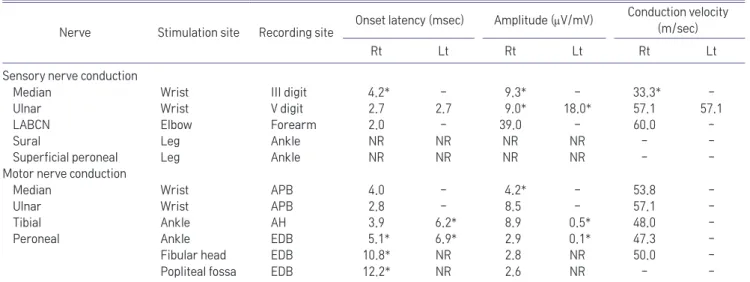
Follow-up NCS were performed in the outpatient district in July, 2017. Clinically, the patient still complained of mild tingling sense in the right second
to fourth finger, but she no longer had hypoesthesia
in the same region. Moreover, the motor power of the extremities showed further recovery as follows: ankle dorsiflexors (right/left), grade 5/4; great toe extensors (right/left), grade 4/4; ankle plantar flexors, grade 4/4;
finger abductors (right/left), grade 4/5. Only few nerves were stimulated during the NCS due to discomfort
of the exam and the patient also refused the needle EMG. The follow-up exam showed improvement in the latencies and amplitudes of several nerves;
the left tibial, right median and bilateral peroneal motor responses that were unobtainable in the initial evaluation showed delayed and small amplitude responses (Table 3).
Table 1. Nerve Conduction Studies
Nerve Stimulation site Recording site Onset latency (msec) Amplitude (mV/mV) Conduction velocity (m/sec)
Rt Lt Rt Lt Rt Lt
Sensory nerve conduction
Median Wrist III digit NR 2.7 NR 20.4 NR 51.9
Ulnar Wrist V digit 2.7 2.9 10.0* 13.4* 51.9 48.3
LABCN Elbow Forearm 1.6 1.3 45.1 30.6 50.0 61.5
Sural Leg Ankle NR NR NR NR - -
Superficial peroneal Leg Ankle NR NR NR NR - -
LFCN ASIS Thigh 1.3 1.5 6.6 10.7 61.5 53.3
MFCN Inguinal area Thigh 1.4 - 10.7 - 57.1 -
Motor nerve conduction
Median Wrist APB 3.4 - 1.8* - 24.6* -
Ulnar Wrist APB 3.3 - 5.1* - 54.4 -
Tibial Ankle AH 4.4 NR 2.2* NR 38.8* NR
Peroneal Ankle EDB NR NR NR NR NR NR
Fibular head Popliteal fossa
TA TA
1.6 3.2
2.0 4.1
4.7 4.6
1.7*
1.5*
40.6 -
43.0 - LABCN: lateral antebrachial cutaneous nerve, LFCN: lateral femoral cutaneous nerve, MFCN: medial femoral cutaneous nerve, ASIS: anterior superior iliac spine, APB: abductor pollicis brevis, ADQ: abductor digiti quintii, AH: abductor hallucis, EDB: extensor digitorum brevis, TA:
tibialis anterior, NR: no response, -: not applicable Abnormal values are represented with asterisk
Table 2. Needle Electromyography
Side Muscle Insertional activity Spontaneous activity Motor unit action potentials Interference pattern
Left Vastus lateralis Normal - Normal Normal
Tibialis anterior - P(+) Polys Reduced
Peroneus longus Normal - Polys Reduced
Gastrocnemius - F&P(++) Polys Reduced
Gluteus maximus Normal - Normal Normal
Tensor fascia lata Biceps femoris (long)
Normal Normal
- -
Normal Normal
Normal Normal
First dorsal interossei - F&P(++) Polys Reduced
Right Vastus lateralis Normal - Normal Normal
Tibialis anterior - F&P(++) Polys Reduced
Peroneus longus Normal - Polys Reduced
Gastrocnemius Normal - Polys Reduced
Biceps femoris (long) Normal - Normal Normal
Triceps brachii Normal - Normal Normal
Pronator teres Normal - Normal Normal
First dorsal interossei - F&P(++) Polys Reduced
F: fibrillation potentials, P: positive sharp waves, -: not applicable
Discussion
As mentioned earlier, ECMO is essential in intensive care unit, sustaining many patients without proper cardiac or respiratory function. The annual number of patients being supported by ECMO is increasing as improvement and fine-tuning of the method pull up the successful weaning ratio as high as 62% after 79
± 57 hours of ECMO support.5,6 VA ECMO requires cannulation of a large artery to deliver oxygenated blood and conventionally carotid artery had been used as the site of insertion for the arterial cannula.
However, distal ligation of the carotid artery could interrupt blood flow to the brain and may increase the risk of stroke if collateral circulation is not sufficient.5 Therefore, common femoral artery (CFA) is often used for ECMO in the current trend to minimize the potential neurologic insult of carotid artery ligation.2,6 Although this decreases the neurologic complications of arterial cannula placement, it may obstruct forward flow to the limbs, placing them at risk for ischemia. In fact, the true incidence of limb ischemia may be underestimated as there are high mortality rates in patients that receive ECMO after cardiac arrest.6
Ischemic neuropathies involving both upper and lower extremities were rarely reported. The abnormal electrodiagnostic findings of our case in both upper and lower extremities, especially in the distal recording sites, suggested possible distal ischemic neuropathy of the limbs. The NCS findings presented a form of axonal loss mainly involving distal portions of the nerves rather than demyelination pattern. Although the abnormal findings of our case were not confined to a single limb, motor and sensory axon loss in distal regions showed similar pattern with the reported cases of IMN.7 For example, distal portions of tibialis anterior and gastrocnemius medial head revealed fibrillation similar to IMN cases, while proximal muscles were relatively spared.
Several other types of acquired neuropathies need to be considered before reaching our conclusion. To begin with, when the patient was referred to us for EMG, the pattern of asymmetric weakness and sensory symptoms could suggest a type of vasculitic neuropathy such as vasculitic mononeuritis multiplex. However, the abnormal NCS findings were not confined to certain affected nerves but rather diffusely found over distal upper and lower motor and sensory nerves. Moreover,
Table 3. Follow-up Nerve Conduction Studies
Nerve Stimulation site Recording site Onset latency (msec) Amplitude (mV/mV) Conduction velocity (m/sec)
Rt Lt Rt Lt Rt Lt
Sensory nerve conduction
Median Wrist III digit 4.2* - 9.3* - 33.3* -
Ulnar Wrist V digit 2.7 2.7 9.0* 18.0* 57.1 57.1
LABCN Elbow Forearm 2.0 - 39.0 - 60.0 -
Sural Leg Ankle NR NR NR NR - -
Superficial peroneal Leg Ankle NR NR NR NR - -
Motor nerve conduction
Median Wrist APB 4.0 - 4.2* - 53.8 -
Ulnar Wrist APB 2.8 - 8.5 - 57.1 -
Tibial Ankle AH 3.9 6.2* 8.9 0.5* 48.0 -
Peroneal Ankle EDB 5.1* 6.9* 2.9 0.1* 47.3 -
Fibular head Popliteal fossa
EDB EDB
10.8*
12.2*
NR NR
2.8 2.6
NR NR
50.0 -
- - LABCN: lateral antebrachial cutaneous nerve, LFCN: lateral femoral cutaneous nerve, MFCN: medial femoral cutaneous nerve, ASIS: anterior superior iliac spine, APB: abductor pollicis brevis, ADQ: abductor digiti quintii, AH: abductor hallucis, EDB: extensor digitorum brevis, NR: no response, -: not applicable
Abnormal values are represented with asterisk
the lab data showed negative perinuclear and
cytoplasmic anti-neutrophil cytoplasmic antibodies (p-ANCA, c-ANCA) which are fairly sensitive and specific in detecting systemic vasculitis. Nevertheless, it is difficult to completely differentiate between vasculitic mononeuritis multiplex and ischemic neuropathy based solely on EMG findings and ANCA results. Unfortunately, further evaluations such as nerve biopsies and immunocytochemistry analysis have not been carried out to support further diagnosis.
Additionally, electrodiagnostic study results could propose the possibility of Guillain Barre Syndrome (GBS) variant such as acute motor and sensory axonal neuropathy (AMSAN). Markedly reduced or absent CMAPs and SNAPs were noted in the NCS findings suggesting axonal injuries of both motor and sensory nerves as in AMSAN. Nonetheless, our NCS findings as well as clinical symptoms were not symmetric on both sides and abnormal spontaneous activities in needle electromyography were not as abundant as they should be in most AMSAN cases. Therefore, despite the fact that the NCS findings in this study involved multiple limbs in both motor and sensory nerves, the results do not strongly indicate AMSAN. Again, absence of additional workups such as cerebrospinal fluid exam and nerve biopsy is a limitation in this study.
Other than the mentioned disease entities, critical illness polyneuropathy (CIP) could be considered as another cause for clinical weakness of the patient since she was treated in Intensive Care Unit (ICU) for nearly ten days. However, the patient was not in septic condition; alert mentality with stable vital signs and absence of multi organ failure were noted throughout the ICU care. Although clinical or electrophysiological abnormalities for CIP are not fully established in the literature, septic conditions are the inclusion criteria for diagnosing CIP.8 Moreover, as previous studies have suggested, abnormal prolongation of CMAP duration and low amplitude occur preferentially with proximal stimulation in CIP which was not compatible with our findings.9 Our case demonstrates similar pattern but
different severity in clinical and electrophysiological findings in four extremities which implies that the pathophysiologic mechanism are likely due to poor perfusion in each extremity rather than from systemic effects of CIP. Further NCS of other extremities might have strengthened the results but the number of stimulations had to be minimized due to patient’s poor cooperation. Accordingly, only the similar pattern of neuropathy was confirmed by examining SNAPs in proximal and distal parts of extremities. The originality of our work lies in its evaluation of the possibility of axonopathy that involves multiple limbs. As ECMO is becoming more in demand by critical care medicine, one must be well aware of such outcome that may ultimately result in more severe damage in patients prone to ischemia.
References
1. Wilbourn AJ, Furlan AJ, Hulley W, Ruschhaupt W: Ischemic monomelic neuropathy. Neurology 1983: 33: 447-451 2. Lafç G, Budak AB, Yener AÜ, Cicek OF: Use of extracorporeal
membrane oxygenation in adults. Heart Lung Circ 2014: 23:
10-23
3. Ko WJ, Lin CY, Chen RJ, Wang SS, Lin FY, Chen YS: Extra- corporeal membrane oxygenation support for adult postcar- diotomy cardiogenic shock. Ann Thorac Surg 2002: 73: 538- 545
4. Smedira NG, Moazami N, Golding CM, McCarthy PM, Apper- son-Hansen C, Blackstone EH, et al: Clinical experience with 202 adults receiving extracorporeal membrane oxygenation for cardiac failure: survival at five years. J Thorac Cardiovasc Surg 2001: 122: 92-102
5. Gander JW, Fisher JC, Reichstein AR, Gross ER, Aspelund G, Middlesworth W, et al: Limb ischemia after common femoral artery cannulation for venoarterial extracorporeal mem- brane oxygenation: an unresolved problem. J Pediatr Surg 2010: 45: 2136-2140
6. Slottosch I, Liakopoulos O, Kuhn E, Deppe AC, Scherner M, Madershahian N, et al: Outcomes after peripheral extracor- poreal membrane oxygenation therapy for postcardiotomy cardiogenic shock: a single-center experience. J Surg Res 2013: 181: e47-e55
7. Levin KH: AAEE case report #19: ischemic monomelic neu-
ropathy. Muscle Nerve 1989: 12: 791-795
8. Latronico N, Bolton CF: Critical illness polyneuropathy and myopathy: a major cause of muscle weakness and paralysis.
Lancet Neurol 2011: 10: 931-941
9. Kramer CL, Boon AJ, Harper CM, Goodman BP: Compound muscle action potential duration in critical illness neuromy- opathy. Muscle Nerve 2018: 57: 395-400