A Case Treated with Extracorporeal Membrane Oxygenation for Disseminated Cytomegalovirus Infection after Liver Transplantation
Jimyung Park, M.D.
1, Yoon Hyun Lee, M.D.
2, Young Rok Choi, M.D.
2, Yeon Joo Lee, M.D.
1, Dong Jung Kim, M.D.
3, Sanghoon Jheon, M.D.
3and Young-Jae Cho, M.D.
1Division of Pulmonary and Critical Care Medicine, Department of Internal Medicine
1, Departments of Surgery
2, Thoracic and Cardiovascular Surgery
3, Seoul National University Bundang Hospital,
Seoul National University College of Medicine, Seongnam, Korea
Cytomegalovirus (CMV) is a clinically important pathogen in immunocompromised patients, especially after organ transplantation. However, there have been several reports of severe CMV infections in immunocompetent patients. This report presents a case of an immunocompetent patient who presented with fulminant hepatitis requiring liver transplantation. Because CMV was detected upon histopathologic review of the explanted liver, it was later assumed that CMV may be the primary cause of hepatitis. However, at the time of transplantation, we did not suspect CMV hepatitis. Following transplantation and initiation of immunosuppression, the patient developed viral sepsis with a disseminated CMV infection. Respiratory failure because of CMV pneumonia worsened despite antiviral therapy, and venovenous extracorporeal membrane oxygenation (ECMO) was initiated.
Although ECMO has been traditionally contraindicated in patients with sepsis, this patient recovered and was successfully weaned off ECMO. CMV should be included in the differential diagnosis of fulminant hepatitis, even in immunocompetent patients, espe- cially when liver transplantation is considered.
Key Words: Cytomegalovirus, Extracorporeal membrane oxygenation, Liver transplantation, Sepsis
중심 단어: 거대세포바이러스, 체외막산소공급, 간이식, 패혈증Received September 13, 2016 Revised May 14, 2017 Accepted May 23, 2017
Corresponding author: Young-Jae Cho
Division of Pulmonary and Critical Care Medicine, Department of Internal Medicine, Seoul National University Bundang Hospital, Seoul National University College of Medicine, 82 Gumi-ro 173beon-gil, Bundang-gu, Seongnam 13620, Korea
Tel: 82-31-787-7058, Fax: 82-31-787-4051 E-mail: [email protected]
INTRODUCTION
Cytomegalovirus (CMV) is an important pathogen that can cause severe disease in immunocompromised patients and it is the single most common viral pathogen complicat- ing organ transplantation. In immunocompromised patients, CMV can cause a wide variety of diseases depending on the organ involved, including esophagitis, enterocolitis, hepatitis,
pneumonia, retinitis, and encephalitis(1).
In contrast, in immunocompetent patients, CMV infection usually presents as a benign, self-limited disease. It most commonly manifests as a heterophile antibody-negative mononucleosis syndrome. However, there have been several reports of severe CMV infection in immunocompetent pa- tients, among which involvement of gastrointestinal tract and central nervous system were the most common(2).
Abnormal liver function tests (LFTs) are frequently asso- ciated with a self-limited CMV infection but fulminant CMV hepatitis is rare(3).
As cell-mediated immunity plays a critical role in the host defense against CMV, the clinical manifestations of CMV infection may worsen if immunosuppressive agents are pre- scribed without the proper diagnosis and treatment of CMV.
Here we report a case of fulminant hepatitis, initially of un-
known origin, requiring liver transplantation. After trans- plantation and histopathologic review of the explanted liver, it was assumed that CMV may be the primary cause of hepatitis. In fact, CMV was not diagnosed before trans- plantation and immunosuppression was initiated without an- tiviral therapy. Consequently, the virus disseminated to the lung causing severe respiratory failure.
Extracorporeal membrane oxygenation (ECMO) is a life support technology that is used for respiratory or cardiac failure refractory to conventional treatment(4). Owing to rapid technical improvements, the use of ECMO has been dramatically increasing over the last decade(5). ECMO was traditionally contraindicated in cases of sepsis or severe in- fection due to the concern for infecting the circuit(6,7). It is still controversial as to whether ECMO can be useful in septic conditions and data on the clinical outcomes of ECMO in viral sepsis, including CMV infection, are espe- cially scarce. Currently, there are no reports on ECMO for CMV infection. In fact, there is mounting evidence that CMV infection leads to a hypercoagulable state and un- provoked thrombosis, which can be problematic during ex- tracorporeal therapy(8).
Although there was limited evidence of its effect, we ini- tiated ECMO in this patient because her respiratory failure progressed to a refractory state, despite the use of antiviral therapy and mechanical ventilation. The patient recovered well and was successfully weaned off ECMO. This case em- phasizes the importance of considering CMV as a cause of fulminant hepatitis when other common etiologies of hep- atitis have been ruled out. In addition, this case proposes a possible role of ECMO in treating severe viral pneumonia in an immunocompromised patient.
CASE REPORT
A 56-year-old woman, without previous liver disease, was admitted for jaundice, which had developed 3 weeks prior to admission. She had been on medication for hypertension and dyslipidemia for years, but denied any history of ex- posure to other new drugs, including herbal medications.
Initial laboratory results revealed abnormal LFTs; the levels of aspartate transaminase, alanine transaminase, and bilirubin were 1,463 IU/L, 801 IU/L, and 38.2 mg/dL, respectively,
and her international normalized ratio for prothrombin time was 2.20. Serological test results for hepatitis A, B, and C virus were all negative. The test results for autoimmune an- tibodies, including antinuclear antibody and antimitochon- drial antibody, were also negative. To determine the cause of hepatitis, a percutaneous liver biopsy was performed and the histopathology showed portal inflammatory cell infiltra- tion and bile duct damage, which are consistent with an acute hepatitis. However, no specific etiology was identified.
In spite of conservative management for acute hepatitis, her liver function showed no improvement. In spite of the lack of clinical evidence, the possibility of autoimmune hep- atitis or toxic hepatitis was considered and high-dose corti- costeroids were administered, but there was no response.
Hepatorenal syndrome rapidly progressed and symptoms of hepatic encephalopathy started to appear. Her urine output decreased, followed by the development of a metabolic acidosis. The hepatic encephalopathy worsened and the pa- tient progressed into a comatose state. At 3 weeks after ad- mission, she was transferred to the medical intensive care unit (ICU) and continuous renal replacement therapy (CRRT) was initiated. At the same time, she was registered for de- ceased donor liver transplantation and placed on the waiting list of candidates.
On the 2nd day of her ICU admission, a matched de- ceased donor was found in the Korean Network for Organ Sharing and the patient underwent liver transplantation.
After transplantation, LFT abnormality and azotemia rap- idly improved to near normal ranges. Her mental status re- covered to an alert state. CRRT was stopped and she was successfully extubated on postoperative day (POD) 2.
For immunosuppression, high doses of corticosteroids (initially 200 mg/day and tapered to 20 mg/day), tacrolimus (initially 2 mg/day and adjusted to target blood trough con- centration around 10 ng/mL), mycophenolate mofetil (1,000 mg/day), and basiliximab (20 mg on the day of trans- plantation, followed by a second dose 4 days after trans- plantation) were administered according to the protocol at our center.
Results of pre-transplantation serological test showed that both recipient and donor were seropositive for CMV. In such cases, our center uses preemptive therapy with serial follow-up with a CMV antigenemia assay (every 2 weeks
Fig. 1. Bilateral lung opacities deve- loped after liver transplantation. (A) As shown in the chest computed tomography image, bilateral consoli- dation was initially seen predomi- nantly in both the upper lobes (pos- toperative day [POD] 5). (B) However, it progressed to involve the entire lung field (POD 7). (C) A simple chest radiograph taken after the initiation of extracorporeal membrane oxyge- nation (ECMO) showing that most of the lung was non-aerated (POD 10). (D) A chest radiograph taken 3 months after weaning off ECMO shows dramatic improvement (POD 120).
until 3 months after transplantation)(9). We typically con- sider antiviral therapy for CMV when the CMV anti- genemia assay shows over 10 infected cells per 200,000 leu- kocytes in the peripheral blood. For prophylaxis of
Pneu- mocystis jirovecii
and fungal infection, we prescribed trime- thoprim/sulfamethoxazole and fluconazole, respectively.On POD 5, she had an acute onset of fever with bilateral lung opacities on chest radiographs (Fig. 1A) and required oxygen therapy for desaturations. As hypoxic respiratory failure developed, LFT abnormalities developed again with a pattern consistent with hepatocellular injury: serum aspar- tate transaminase and alanine transaminase levels were ele- vated up to 801 and 343 IU/L, respectively. Her fever per- sisted and her blood pressure began to drop, requiring nor- epinephrine and dopamine infusion. Blood cultures were ob- tained and we empirically prescribed broad spectrum anti- biotics. However, no microorganism was identified on blood culture. Although CMV antigenemia assay is typically per- formed after 2 weeks of transplantation at our center, it was performed earlier than planned for this patient in order to
rule out a CMV infection.
On POD 7, since there was no response to empirical anti- biotic therapy, we performed a biopsy of the transplanted liver in order to identify the cause of the newly deteriorat- ing liver function. A type 1 respiratory failure also rapidly progressed with no improvement in chest radiographs (Fig.
1B) and mechanical ventilation was restarted. After in- tubation, a bronchoscopy was performed in order to search for infectious causes of respiratory failure. We examined the bronchoalveolar lavage (BAL) fluid for several mic- roorganisms. On that day, the result of CMV antigenemia assay showed 235 infected cells per 200,000 leukocytes in the peripheral blood and we started an induction dose of ganciclovir (5 mg/kg every 12 hours).
The liver biopsy specimen, which was obtained on POD 7, showed multifocal necrosis with some hepatocytic intra- nuclear cytomegaloviral inclusions and an immunohisto- chemical staining for CMV was positive (Fig. 2). From the BAL fluid, polymerase chain reaction for CMV and CMV culture were found to be positive as well. We did not per-
Fig. 2. (A) Histopathology of a liver biopsy specimen, which was obtained 7 days after transplantation, showing the microabscess with hepatocytic intranuclear cytomegaloviral inclusion (HE stain, ×400). (B) Immunohistochemical staining for cytomegalovirus is positive (×400).
form a transbronchial lung biopsy because the patient’s ven- tilatory status was unstable. Other than CMV, neither bac- teria nor fungus was identified on the examination of the BAL fluid. Based on the results of the above tests, we diag- nosed the patient with disseminated CMV infection involv- ing the lung and liver(10).
Although ganciclovir was started, there was no immediate clinical improvement. Pneumonia did not subside rapidly and hypoxia was not corrected on the mechanical ventilator.
Hypoxia was refractory to all attempted therapies including lung-protective ventilation, high positive end-expiratory pressure (PEEP), and inhaled nitric oxide. Arterial blood gas analysis showed an arterial partial oxygen pressure (PaO2) of 55 mmHg with a fraction of inspired oxygen (FiO2) of 100%, PEEP of 12 cm H2O, and inhalation of 20-ppm nitric oxide. Although she was hypotensive, echo- cardiography showed preserved cardiac function with an ejection fraction of 63%. Therefore, we decided to initiate venovenous ECMO for refractory respiratory failure on POD 10.
Two 21-French venous cannulas (Bio-Medicus, Medtronic Inc., Minneapolis, MN, USA) were placed into the right and left femoral veins for inflow and outflow, respectively (Fig.
1C). We used the PLS system (MAQUET GmbH, Rastatt, Germany). The procedure was performed at the bedside in the ICU under maximal sterile barrier precautions. ECMO
was initiated with a circuit flow of 4.5 L/min and a sweep gas flow of 3 L/min of oxygen (100% FiO2). After ECMO initiation, the ventilator settings were changed to the mini- mum levels. The PEEP was 10 cm H2O and the peak in- spiratory pressure was 25 cm H2O, resulting in a tidal vol- ume ranging from 150 to 200 mL. The FiO2 setting of the ventilator was still maintained at 100% because of the hypoxia. Resultant arterial blood gas analysis revealed a PaO2 of 75 mmHg and arterial partial carbon dioxide pres- sure (PaCO2) of 42 mmHg. Her oxygen saturation remained in the range of 90% to 95% as estimated using pulse oxi- metry. We started anticoagulation therapy with heparin, but stopped after 5 days because of a prolonged prothrombin time with LFT aggravation. Subsequently, we maintained ECMO without any anticoagulation.
We continued the administration of ganciclovir and mini- mized the level of immunosuppression. On POD 16, 6 days after the initiation of ECMO, a negative result on the CMV antigenemia assay was achieved. Her radiographic findings and LFTs also began to improve. On POD 23, 13 days after ECMO, given the dramatic improvement in her radiographic findings and oxygenation status, we decided to wean her off ECMO. We changed the sweep gas flow to zero and the pa- tient’s oxygenation status was well maintained with mechan- ical ventilator support only (FiO2 70%, PEEP 7 cm H2O, peak inspiratory pressure 23 cm H2O). She was successfully
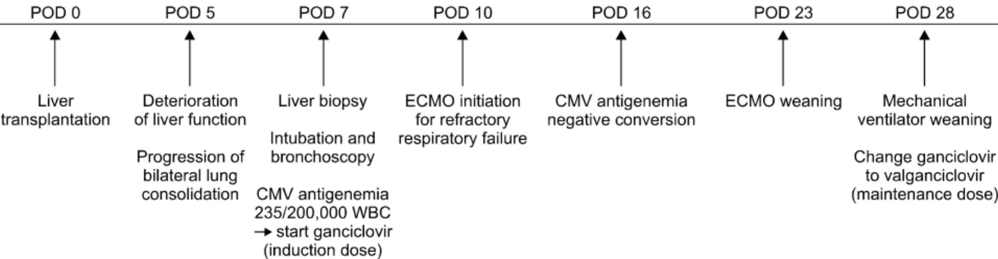
Fig. 3. A timeline of clinically important diagnostic and therapeutic procedures. Abbreviations: POD, postoperative day; CMV, cytomegalovirus; WBC, white blood cell; ECMO, extracorporeal membrane oxygenation.
weaned off ECMO, and on POD 28, 5 days after weaning from ECMO, was also taken off the mechanical ventilator.
After a 3-week course of an induction dose of ganciclovir, antiviral therapy was changed to a maintenance dose of val- ganciclovir (900 mg daily). She was discharged and the maintenance antiviral therapy was continued for 3 months.
Eighteen months after transplantation, she was doing well with no rejection. Follow-up radiographs showed no defini- tive sequelae (Fig. 1D).
After the patient recovered, we investigated the explanted liver again to elucidate the primary cause of initial ful- minant hepatitis. The histopathology showed submassive hepatic necrosis and immunohistochemistry revealed a few cells that were positive for CMV. Therefore, in this case, it can be assumed that the delayed diagnosis of the initial CMV hepatitis and liver transplantation without proper anti- viral therapy resulted in CMV dissemination. A brief time- line of the clinically important diagnostic and therapeutic procedures is shown in Fig. 3.
DISCUSSION
CMV is an important pathogen in immunocompromised patients and critically ill patients. It is also the single most common viral pathogen complicating organ transplantation, including liver transplantation(11). However, CMV can also cause severe diseases in immunocompetent patients, as illus- trated by this case. In a previous systematic review about severe CMV infection in immunocompetent patients, colitis was the most common manifestation and hepatitis was rare(2). However, there have been a few reports of ful-
minant CMV hepatitis(12,13). Since CMV was only de- tected in the explanted liver, but not in the initial percuta- neous liver biopsy, it is not clear if the initial hepatic failure resulted solely from a CMV infection. Because empirical corticosteroids for a presumed autoimmune hepatitis were administered before transplantation, it might have caused a reactivation of the CMV. However, since there was no clin- ical evidence of an autoimmune hepatitis and the patient de- nied any history suspicious of toxic hepatitis, we assumed that initial culprit was CMV.
After organ transplantation, an allograft is the most com- mon site for organ involvement of CMV. For example, CMV hepatitis is the most common manifestation of a CMV infection after liver transplantation(11). However, the in- volvement of multiple organs is possible, as illustrated by this case. In this case, although the infected liver was ex- planted, immunosuppression after transplantation was thought to have resulted in the dissemination of CMV. In addition to immunosuppression, combined renal failure and cortico- steroid therapy before transplantation might have had a synergistic effect, impairing cell-mediated immunity signifi- cantly.
In this case, we used venovenous ECMO for the treatment of respiratory failure due to CMV pneumonia. Despite the increased use of ECMO in critically ill patients, there is a lack of evidence regarding whether ECMO can be useful in patients with sepsis. Historically, sepsis was considered a contraindication to ECMO(6,7). However, sepsis is a com- mon cause of acute respiratory distress syndrome and shock, requiring extracorporeal therapy. The reported outcomes of ECMO in adult patients with sepsis are variable(14-16). It
is postulated that venoarterial ECMO could be helpful in sepsis patients with severe cardiac dysfunction, but there is still minimal experience with adult patients(16). Further- more, the current data available are mostly limited to bacte- rial sepsis and there are few reports regarding ECMO in se- vere viral infection. The pathophysiology of viral sepsis is less certain than bacterial sepsis and the role of ECMO in viral sepsis is unclear yet(17).
In our case, the patient was successfully weaned from ECMO without significant complications and, as far as we know, this is the first report of the use of ECMO for the treatment of CMV pneumonia. It is well-known that CMV infection leads to a hypercoagulable state and there have been many reports of thrombosis associated with a CMV in- fection(8,18). Although the role of anticoagulation may be less important in venovenous ECMO compared to venoarte- rial ECMO, thromboembolism remains a major concern in ECMO. Therefore, in theory, CMV infection may not be a good indication for ECMO. In this case, there was no clin- ically significant thromboembolism or circuit obstruction even though anticoagulation was suspended early. Thrombo- sis might have been prevented because of the concomitant coagulopathy caused by the liver dysfunction in our patient.
Despite the major technological advancements, ECMO is still associated with considerable complications and costs. It is necessary to choose the appropriate patients who may benefit from ECMO. Although patients with viral pneumo- nia were reported to have a favorable prognosis when ECMO was used, the evidence is largely limited to influenza pneumonia and data pertaining to other viral pneumonias are scarce(19). More data are needed to establish the role of ECMO in viral pneumonia(20). On the other hand, an immunocompromised state was reported to be significantly associated with poor outcomes in ECMO(19). However, ECMO can be life-saving even if the patient is immunocom- promised, as in our case, when a high level of expertise and intensive support are available.
In conclusion, this report presents a case of fulminant CMV hepatitis, requiring liver transplantation, which was followed by a disseminated infection involving the lung, af- ter transplantation. CMV pneumonia progressed to a re- fractory respiratory failure, but it was successfully treated with venovenous ECMO and antiviral therapy. We suggest
that CMV should be suspected as a possible cause of hep- atitis when other common etiologies are ruled out and espe- cially when transplantation is considered. In addition, as shown in this case, ECMO can be a life-saving tool even for immunocompromised patients.
![Fig. 1. Bilateral lung opacities deve- deve-loped after liver transplantation. (A) As shown in the chest computed tomography image, bilateral consoli-dation was initially seen predomi-nantly in both the upper lobes (pos-toperative day [POD] 5)](https://thumb-ap.123doks.com/thumbv2/123dokinfo/5443520.651232/3.918.88.622.647.1129/bilateral-opacities-transplantation-computed-tomography-bilateral-initially-toperative.webp)