Update on the Extracorporeal Life Support
Jin-Won Huh, M.D., Ph.D.
Department of Pulmonary and Critical Care Medicine, Asan Medical Center, University of Ulsan College of Medicine, Seoul, Korea
Extracorporeal life support (ECLS) is a type of cardiopulmonary bypass. It is an artificial means of supplying oxygen and removing CO
2on behalf of damaged lungs while patients are recovering from underlying diseases. Recently, the use of ECLS is rapidly increasing as this machine becomes smaller, less invasive and easier to use. In addition, the improvement of clinicians’ technique and outcome is increasing their application to patients with acute respiratory distress. In this regard, the purpose of this review is to introduce the physiological principles, risk factors, and advantages of ECLS, clinical rationale for using ECLS, ventilatory strategy during ECLS, which are still causing different opinions, the weaning from ECLS, and the use of anticoagulant.
Keywords: Respiratory Distress Syndrome, Adult; Extracorporeal Membrane Oxygenation
genation and CO
2removal are determined by three factors:
extracorporeal blood flow rate controlled by the centrifugal- pump speed, sweep-gas flow rate controlled by a flow meters, and oxygen tension within the sweep gas controlled by a gas blender (Table 2)
3.
The ECLS strategy which is mostly applied to ARDS pa- tients is VV ECMO, but a switch to veno-arterial ECMO can be considered if reduced cardiac function is accompanied or hypoxia progresses even during the use of VV ECMO
4.
As extracorporeal CO
2removal (ECCO
2R) requires low blood flow rates (1–2 L/min), small cannulas, and less anti- coagulation to remove CO
2, it is more convenient to deal with than ECMO
5. Because of low blood flow rates, oxygen is sup- plied by a patient’s own lungs. As another type, the pumpless arteriovenous extracorporeal circuit is also used. Here, extra- corporeal blood flows are caused by the native arteriovenous pressure gradient (≥60 mm Hg)
6.
Considerations in Adult Patients with Respiratory Failure
There are no standardized criteria for the application of ECLS. However, it is mostly applied for rescue therapy on refractory hypoxia or hypercapnia or for ultra-protective ven- tilator strategies for the prevention of ventilator-induced lung injury (VILI). High ECMO flow rates (3–7 L/min) are required to improve oxygenation, and low flow rates (500–1,500 mL/
min) are sufficient to remove CO
2effectively. Indication of Copyright © 2015
The Korean Academy of Tuberculosis and Respiratory Diseases.
All rights reserved.
What Is Extracorporeal Life Support?
Extracorporeal life support (ECLS), in particular, veno-ve- nous (VV) extracorporeal membrane oxygenation (ECMO) is currently used as rescue therapy on patients with severe acute respiratory distress syndrome (ARDS) or severe hypoxia. Over the last five years, bridge therapy using ECLS has shown good clinical outcomes
1,2.
The basic principle of ECLS is that while a pump (from semi-occlusive roller-head device to centrifugal pump) drives blood flow through an oxygenator (from silicone membrane to polymethylpentene fibers) via the extracorporeal circuit, the blood interacts with constant flow of oxygen at a specific speed using sweep-gas flows (Table 1)
3. Extracorporeal oxy-
Address for correspondence: Jin-Won Huh, M.D., Ph.D.
Department of Pulmonary and Critical Care Medicine, Asan Medical Center, University of Ulsan College of Medicine, 88 Olympic-ro 43-gil, Songpa-gu, Seoul 138-736, Korea
Phone: 82-2-3010-3985, Fax: 82-2-3010-6968 E-mail: [email protected]
Received: Jan. 30, 2015 Revised: Feb. 17, 2015 Accepted: Feb. 23, 2015
cc
It is identical to the Creative Commons Attribution Non-Commercial
License (http://creativecommons.org/licenses/by-nc/4.0/).
ECLS should be decided after considering the risk-benefit ra- tio by multidisciplinary discussions.
1. Extracorporeal membrane oxygenation
Factors deciding the application of ECLS in patients with re-
spiratory failure are the oxygenation index, PaO
2/FiO
2, Murray score, and refractory hypercapnia with acidosis (Table 3).
In 2009 H1N1 influenza epidemic, many centers applied ECMO to patients with severe ARDS and refractory hypoxia.
Australia and New Zealand Extracorporeal Membrane Oxy- genation (ANZ ECOMO) Influenza Investigators
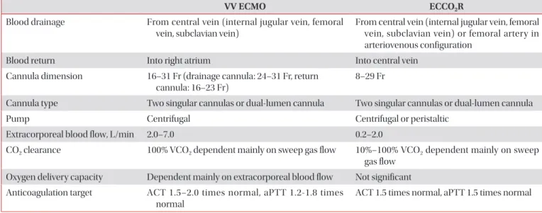
1reported a Table 1. ECMO vs. ECCO
2R
VV ECMO ECCO
2R
Blood drainage From central vein (internal jugular vein, femoral vein, subclavian vein)
From central vein (internal jugular vein, femoral vein, subclavian vein) or femoral artery in arteriovenous configuration
Blood return Into right atrium Into central vein
Cannula dimension 16–31 Fr (drainage cannula: 24–31 Fr, return cannula: 16–23 Fr)
8–29 Fr
Cannula type Two singular cannulas or dual-lumen cannula Two singular cannulas or dual-lumen cannula
Pump Centrifugal Centrifugal or peristaltic
Extracorporeal blood flow, L/min 2.0–7.0 0.2–2.0
CO
2clearance 100% VCO
2dependent mainly on sweep gas flow 10%–100% VCO
2dependent mainly on sweep gas flow
Oxygen delivery capacity Dependent mainly on extracorporeal blood flow Not significant Anticoagulation target ACT 1.5–2.0 times normal, aPTT 1.2-1.8 times
normal
ACT 1.5 times normal, aPTT 1.5 times normal
Adopted from Del Sorbo L et al. Lancet Respir Med 2014;2:154-64, with permission of Elsevier
3.
ECMO: extracorporeal membrane oxygenation; ECCO
2R: extracorporeal CO
2removal; VV: veno-venous; VCO
2: CO
2production; ACT:
activated clotting time; aPTT: activated partial thromboplastin time.
Table 2. Characteristics of gas exchange and hemodynamic support during ECLS Factors determining the
oxygenation in the ECLS circuit
Volume of blood crossing the oxygenator over time (blood flow)
Arterial oxygen saturation before crossing the artificial lung Hemoglobin concentration
Fraction of delivered oxygen in the sweep gas Diffusion of oxygen through the oxygenator Clearance of CO
2Sweep-gas flow
Total surface area of the artificial lung
Systemic oxygen delivery VV ECMO: ratio of ECLS blood flow to cardiac output, ECLS blood flow, recirculation of blood in ECLS circuit
VA ECMO: ratio between ECLS blood flow and residual intrapulmonary blood flow, ECLS blood flow, maximum oxygenation, residual intrapulmonary blood flow
Extracorporeal CO
2removal: provides insufficient oxygenation of the blood
Systemic CO
2elimination VV or VA ECMO: potentially eliminates entire CO
2production because the ECLS blood flow is enough Extracorporeal CO
2removal: usually needs high (>8 L/min) sweep-gas flow to remove CO
2Hemodynamic support VV ECMO and extracorporeal CO
2removal: no
VA ECMO: might replace lung and heart function by bypassing the cardiac out Adopted from Del Sorbo L et al. Lancet Respir Med 2014;2:154-64, with permission of Elsevier
3.
ECLS: extracorporeal life support; VV ECMO: veno-venous extracorporeal membrane oxygenation; VA: veno-arterial.
Table 3. VV ECMO for rescue treatment in patients with acute respiratory distress syndrome
3Indication Contraindication
REVA
9PaO
2/FiO
2<50 despite PEEP 10–20 cm H
2O and FiO
2>80%; P
plat>35 cm H
2O despite the attempt to reduce TV <4 mL/kg PBW
Presence of severe comorbidities and multiorgan failure (SOFA score >15)
ANZ ECMO
1PaO
2/FiO
2<60; PaCO
2>100 mm Hg with PaO
2/ FiO
2<100
Irreversible CNS condition; cirrhosis with ascites, encephalopathy, or history of variceal bleeding; active and rapidly fatal malignant disease; HIV infection; weight >120 kg; pulmonary hypertension; cardiac arrest
ECMOnet
7Oxygenation index >30; PaO
2/FiO
2<70 with PEEP ≥15 cm H
2O for patients already admitted to an ECMO center; pH <7.25 for ≥2 hr; hemodynamic instability
Intracranial bleeding or other major contraindication to anticoagulation; previous severe disability; poor prognosis because of underlying disease; mechanical ventilation >7 days
CESAR
2Potentially reversible respiratory failure;
Murray score ≥3; pH <7.2 despite optimum conventional treatment
PIP >30 cm H
2O or FiO
2>80%; mechanical ventilation >7 days;
intracranial bleeding; contraindication to limited heparinization;
contraindication to continuation of active treatment EOLIA
(NCT01470703)
PaO
2/FiO
2<50 with FiO
2>80% for 3 hr, des- pite optimum mechanical ventilation and adjunctive treatment; PaO
2/FiO
2<80 with FiO
2>80% for 6 hr, despite optimum mecha- nical ventilation and adjunctive treatment;
pH <7.25 for 6 hr (RR increased to 35 beats per minute) with mechanical ventilation adjusted to keep P
plat<32 cm H
2O
Mechanical ventilation ≥7 days; age <18 yr; pregnancy; weight
>1 kg/cm; BMI >45 kg/m
2; chronic respiratory insufficiency treated oxygen therapy of long duration and/or long- term respiratory assistance; history of heparin-induced thrombocytopenia; malignant disease with 5-year fatal prognosis; patient moribund; SAPS II >90; non-drug-induced coma following cardiac arrest; irreversible CNS pathology;
decision to limit therapeutic interventions; unable to cannulate Adopted from Del Sorbo L et al. Lancet Respir Med 2014;2:154-64, with permission of Elsevier
3.
VV ECMO: veno-venous extracorporeal membrane oxygenation; PaO
2: arterial partial pressure of O
2; FiO
2: fraction of inspired oxygen;
PEEP: positive end-expiratory pressure; TV: tidal volume; PBW: predicted body weight; SOFA: sequential organ failure assessment score;
CNS: central nervous system; HIV: human immunodeficiency virus; RR: respiratory rate; BMI: body mass index; SAPS II: Simplified Acute Physiology Score.
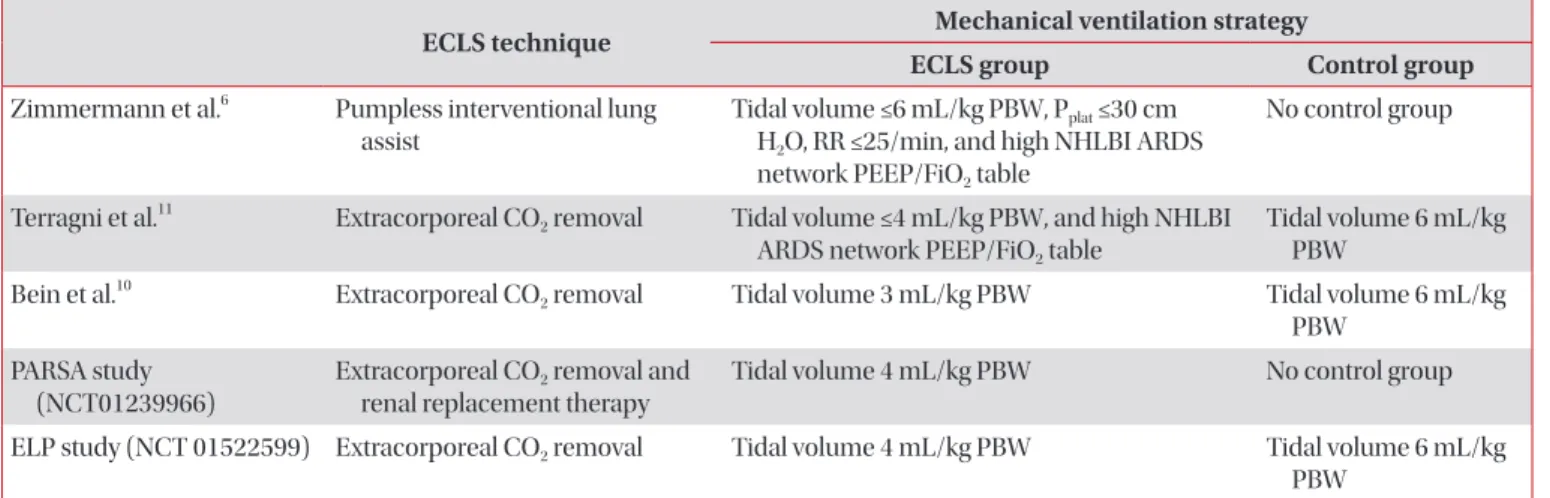
Table 4. Clinical studies of ECLS to prevent ventilator-induced lung injury
3ECLS technique Mechanical ventilation strategy
ECLS group Control group
Zimmermann et al.
6Pumpless interventional lung assist
Tidal volume ≤6 mL/kg PBW, P
plat≤30 cm H
2O, RR ≤25/min, and high NHLBI ARDS network PEEP/FiO
2table
No control group
Terragni et al.
11Extracorporeal CO
2removal Tidal volume ≤4 mL/kg PBW, and high NHLBI ARDS network PEEP/FiO
2table
Tidal volume 6 mL/kg PBW
Bein et al.
10Extracorporeal CO
2removal Tidal volume 3 mL/kg PBW Tidal volume 6 mL/kg PBW
PARSA study (NCT01239966)
Extracorporeal CO
2removal and renal replacement therapy
Tidal volume 4 mL/kg PBW No control group
ELP study (NCT 01522599) Extracorporeal CO
2removal Tidal volume 4 mL/kg PBW Tidal volume 6 mL/kg PBW
Adopted from Del Sorbo L et al. Lancet Respir Med 2014;2:154-64, with permission of Elsevier
3.
ECLS: extracorporeal life support; PBW: predicted bodyweight; P
plat: inspiratory plateau pressure; RR: respiratory rate; NHLBI: National Heart,
Lung, and Blood Institute; ARDS: acute respiratory distress syndrome; PEEP: positive end-expiratory pressure; FiO
2: fraction of inspired
oxygen.
survival rate of 75% in the ECMO treatment group. The Ital- ian ECMO Network also showed a survival rate of 68% in the ECMO treatment group
7. The Swine Flu Triage (SWiFT) study, done in the UK showed the lower in-hospital mortality in the ECMO treatment group (24% vs. 53%, p=0.006)
8. In the CESAR trial, severe ARDS patients also showed the higher survival rate in the ECMO treatment group (63% vs. 47%, p=0.03)
2.
The above results suggested that the implementation of protective mechanical ventilation during ECMO can improve the prognosis.
EOLIA (NCT01470703) should help to define the clinical ef- ficacy of VV ECMO in severe ARDS patients.
2. Extracorporeal CO
2removal
Recent studies reported that the application of ECCO
2R in ARDS patients can reduce the lung injury as it enables the ultraprotective strategies of mechanical ventilation (Table 4). Zimmermann et al.
6reported that when pumpless AV ECLS was applied to 51 patients with ARDS, low tidal volume ventilation could be maintained along with the continuous removal of CO
2, and the survival rate was 50%.
A randomized, controlled study was done to compare an ultra-protective mechanical ventilation (3 mL/kg predicted body weight [PBW] with Pumpless AV ECLS) with low tidal volume ventilation (6 mL/kg PBW) strategies in 79 patients with ARDS. While the two groups did not differ for in-hospital mortality, within the patient group with PF ratio <200, the ultra-protective group showed an improved survival
10. At pres- ent, studies on the efficacy of very low tidal volumes ventila- tion strategies during ECCO
2R are working in progress.
Controversies
1. Mechanical ventilation strategies
For minimizing VILI, the ventilator settings during VV ECMO should be maintained at low levels to enable the pre- vention of atelectasis while keeping the alveoli open. How- ever, there are no specific recommendations other than the maintenance of positive end-expiratory pressure (PEEP) at 10 cm H
2O or above. As the injured lungs contribute little to oxygenation, lung recruitment using PEEP while maintaining minimal tidal volumes might accelerate lung healing or opti- mise cardiopulmonary function
12,13.
In the CESAR trial, lung rest was induced by limitation of the peak inspiratory pressure to 20 cm H
2O with PEEP 10 cm H
2O, 10 breaths per minute, and FiO
2of 30%. Another study also showed positive outcomes in patients who maintained a mean plateau pressure of 25 cm H
2O
9.
After the acute phase of the illness, mechanical ventilation with spontaneous breathing should be considered to reduce the use of sedatives and to improve the diaphragmatic func- tion
14,15.
2. Tracheostomy
In the case of applying ECLS due to severe ARDS, mechani- cal ventilation for a long period of time is predicted. Therefore, the early tracheostomy might be considered. The use of anti- coagulants during ECLS is not the contraindication for trache- ostomy. In a recent study, a tracheostomy with the percutane- ous dilatational technique done by experienced physician is safe with a brief interruption of anticoagulation. In this study,
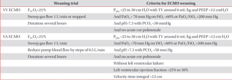
Table 5. Weaning from ECMO
Weaning trial Criteria for ECMO weaning
VV ECMO F
ECO
2=21% P
plat<25 to 30 cm H
2O with TV around 6 mL/kg and PEEP <12 cmH
2O Sweep gas flow 1 L/min or stopped And PaO
2> 70 mm Hg on FiO
2<60% or PaO
2/FiO
2>200 mm Hg Duration: several hours And pH>7.3 with PCO
2<50 mmHg
And no acute cor pulmonale
VA ECMO F
ECO
2=21% P
plat<25 to 30 cm H
2O with TV around 6 mL/kg and PEEP <12 cm H
2O Sweep gas flow 1 L/min And PaO
2>70 mm Hg on FiO
2<60% or PaO
2/FiO
2>200 mm Hg Reduce pump blood flow by steps of 0.5 L/min And pH >7.3 with PCO
2<50 mm Hg
Duration: several hours And no acute cor pulmonale Without left ventricular failure:
Left ventricular ejection fraction >25% to 30%
Velocity-time integral >12 cm
Adopted from Richard C et al. Ann Intensive Care 2014;4:15, according to the Creative Commons License
17.
ECMO: extracorporeal membrane oxygenation; VV: veno-venous; F
ECO
2: oxygen fraction delivered by the extracorporeal circuit; P
plat: plateau pressure; TV: tidal volume; PEEP: positive end-expiratory pressure; PaO
2: arterial partial pressure of O
2; FiO
2: fraction of inspired oxygen;
PCO
2: partial pressure of CO
2; VA: venous-arterial.
no major complications such as death were observed
16. 3. Weaning from ECLS (Table 5)
17When mechanical ventilation settings is acceptable (tidal volume <6 mL/Kg PBW, plateau pressure <30 cm H
2O, PEEP
<12 cm H
2O, FiO
2<60%) and respiratory mechanics, gas ex- changes, and radiographic findings are improved, weaning from ECLS can be considered. Before weaning, the existence of acute cor pulmonale should be identified. Two main strate- gies of weaning can be used: reducing sweep-gas flow rates or reducing extracorporeal blood-flow rates. Alternatively, wean- ing of mechanical ventilation may be considered earlier than weaning from ECLS
18,19.
4. Sedation
While deep sedation and neuromuscular blockade might be required in the initial stages to relieve symptoms and re- duce oxygen consumption, patients should be kept awake to actively participate in rehabilitation therapy during ECLS. In addition, early mobilization could suppress the progression of weakness and reduce the incidence of delirium.
The indication for the use of awake ECMO, instead of inva- sive mechanical ventilation is not confirmed in patients with ARDS refractory to non-invasive ventilation. However, the use of awake ECMO as a bridge before lung transplantation has shown promising results
20-23. Mechanical ventilation and seda- tion might worsen outcomes before and after the transplanta- tion. Awake ECMO enables patients to communicate, eat, and walk and improves physical and physiological conditions.
5. Technological advances
The first technological advances in this field may be the pro- duction of bicaval dual-lumen cannulas
24. This cannula is in- serted via the right internal jugular vein, and then drains blood from the superior and inferior vena cava through one lumen and returns blood into the right atrium through a second lu- men. Only one cannulation enables patients to receive inten- sive physiotherapy more conveniently. Second, reduction in the size of ECLS equipment has enabled patients receiving ECLS to transfer and mobilise
25.
6. Anticoagulantion and transfusion
Although the ECLS circuits are engineered with biocompat- ible materials, the systemic anticoagulants are still required to prevent thrombotic complication. Unfractionated heparin is most commonly used and monitoring is performed using activated partial thromboplastin time (1.2–1.5 times control), anti-Xa activity (0.2–0.4 IU/mL), or the activated clotting time.
When heparin-induced thrombocytopenia is suspected, arg-
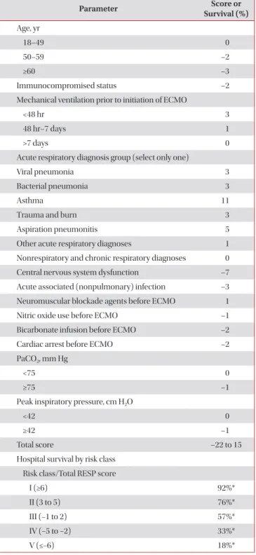
Table 6. RESP score
Parameter Score or
Survival (%)