Extended Use of Extracorporeal Membrane Oxygenation for Acute Respiratory Distress Syndrome: A Retrospective Multicenter Study
Won-Young Kim, M.D., Ph.D.
1,* , SeungYong Park, M.D., Ph.D.
2,* , Hwa Jung Kim, M.D., Ph.D.
3, Moon Seong Baek, M.D.
4, Chi Ryang Chung, M.D., Ph.D.
5, So Hee Park, M.D.
6, Byung Ju Kang, M.D.
7, Jin Young Oh, M.D., Ph.D.
8, Woo Hyun Cho, M.D., Ph.D.
9, Yun Su Sim, M.D., Ph.D.
10, Young-Jae Cho, M.D., Ph.D.
11, Sunghoon Park, M.D., Ph.D.
12, Jung-Hyun Kim, M.D., Ph.D.
13and Sang-Bum Hong, M.D., Ph.D.
14*Author affiliations appear at the end of this article.
Background: Beyond its current function as a rescue therapy in acute respiratory distress syndrome (ARDS), extracorporeal membrane oxygenation (ECMO) may be applied in ARDS patients with less severe hypoxemia to facilitate lung protective ventilation. The purpose of this study was to evaluate the efficacy of extended ECMO use in ARDS patients.
Methods: This study reviewed 223 adult patients who had been admitted to the intensive care units of 11 hospitals in Korea and subsequently treated using ECMO. Among them, the 62 who required ECMO for ARDS were analyzed.
The patients were divided into two groups according to pre-ECMO arterial blood gas: an extended group (n=14) and a conventional group (n=48).
Results: Baseline characteristics were not different between the groups. The median arterial carbon dioxide tension/
fraction of inspired oxygen (FiO
2) ratio was higher (97 vs. 61, p<0.001) while the median FiO
2was lower (0.8 vs. 1.0, p<0.001) in the extended compared to the conventional group. The 60-day mortality was 21% in the extended group and 54% in the conventional group (p=0.03). Multivariate analysis indicated that the extended use of ECMO was independently associated with reduced 60-day mortality (odds ratio, 0.10; 95% confidence interval, 0.02–0.64; p=0.02).
Lower median peak inspiratory pressure and median dynamic driving pressure were observed in the extended group 24 hours after ECMO support.
Conclusion: Extended indications of ECMO implementation coupled with protective ventilator settings may improve the clinical outcome of patients with ARDS.
Keywords: Extracorporeal Membrane Oxygenation; Respiratory Distress Syndrome, Adult; Respiration, Artificial;
Retrospective Studies; Multicenter Studies as Topic
Address for correspondence: Sang-Bum Hong, M.D., Ph.D.
Department of Pulmonary and Critical Care Medicine, Asan Medical Center, University of Ulsan College of Medicine, 88 Olympic-ro 43-gil, Songpa-gu, Seoul 05505, Korea
Phone: 82-2-3010-3893, Fax: 82-2-2045-4039, E-mail: [email protected]
*Won-Young Kim and SeungYong Park contributed equally to this work.
Received: Jul. 24, 2018, Revised: Sep. 20, 2018, Accepted: Oct. 25, 2018, Published online: Feb. 28, 2019
cc It is identical to the Creative Commons Attribution Non-Commercial License (http://creativecommons.org/licenses/by-nc/4.0/).
Copyright © 2019
The Korean Academy of Tuberculosis and Respiratory Diseases.
Introduction
Acute respiratory distress syndrome (ARDS) is character- ized by lung injury caused by either direct or indirect insults and leads to severe respiratory failure that is refractory to conventional oxygen therapy
1,2. Hospital mortality in patients with severe ARDS ranges from 45% to 60%
2-4. Mechanical ventilation is the mainstay of ARDS therapy. However, over- distention and cyclic alveolar recruitment and de-recruitment during ventilation may further damage lungs and increase in- flammatory mediators, eventually resulting in multiple organ failure and death
5. Lung protective ventilation strategies using low tidal volume and higher levels of positive end-expiratory pressure (PEEP) are widely accepted approaches
6,7, although rescue therapies may still be required in refractory cases.
Extracorporeal membrane oxygenation (ECMO) can provide adequate blood carbon dioxide removal and oxy- genation, allowing a reduction in mechanical ventilation and minimization of ventilator-induced lung injury (VILI). Several clinical trials in ARDS have shown positive results of veno- venous-ECMO
3,8,9. Although there are no absolute criteria for ECMO initiation in ARDS, suggested indications include se- vere hypoxemia (arterial carbon dioxide tension [PaO
2]/frac- tion of inspired oxygen [FiO
2] ratio <80 on FiO
2>0.9), uncom- pensated hypercapnia with acidemia, or excessively elevated end-inspiratory plateau pressures despite standard ventilator management
10. Venovenous-ECMO may be applied in ARDS patients with less severe hypoxemia in whom it might allow
“lung rest” by lowering airway pressures and tidal volume rather than improving oxygenation, considering that there is a linear relationship between mortality and plateau pressure, even at less than 30 cm H
2O
11. Moreover, lung hyperinflation occurs in approximately 30% of ARDS patients ventilated us- ing the protective ARDS Network strategy
12.
To date, there are limited data on the impact of initiation of ECMO in patients with less severe forms of ARDS. Accord- ingly, the aim of this study was to compare the clinical char- acteristics of patients who underwent ECMO for ARDS with less severe hypoxemia (namely “extended indications”) versus conventional indications.
Materials and Methods
1. Study design and patient selection
This study was a retrospective analysis of a prospective mul- ticenter cohort. The cohort was composed of critically ill adult patients who were at least 18 years old, received ECMO thera- py, and were admitted to one of the intensive care units (ICUs) of the 11 participating tertiary or referral hospitals of Korea from January 2014 to December 2015. From this cohort, we included in our analysis patients who required ECMO sup-
port for ARDS. The exclusion criteria were as follows: received lung transplantation (either bridge to transplant or destina- tion therapy), cardiopulmonary resuscitation before ECMO, ECMO transferred from other hospital, venoarterial-ECMO, acute respiratory diagnosis other than ARDS, and incomplete data for analysis. According to the pre-ECMO arterial blood gas, patients were divided into extended (PaO
2/FiO
2ratio ≥80 without uncompensated respiratory acidosis) and conven- tional (PaO
2/FiO
2ratio <80 on FiO
2>0.9 and/or arterial carbon dioxide tension [PaCO
2] ≥80 mm Hg with pH <7.15) groups.
The PaO
2/FiO
2cutoff of 80 was selected based on recently proposed criteria for initiating ECMO in ARDS patients
10. The primary study outcome was 60-day mortality. Secondary outcomes included ECMO duration, ECMO weaning failure rate, mechanical ventilation weaning success rate, mechanical ventilation-free days at day 60, and 30- and 90-day mortality.
We also analyzed factors associated with 60-day mortality, including extended ECMO use. The local institutional review board or independent ethics committee of each hospital ap- proved the study protocol (Institutional Review Board of Asan Medical Center, No. 2016-0269). Written informed consent was waived due to the observational nature of the study, and the patient records were anonymized and de-identified prior to analysis.
2. Data collection and definitions
Baseline demographic and clinical characteristics were
collected and included age, sex, body mass index, immune
status, etiologies of ARDS, dates of hospital and ICU admis-
sion, date of initiation of mechanical ventilation and ECMO,
and treatment prior to ECMO. Acute Physiology and Chronic
Health Evaluation (APACHE) II
13and Sequential Organ Fail-
ure Assessment (SOFA)
14scores were also collected at the
time of ICU admission and ECMO initiation, respectively. The
severity of ARDS before ECMO initiation was assessed by
the PRedicting dEath for SEvere ARDS on VV-ECMO (PRE-
SERVE) score
15and the Respiratory ECMO Survival Predic-
tion (RESP) score
16as previously described. The pre-ECMO
variables included rescue therapies (neuromuscular blocker,
inhaled nitric oxide, or prone positioning), ventilator settings,
and arterial blood gas. Ventilator settings included PEEP, peak
inspiratory pressure, dynamic driving pressure (the difference
between peak inspiratory pressure and PEEP)
17, tidal volume,
respiratory rate, and FiO
2, which were determined at base-
line (before ECMO application) and at 4 hours and 24 hours
thereafter. ARDS was diagnosed by consensus definition
2. An
immunocompromised status was diagnosed if there was an
underlying disease that affected the immune system (chronic
liver disease, chronic kidney disease, human immunodefi-
ciency virus infection, or malignancy) or if immunosuppres-
sive therapy was being administered at the time of ECMO
initiation. Steroid use was defined as corticosteroid adminis-
tration within 14 days of ECMO initiation.
3. Statistical analysis
Continuous variables are presented as median and inter- quartile range or as mean±standard deviation, whereas cat- egorical variables are presented as percentages. Continuous variables were compared using a Mann–Whitney U test or Student’s t test. Categorical variables were compared using a chi-square or Fisher exact test. Binary logistic regression was used to identify factors predicting 60-day mortality. Variables with p-values <0.20 in the univariate analysis were included in the multivariate analysis by using stepwise backward selec- tion procedures. Highly correlated variables were identified to prevent multicollinearity. Model discrimination was assessed with the area under the receiver operating characteristic curve, and model calibration was assessed with the Hosmer- Lemeshow test. A Cox proportional hazards regression model with stepwise backward selection procedures was also ap- plied. The Kaplan–Meier curve was rendered to compare 60- day survival among the groups. All tests of significance were two-tailed, and p-values of <0.05 were considered significant.
All analyses were performed using SPSS version 18.0 for Win- dows (SPSS Inc., Chicago, IL, USA).
Results
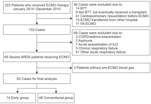
There were 223 patients in the initial cohort; 161 were not included for the following reasons: met specific exclusion criteria (n=90); ECMO provided for an acute respiratory diag-
nosis other than ARDS (n=68); and pre-ECMO arterial blood gas unavailable for analysis (n=3). Our study consisted of 62 ARDS patients who received ECMO as a rescue therapy.
There were 14 patients (23%) in the extended group and 48 (77%) in the conventional group (Figure 1).
The baseline characteristics of the study patients are shown in Table 1. The percentage of male patients was significantly higher in the extended than in the conventional group, but there were no differences in age or body mass index. The proportion of immunocompromised patients was also similar between the two groups. In both groups, the main etiology of ARDS was bacterial pneumonia, followed by viral pneumonia.
There were no differences between the groups in baseline APACHE II and SOFA scores, treatment prior to ECMO, and pre-ECMO rescue therapies. There was no difference between the groups in time from intubation to ECMO cannulation. The median PRESERVE and RESP scores were similar between the two groups (4 [3–6] vs. 5 [4–6], p=0.50; and 3 [0–5] vs. 2 [1–4], p=0.33, respectively). Compared with the conventional group, the extended group had a significantly higher median PaO
2/FiO
2ratio (97 [88–112] vs. 61 [53–70], p<0.001) and lower median FiO
2(0.8 [0.7–1.0] vs. 1.0 [1.0–1.0], p<0.001).
Other ventilator settings before ECMO support were not sig- nificantly different between the two groups.
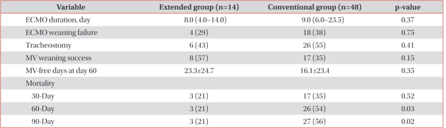
Clinical outcomes of the study patients are shown in Table 2. The primary outcome, 60-day mortality, was observed in three of the 14 patients (21%) in the extended group and 26 of the 48 patients (54%) in the conventional group (p=0.03).
In addition, 90-day mortality was significantly lower in the extended group (3/14, 21%) than in the conventional group (27/48, 56%) (p=0.02), although the 30-day mortality rate
Figure 1. Illustration of a study flow dia- gram. ECMO: extracorporeal membrane oxygenation; BTT: bridge to transplant;
VA: venoarterial; COPD: chronic obstruc-
tive pulmonary disease; ILD: interstitial
lung disease; ARDS: acute respiratory
distress syndrome.
was comparable between the groups. The median duration of ECMO was 8.0 (4.0–14.0) days in the extended group and 9.0 (6.0–23.5) days in the conventional group (p=0.37). There were no differences between the groups in the ECMO wean-
ing failure rate and tracheostomy rate. Both the mechanical ventilation weaning success rate and mechanical ventilation- free days at day 60 tended to be more favorable in the extend- ed than in the conventional group, although the results were Table 1. Baseline characteristics of the study patients
Variable Extended group (n=14) Conventional group (n=48) p-value
Age, yr 50 (35–61) 59 (46–65) 0.21
Male sex 14 (100) 31 (65) 0.007
Body mass index, kg/m
224.9 (21.3–26.9) 23.3 (20.6–25.0) 0.19
Immunocompromised 2 (14) 9 (19) >0.99
ARDS etiology 0.57
Viral pneumonia 4 (29) 13 (27)
Bacterial pneumonia 8 (57) 32 (67)
Trauma/burn 2 (14) 3 (6)
APACHE II score at ICU admission 20 (12–25) 19 (14–26) 0.96
SOFA score at ECMO initiation 13 (11–16) 11 (7–14) 0.07
Pre-ECMO renal replacement therapy 3 (21) 10 (21) >0.99
Pre-ECMO steroids 2 (14) 6 (13) >0.99
Pre-ECMO bicarbonate infusion 2 (14) 8 (17) >0.99
Pre-ECMO rescue therapy
Neuromuscular blocker 10 (71) 35 (73) >0.99
Inhaled nitric oxide 6 (43) 12 (25) 0.32
Prone positioning 6 (43) 22 (46) 0.84
Time between MV-ECMO, day 1.5 (1.0–5.0) 1.0 (0–3.5) 0.43
PRESERVE score 4 (3–6) 5 (4–6) 0.50
RESP score 3 (0–5) 2 (1–4) 0.33
Pre-ECMO ventilator settings
Positive end-expiratory pressure, cm H
2O 10 (10–12) 10 (8–12) 0.60
Peak inspiratory pressure, cm H
2O 28 (27–30) 28 (23–30) 0.67
Dynamic driving pressure, cm H
2O 17 (15–20) 16 (14–20) 0.58
Tidal volume, mL 436 (350–512) 390 (300–600) 0.83
Respiratory rate, breaths/min 23 (20–28) 22 (16–27) 0.37
FiO
20.8 (0.7–1.0) 1.0 (1.0–1.0) <0.001
Pre-ECMO arterial blood gas
pH 7.29 (7.23–7.40) 7.26 (7.17–7.33) 0.15
PaCO
2, mm Hg 47 (40–50) 52 (38–66) 0.17
PaO
2, mm Hg 84 (67–94) 60 (53–69) <0.001
PaO
2/FiO
297 (88–112) 61 (53–70) <0.001
Bicarbonate, mEq/L 22.1 (18.7–25.0) 22.0 (18.1–26.7) 0.87
Values are presented as median (interquartile range) or number (%).
ARDS: acute respiratory distress syndrome; APACHE: Acute Physiology and Chronic Health Evaluation; ICU: intensive care unit; SOFA:
Sequential Organ Failure Assessment; ECMO: extracorporeal membrane oxygenation; MV: mechanical ventilation; PRESERVE: PRedicting
dEath for SEvere ARDS on VV-ECMO; RESP: Respiratory ECMO Survival Prediction; FiO
2: fraction of inspired oxygen; PaCO
2: arterial carbon
dioxide tension; PaO
2: arterial oxygen tension.
not statistically significant. The Kaplan–Meier survival curves of the study patients are shown in Figure 2.
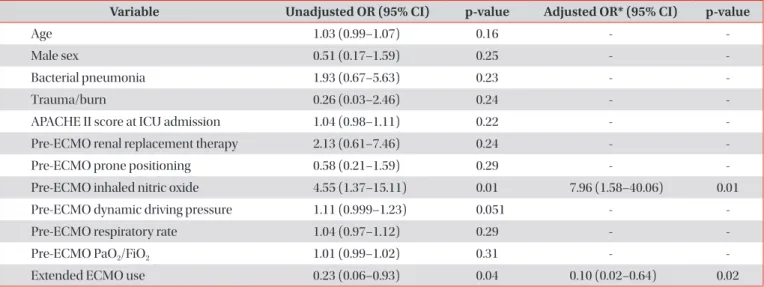
The results of univariate and multivariate analyses of risk factors predicting 60-day mortality are shown in Table 3. Mul- tivariate analysis indicated that inhaled nitric oxide as pre- ECMO rescue therapy was significantly associated with mor- tality. Conversely, extended ECMO use was a protective factor (odds ratio, 0.10; 95% confidence interval, 0.02–0.64; p=0.02).
This model had acceptable discrimination (area under the receiver operating characteristic curve=0.72) and calibration (Hosmer and Lemeshow chi-square=1.10; p=0.58). Consis- tently, inhaled nitric oxide and extended ECMO use were in- dependently associated with “time to 60-day mortality” using Cox proportional hazards modeling (Table 4).
The changes in ventilator settings between the study groups during the 24-hour study period are shown in Figure 3 and Supplementary Table S1. After ECMO support, tidal volume, respiratory rate, and FiO
2were decreased, but there were no significant differences between the extended and convention- al groups until 24 hours. Both peak inspiratory pressure and dynamic driving pressure were decreased after ECMO initia- tion. The extended group had significantly lower median peak inspiratory pressure after 24-hour ECMO support (20 [18–20]
cm H
2O vs. 22 [20–24] cm H
2O, p=0.006). The extended group also tended to have lower median dynamic driving pressure after 24-hour ECMO support (10 [10–12] cm H
2O vs. 12 [10–
15] cm H
2O, p=0.13).
Discussion
The present study revealed a lower mortality rate in patients who received ECMO for ARDS with an extended indication, and extended ECMO use itself was positively associated with survival. The 54% 60-day mortality in the conventional group (where the median PRESERVE and RESP scores at ECMO
initiation were 5 and 2, respectively) is comparable with the 43%–46% mortality rates reported in previous studies of similar ECMO-treated ARDS patients
15,16. However, despite comparable severity at ECMO initiation, mortality was lower than expected in the extended group. In the extended group, lower median peak inspiratory pressure and median dynamic driving pressure were observed within 24 hours after ECMO initiation. These findings suggest that extended indications of ECMO implementation than conventional indications ac- companied by an additional “lung rest” may improve the clini- cal outcome. To our knowledge, this is one of the few studies to evaluate the role of extended ECMO use in patients with ARDS.
Lung protective ventilator settings that keep tidal volume and plateau pressures within narrow limits should be used in ARDS patients to improve outcomes and prevent the develop- ment of VILI
18. However, when there is dramatic hypoxemia and/or profound acidosis with a high degree of hypercapnia,
Table 2. Clinical outcomes of the study patients
Variable Extended group (n=14) Conventional group (n=48) p-value
ECMO duration, day 8.0 (4.0–14.0) 9.0 (6.0–23.5) 0.37
ECMO weaning failure 4 (29) 18 (38) 0.75
Tracheostomy 6 (43) 26 (55) 0.41
MV weaning success 8 (57) 17 (35) 0.15
MV-free days at day 60 23.3±24.7 16.1±23.4 0.35
Mortality
30-Day 3 (21) 17 (35) 0.52
60-Day 3 (21) 26 (54) 0.03
90-Day 3 (21) 27 (56) 0.02
Values are presented as median (interquartile range), number (%), or mean±standard deviation.
ECMO: extracorporeal membrane oxygenation; MV: mechanical ventilation.
Figure 2. Kaplan–Meier survival curves of the patients under study.
ECMO: extracorporeal membrane oxygenation.
these “protective” mechanical ventilation strategies may not be possible. In addition, even if the above-mentioned variables are kept within the defined limits, the patient might not be ventilated in a lung protective manner
11,12. Rescue therapies such as ECMO may be used timely or even immediately to resolve potentially deleterious conditions, although defined thresholds of mechanical ventilation that characterize the status under which the therapy is to be applied are required.
However, a clear cutoff that defines severe or life-threatening hypoxemia is still an ongoing matter of debate
19, and evidence supporting indications or contraindications to ECMO initia-
tion in ARDS is scarce. A recent international multicenter randomized ECMO to Rescue Lung Injury in Severe ARDS (EOLIA) trial tested the efficacy of early venovenous-ECMO in patients with severe ARDS with conventional mechani- cal ventilation with prone positioning
20. The analysis of the primary end point (60-day mortality) showed no significant benefit of early ECMO, although the 28% rate of crossover to ECMO among patients with refractory hypoxemia in the con- ventional group (43% of them survived) may have diluted the potential effect of ECMO. Meanwhile, patients who were en- rolled in this trial were very hypoxemic (PaO
2/FiO
2ratio <80) Table 3. Univariate and multivariate analysis of factors associated with 60-day mortality
Variable Unadjusted OR (95% CI) p-value Adjusted OR* (95% CI) p-value
Age 1.03 (0.99–1.07) 0.16 - -
Male sex 0.51 (0.17–1.59) 0.25 - -
Bacterial pneumonia 1.93 (0.67–5.63) 0.23 - -
Trauma/burn 0.26 (0.03–2.46) 0.24 - -
APACHE II score at ICU admission 1.04 (0.98–1.11) 0.22 - -
Pre-ECMO renal replacement therapy 2.13 (0.61–7.46) 0.24 - -
Pre-ECMO prone positioning 0.58 (0.21–1.59) 0.29 - -
Pre-ECMO inhaled nitric oxide 4.55 (1.37–15.11) 0.01 7.96 (1.58–40.06) 0.01
Pre-ECMO dynamic driving pressure 1.11 (0.999–1.23) 0.051 - -
Pre-ECMO respiratory rate 1.04 (0.97–1.12) 0.29 - -
Pre-ECMO PaO
2/FiO
21.01 (0.99–1.02) 0.31 - -
Extended ECMO use 0.23 (0.06–0.93) 0.04 0.10 (0.02–0.64) 0.02
*Variables with p-values of <0.20 in the univariate analysis were included in the multivariate analysis by using stepwise backward selection procedures.
OR: odds ratio; CI: confidence interval; APACHE: Acute Physiology and Chronic Health Evaluation; ICU: intensive care unit; ECMO: extracor- poreal membrane oxygenation; PaO
2: arterial oxygen tension; FiO
2: fraction of inspired oxygen.
Table 4. Cox proportional hazards regression model with 60-day mortality as outcome
Variable Unadjusted HR (95% CI) p-value Adjusted HR* (95% CI) p-value
Male sex 0.56 (0.26–1.21) 0.14 - -
APACHE II score at ICU admission 1.03 (0.99–1.07) 0.20 - -
Pre-ECMO renal replacement therapy 1.84 (0.81–4.16) 0.14 - -
Pre-ECMO prone positioning 0.62 (0.29–1.31) 0.21 - -
Pre-ECMO inhaled nitric oxide 3.24 (1.55–6.77) 0.002 4.19 (1.87–9.36) <0.001
Pre-ECMO bicarbonate infusion 1.72 (0.70–4.23) 0.24 - -
Pre-ECMO dynamic driving pressure 1.07 (1.003–1.13) 0.04 - -
Pre-ECMO respiratory rate 1.03 (0.97–1.09) 0.28 - -
Pre-ECMO PaO
2/FiO
21.01 (0.999–1.02) 0.09 1.01 (1.01–1.02) 0.001
Extended ECMO use 0.36 (0.11–1.18) 0.09 0.19 (0.06–0.66) 0.009
*Variables with p-values of <0.20 were included in the multivariate analysis using stepwise backward selection procedures.
HR: hazard ratio; CI: confidence interval; APACHE: Acute Physiology and Chronic Health Evaluation; ICU: intensive care unit; ECMO: extra-
corporeal membrane oxygenation; PaO
2: arterial oxygen tension; FiO
2: fraction of inspired oxygen.
and the trial did not evaluate patients with less severe forms of ARDS.
The use of ECMO in ARDS patients with less severe hy- poxemia may be beneficial for the following reasons. First, venovenous-ECMO with an “ultraprotective” mechanical ventilation strategy (tidal volume reduction to below 6 ml/kg of predicted body weight to achieve a plateau pressure less than 25 cm H
2O) may further reduce VILI and mortality in patients mechanically ventilated for ARDS
21,22. In addition, venovenous-ECMO may reduce the effect of acute lung injury on right ventricular dysfunction by reducing hypoxic pul- monary vasoconstriction and unloading the right ventricle
23. Second, modern ECMO devices are simpler, safer, and require less anticoagulation and it is now possible to support patients for weeks
24. Third, patients with ECMO can be awake, facilitat- ing rehabilitation and decreasing weakness and decondition- ing. Several studies indicate that early rehabilitation in ECMO patients may improve survival, reduce mechanical ventilation duration, shorten ICU length of stay, and improve functional recovery
25,26. Fourth, ECMO might improve long-term quality- of-life by improving blood oxygenation in severely hypoxemic ARDS patients. In fact, ARDS patients treated with ECMO
3,15showed comparable or better health-related quality-of-
life scores than patients with less severe ARDS treated with conventional management
27. Lastly, several studies have identified duration of mechanical ventilation prior to ECMO initiation and low pre-ECMO respiratory system compliance as factors strongly associated with mortality in severe ARDS patients receiving ECMO
15,16,28.
Several studies have described the impact of different venti- lator settings in ARDS patients undergoing ECMO
17,22,28, with lower PEEP levels and higher driving pressure independently associated with mortality. In this study, we observed changes in ventilator settings within 24 hours of ECMO initiation.
Thus, the extended group had lower peak inspiratory pressure and dynamic driving pressure after 24-hour ECMO support.
We used the difference between the peak inspiratory pres- sure and PEEP to calculate the “dynamic” driving pressure
17because most patients were on pressure-controlled ventila- tion during the evaluation of ventilator settings. Moreover, our multivariate logistic regression and Cox proportional hazards modeling demonstrated that the extended group was independently associated with 60-day survival. Based on our findings, implementation of ECMO with extended indications accompanied by protective ventilation settings may affect the clinical outcome in ARDS patients.
Figure 3. Serial changes in tidal volume (A), respiratory rate (B), positive end-expiratory pressure (PEEP) (C), peak inspiratory pressure (PIP)
(D), dynamic driving pressure (E), and fraction of inspired oxygen (FiO
2) (F) in the extended group (dark line) and the conventional group
(gray line) during the 24-hour study period. Data is presented as a median value (interquartile range). *p<0.05.
†p=0.13. ECMO: extracorporeal
membrane oxygenation.
This multicenter study had several limitations of note.
First, the study was retrospective and underpowered. Its non- randomized design was prone to selection bias and precluded any inference of causality regarding the association between extended ECMO use and outcome. We also could not adjust for unmeasured confounders, and a matching process be- tween the two groups was not feasible due to a relatively small sample size used for the extended group compared with that of conventional group. In addition, the multiple confounding factors may have affected the clinical outcome, despite the similar baseline characteristics of the study patients in the extended and conventional groups. Second, one could argue that the better outcomes in the extended group could have been related with the better oxygenation status of patients, considering that several studies identified oxygenation criteria as a predictor of mortality in ARDS
2,29. However, the multivari- ate analysis showed no relation between PaO
2/FiO
2ratio and mortality. The Cox regression analysis even showed a positive association between increased PaO
2/FiO
2ratio and mortality, although it is difficult to explain such a phenomenon. More- over, pre-ECMO ventilator settings in the extended group were comparable to those in the conventional group, and most patients in the extended group had already been managed with other rescue therapies before ECMO initiation. Third, ventilator settings were collected only at baseline, 4 hours, and 24 hours after ECMO application. We do not know if specific ventilator strategies after day 1 would have changed the pa- tient outcomes. Lastly, criteria for assessing ECMO for ARDS, mechanical ventilation on ECMO, and weaning from ECMO were not standardized among centers. In addition, there was a lack of data on some of the treatment modalities (level of sedation, ECMO settings, anticoagulation, transfusion, or re- habilitation) and complications (either directly related to the ECMO circuit or patient-related) because these data were not collected due to the purposes of the initial cohort. It remains possible that the two groups were not similarly treated, and this may have affected the treatment outcome.
In conclusion, our comparison of extended indications of ECMO implementation with conventional indications sug- gests that extended ECMO use accompanied by protective ventilation strategies may improve the clinical outcome for patients with ARDS.
*Author affiliations
1
Division of Pulmonary, Allergy and Critical Care Medicine, Department of Internal Medicine, Pusan National University, School of Medicine, Busan,
2Department of Internal Medicine, Chonbuk National University Medical School, Jeonju,
3Depart- ment of Clinical Epidemiology and Biostatistics, Asan Medical Center, University of Ulsan College of Medicine, Seoul,
4De- partment of Pulmonary, Allergy, and Critical Care Medicine,
Hallym University Dongtan Sacred Heart Hospital, Hwaseong,
5
Department of Critical Care Medicine, Samsung Medical Center, Sungkyunkwan University School of Medicine, Seoul,
6
Department of Pulmonary and Critical Care Medicine, Kyung Hee University Hospital at Gangdong, Kyung Hee University School of Medicine, Seoul,
7Department of Internal Medi- cine, Ulsan University Hospital, University of Ulsan College of Medicine, Ulsan,
8Division of Pulmonology and Critical Care Medicine, Dongguk University Ilsan Hospital, Dongguk University College of Medicine, Goyang,
9Department of Pul- monology and Critical Care Medicine, Pusan National Uni- versity Yangsan Hospital, Yangsan,
10Division of Pulmonary and Critical Care Medicine, Department of Medicine, Hallym University Kangnam Sacred Heart Hospital, Seoul,
11Division of Pulmonary and Critical Care Medicine, Department of In- ternal Medicine, Seoul National University Bundang Hospital, Seoul National University College of Medicine, Seongnam,
12