Portal Vein Thrombosis in Minimal Change Disease
Gyuri Kim, Jung Yeon Lee, Su Jin Heo, Yoen Kyung Kee, Seung Hyeok Han
Department of Internal Medicine, Yonsei University College of Medicine, Seoul, Korea
Introduction
Thromboembolic events are considered to be critical com- plications of nephrotic syndrome. Much evidence suggests that nephrotic syndrome is associated with a hypercoagulable state.
Altered coagulation factor profiles and increased viscosity due to volume depletion may play a role in thrombus formation.
The renal vein and deep vein of the lower limbs are most fre- quently involved, which often lead to acute kidney injury and pulmonary embolism, respectively. The portal vein, however, is a rare site of thrombus formation in patients with nephrotic syndrome and may be misdiagnosed when physicians assess abdominal pain in these patients. This report describes a case of histologically proven minimal change disease (MCD) com- plicated by portal vein thrombosis.
Case
A 26-year-old male visited our hospital due to sudden nau-
sea, vomiting, and epigastric pain. Five months before the visit, he was diagnosed with MCD by renal biopsy. Complete remis- sion was achieved after treatment with oral corticosteroids at an initial dose of 1 mg/kg/day, followed by a slow taper over the next four months. Blood pressure was 101/76 mmHg, heart rate 72/min, respiratory rate 16/min, and body temperature 37.4oC. On physical examination, the abdomen was tense and rigid. There was direct tenderness in the epigastric abdominal area without rebounding tenderness. Pretibial pitting edema and ascites were also noted, but his mouth and tongue were dry.
Initial laboratory tests showed the following values: hemoglobin, 20.2 g/dL; hematocrit, 58.6%; WBC, 18,890/μL; platelet, 152,000/μL; prothrombin time, 0.96 international normalized ratio (INR); activated partial thromboplastin time, 29.9 seconds;
serum creatinine, 1.39 mg/dL; estimated glomerular filtration rate (eGFR), 61.8 mL/min/1.73 m2 [eGFR modification of diet in renal disease study (MDRD) (mL/min/1.73 m2)=175×(serum creatinine)-1.154×(age)-0.203×(0.742 if female)]; blood urea nitrogen, 13.9 mg/dL; sodium, 139 mEq/L; potassium, 3.3 Among the possible venous thromboembolic events in nephrotic syndrome, renal
vein thrombosis and pulmonary embolism are common, while portal vein thrombosis (PVT) is rare. This report describes a 26-year-old man with histologically proven mini- mal change disease (MCD) complicated by PVT. The patient presented with epigastric pain and edema. He had been diagnosed with MCD five months earlier and achieved complete remission with corticosteroids, which were discontinued one month before the visit. Full-blown relapsing nephrotic syndrome was evident on laboratory and clini- cal findings, and an abdominal computed tomography revealed PVT. He immediately received immunosuppressants and anticoagulation therapy. An eight-week treatment resulted in complete remission, and a follow-up abdominal ultrasonography showed disappearance of PVT. In conclusion, PVT is rare and may not be easily diagnosed in patients with nephrotic syndrome suffering from abdominal pain. Early recognition of this rare complication and prompt immunosuppression and anticoagulation therapy are encouraged to avoid a fatal outcome. (Ewha Med J 2014;37(2):131-135)
Received October 22, 2013, Accepted January 6, 2014 Corresponding author Seung Hyeok Han
Department of Internal Medicine, Yonsei University College of Medicine, 50 Yonsei-ro, Seodaemun-gu, Seoul 120-752, Korea Tel: 82-2-2228-1975, Fax: 82-2-393-6884 E-mail: hansh@yuhs.ac
Key Words
Minimal change disease; Portal vein thrombosis; Anticoagulants; Proteinuria
mEq/L; chloride, 104 mEq/L; total protein, 4.4 g/dL; albumin, 1.7 g/dL; cholesterol, 472 mg/dL; alkaline phosphate, 58 IU/
L; aspartate aminotransferase, 25 IU/L; alanine aminotransfer- ase, 21 IU/L; amylase, 39 U/L; and lipase, 18 U/L. The spot urine protein/urine creatinine ratio (UPCR) was 17.20 and 24- hr proteinuria was 25.3 g/day. Random urine sodium was 60 mEq/L and fractional excretion of sodium (FENa) was 0.99%.
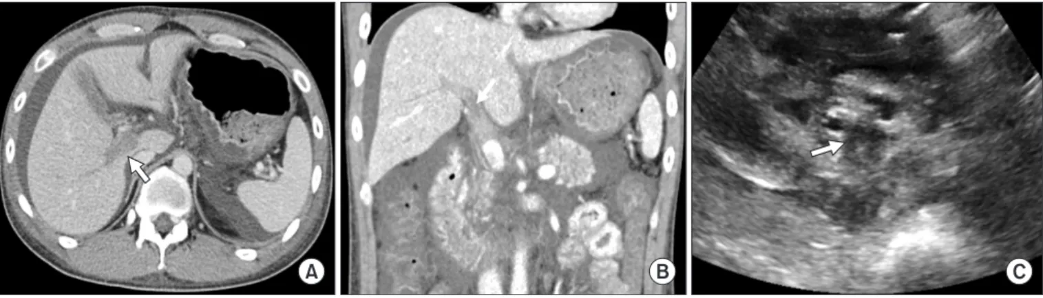
Chest X-ray films showed bilateral pleural effusion and mild pulmonary edema. Abdominal computed tomography (CT) and ultrasonography performed on admission showed thrombosis involving the main and right portal vein without cavernous trans- formation, suggesting an acute phase of thrombosis (Fig. 1). The patient immediately received anticoagulation therapy with intra- venous heparin infusion, followed by oral warfarin. We adjusted the warfarin dose to maintain a target INR between 2.0 and 3.0. In addition, high-dose oral prednisolone at a dose of 1 mg/
kg/day was administered to treat relapse of nephrotic syndrome.
A two-week corticosteroid treatment failed to significantly reduce proteinuria (19 g/day) and the patient was not toler- ant to the side effects of steroid, thus cyclosporine at 5 mg/kg/
day was added. One month later, 24-hr proteinuria decreased to 0.52 g/day and kidney function recovered (eGFR>90 mL/
min/1.73 m2). Complete remission was achieved after six weeks of cyclosporine use. Follow-up abdominal ultrasonography eight weeks after the onset of portal vein thrombosis showed a patent portal vein without thrombus (Fig. 2). Anticoagulation therapy was maintained for one additional month and was then discon- tinued. Cyclosporine at a reduced dose of 2.0 mg/kg/day was maintained during the following 24 months and was tapered to 1.0 mg/kg/day. The patient has remained in remission without
thromboembolic events.
Discussion
Nephrotic syndrome increases the risk of developing venous thrombosis in approximately 25% of patients [1]. The mecha- nisms underlying such a hypercoagulable state are deficiency of endogenous antithrombotic factors, including antithrombin III and proteins C and S due to urinary loss; increased formation of factors that promote thrombosis including factor V and VIII, von Willebrand factor, fibrinogen, and plasminogen activator in- Fig. 1. Imaging studies for portal vein thrombosis. (A) Transverse and (B) coronal views of an abdominal computed tomography scan clearly show portal vein thrombosis (arrow). (C) Thrombi are also noted in an abdominal ultrasonography (arrow).
Fig. 2. Follow-up abdominal ultrasonography eight weeks after the onset of portal vein thrombosis. There is no evidence of thrombi in portal vein, suggesting resolution of portal vein thrombosis after complete remission was achieved.
hibitor 1; and impaired thrombolytic activity, such as decreased plasminogen levels [1-4]. In addition, hypovolemia-induced hyperviscosity, hypercholesterolemia, hypoalbuminemia, and the use of corticosteroid are also associated with the development of thromboembolic events [5-7].
Thromboembolic events most commonly involve the renal vein, deep vein of the lower limbs in patients with the greatest risk seen in membranous glomerulopathy, menbranoproliferative glomerulonephritis, and minimal change disease among subtypes of nephrotic syndrome [2,3]. Portal vein thrombosis is rare.
A review of the literature identified nine cases of portal vein thrombosis to date (Table 1) [8-16]. All occurred in the setting
of severe hypoalbuminemia (1.5 to 2.1 g/dL), heavy proteinuria (3.46 to 25 g/day), high ratio of proteinuria to serum albumin, hypercholesterolemia (360 to 549 mg/dL), and polycythemia (48% to 56%) secondary to hypovolemia when nephrotic syn- drome was first diagnosed or relapsed. Most cases of portal vein thrombosis occurred in MCD. Seven cases (77.8%) were histologically proven by renal biopsy, while the others were clin- ically presumed to have MCD due to a rapid response to steroid therapy, but renal biopsy was not performed. In line with these findings, our patient clearly had a relapse of MCD and exhib- ited a markedly hypovolemic status presenting polycythemia (58.6%) before the use of diuretics and full-blown nephrotic Table 1. Nine cases of portal vein thrombosis in nephrotic syndrome reported in the literature
Reference Age
(yr) Sex Onset Clinical
presentation Involvement of thrombosis
Serum albumin
(g/mL)
Serum cholesterol
(mg/dL)
Urine protein (g/day)
Hb (g/dL)
(Hct%) Renal biopsy
[8] 27 F At
relapse
Abdominal pain, ascites
Portal vein, hepatic vein, splenic vein
1.8 Unknown 25 19.4 (56) MCD
[9] 41 M At
relapse
Abdominal pain, ascites, nausea, vomiting
Splenomes- enteric portal axis, IVC, renal veins
1.7 452 7 16.6 MCD
[10] 44 M At
diagnosis
Abdominal pain, vomiting, ascites
Portal vein, splenic vein
1.9 360 10 17.0 (53) Not
done (MCD)
[11] 51 F At
diagnosis
Asymptomatic Main portal trunk, left renal vein, splenic vein
1.7 549 19.7 18.3 (55) MCD
[12] 45 M At
diagnosis
Abdominal pain, distension
Portal vein 1.6 491 5.5 15.2 (48) MCD
[13] 52 M At
diagnosis
Abdominal pain, distension
Portal vein, SMV 1.7 502 3.5 16.1 Not
done (MCD)
[14] 53 M At
relapse
Abdominal pain Portal vein, SMV 2.1 Unknown 5.29 17.3 (50) MCD
[15] 19 M At
diagnosis
Abdominal pain Portal vein, splenic vein, SMV
1.7 372 3.46 16.6 (49) MCD
[16] 18 M At
diagnosis
Abdominal pain Portal vein, splenic vein, SMV
1.5 465 15.3 15.7 MCD
Our case 26 M At
relapse
Abdominal pain, nausea, vomiting, ascites
Portal vein 1.7 472 25.3 20.2 (59) MCD
MCD, minimal change disease; IVC, inferior vena cava; SMV, superior mesenteric vein.
features such as heavy proteinuria (25.3 g/day), hypoalbumin- emia (1.7 g/dL), high ratio of proteinuria to serum albumin, and hypercholesterolemia (472 mg/dL). However, why portal vein thrombosis is more common in MCD remains unclear be- cause these features are common in all subtypes of nephrotic syndrome. The more severe clinical nephrotic features combined with hyperviscosity secondary to hypovolemia might contribute to the increased tendency for thromboembolism irrespective of nephrotic syndrome subtype. Further studies are required to delineate whether MCD is more predisposing to portal vein thrombosis than other subtypes.
Portal venous thrombosis is a rare condition, thus it cannot be easily diagnosed when patients with nephrotic syndrome present with abdominal pain. Conventionally, gastrointestinal manifesta- tions such as abdominal pain, nausea, and vomiting are consid- ered as ‘nephrotic crisis.’ Although the mechanisms responsible for this are not clear, severe volume depletion associated with full-blown nephrotic status can cause bowel ischemia. This con- dition can be treated by adequate intravenous fluid resuscitation.
However, in cases of portal vein thrombosis, this therapy alone is not sufficient, and persistently unresolved portal hypertension may lead to a fatal outcome. Therefore, portal vein thrombosis should be considered in the differential diagnosis when assessing abdominal pain in patients with nephrotic syndrome. In addi- tion, imaging studies such as an abdominal ultrasonography or CT scan should be encouraged to detect this rare complication.
Spontaneous recanalization after portal vein thrombosis is un- likely, but recanalization occurs in about 40% of patients treated with anticoagulation therapy, which can prevent further throm- bosis and development of portal hypertension and its complica- tions [17,18]. Timing of the initiation of anticoagulation therapy is important because the probability of recanalization decreases from 69% when anticoagulation is initiated within the first week after diagnosis to 25% when initiated in the second week [18,19]. Although there is currently no pre-set guideline on an- ticoagulation therapy in patients with nephrotic syndrome com- plicated by thromboembolic events, immediate anticoagulation treatment with unfractionated or low molecular heparin and oral warfarin is recommended in symptomatic portal vein throm- bosis. Also, anticoagulant therapy should be maintained for the duration of nephrotic syndrome as long as the thromboembolic risk persists [3]. Furthermore, achieving remission of nephrotic syndrome with immunosuppression is of paramount importance
to recover the altered coagulation profiles.
In conclusion, portal vein thrombosis is rare and may not be easily diagnosed in patients with nephrotic syndrome suffering from abdominal pain. Early recognition of this rare complica- tion and prompt anticoagulation therapy and immunosuppression are emphasized to avoid a fatal outcome.
References
1. Kerlin BA, Ayoob R, Smoyer WE. Epidemiology and pathophysi- ology of nephrotic syndrome-associated thromboembolic dis- ease. Clin J Am Soc Nephrol 2012;7:513-520.
2. Singhal R, Brimble KS. Thromboembolic complications in the nephrotic syndrome: pathophysiology and clinical manage- ment. Thromb Res 2006;118:397-407.
3. Glassock RJ. Prophylactic anticoagulation in nephrotic syn- drome: a clinical conundrum. J Am Soc Nephrol 2007;18:2221- 2225.
4. Loscalzo J. Venous thrombosis in the nephrotic syndrome. N Engl J Med 2013;368:956-958.
5. Ozsoylu S, Strauss HS, Diamond LK. Effects of corticosteroids on coagulation of the blood. Nature 1962;195:1214-1215.
6. Bostom AG, Shemin D. Abnormalities of lipoprotein metabo- lism in the nephrotic syndrome. N Engl J Med 1991;324:697-698.
7. Fahal IH, McClelland P, Hay CR, Bell GM. Arterial thrombosis in the nephrotic syndrome. Postgrad Med J 1994;70:905-909.
8. Woolf AS, Street PR, Walmsley KM, Cohen SL. Portal vein thrombosis in the nephrotic syndrome. Nephrol Dial Transplant 1989;4:581-582.
9. De Luca M, Dugo M, Arduini R, Liessi G. Acute venous throm- bosis of spleno-mesenteric portal axis: an unusual localization of thromboembolism in the nephrotic syndrome. Am J Nephrol 1991;11:260-263.
10. Plaisier EM, Legallicier B, Faintuch JM, Ronco PM. Acute portal vein thrombosis at the onset of a nephrotic syndrome. Nephrol Dial Transplant 1996;11:696-698.
11. Etoh Y, Ohsawa I, Fujita T, Fuke Y, Endo M, Ohi H, et al. Nephrot- ic syndrome with portal, splenic and renal vein thrombosis. A case report. Nephron 2002;92:680-684.
12. Varghese J, Mathew A, Seethalekshmy NV, Kurian G, Unni VN.
Isolated portal vein thrombosis in nephrotic syndrome. Indian J Nephrology 2007;17:26-28.
13. Sun L, Xu C. Portal vein thrombosis as the first sign of nephrotic syndrome. Nat Clin Pract Nephrol 2008 ;4:342-345.
14. Bian F, Ge QM, Jiang GR, Wen LP. Cavernous transformation of the portal vein in nephrotic syndrome. Vasa 2011;40:323-326.
15. Wang YC, Chuang FR, Lee WC, Chen TC, Ko SF, Wang IK, et al.
Low-molecular-weight heparin successfully used to treat a ne- phrotic patient complicated by superior mesenteric vein throm- bosis and portal vein thrombosis. Med Princ Pract 2011;20:196- 199.
16. Wang J, Fan Q, Chen Y, Dong X, Zhang Y, Feng J, et al. A case re-
port of minimal change nephrotic syndrome complicated with portal, splenic and superior mesenteric vein thrombosis. Clin Nephrol 2012;77:505-509.
17. Sogaard KK, Astrup LB, Vilstrup H, Gronbaek H. Portal vein thrombosis; risk factors, clinical presentation and treatment.
BMC Gastroenterol 2007;7:34.
18. Turnes J, Garcia-Pagan JC, Gonzalez M, Aracil C, Calleja JL,
Ripoll C, et al. Portal hypertension-related complications after acute portal vein thrombosis: impact of early anticoagulation.
Clin Gastroenterol Hepatol 2008;6:1412-1417.
19. Condat B, Pessione F, Helene Denninger M, Hillaire S, Valla D.
Recent portal or mesenteric venous thrombosis: increased rec- ognition and frequent recanalization on anticoagulant therapy.
Hepatology 2000;32:466-470.