Introduction
Adiponectin is expressed exclusively by differentiated adipocytes, and its expression is induced during adipocyte differentiation [1]. Adiponectin is also expressed in the kidney, and it has been found on the endothelium and smooth muscle cells of intrarenal arteries/arterioles and on the endothelium of glomerular and peritubular capil- laries in normal kidneys [2]. Adiponectin was shown to be related to renal disease [3-6].
CTRP1 is an adiponectin paralog [7] that is expressed ubiquitously, most prominently in vascular tissues [8].
CTRP1 is also expressed in the kidney. Relatively large amout of CTRP1 transcripts were detected in the mouse kidney using a semiquantitative RT-PCR method [7]. How- ever, the localization of CTRP1 expression and its role in the kidney remain unclear. The primary goal of this study was to localize CTRP1 expression and to investigate its function in the kidney.
Materials and Methods
1. Experimental animals
Five adult male C57/BL6 and five male FGS/Nga [9,10]
mice were maintained under standard laboratory con- ditions on a 12-h light/dark cycle, with free access to food and water.
2. Northern blot analysis for CTRP1
Total RNA was obtained from the kidneys of adult male
Podocyte-specific Expression of C1q/TNF-α Related Protein 1 in Mice
Keun-Ja Cho
1,2, Young Yang
3, Young Ho Lee
4,5, Sooil Kim
4,51Department of Emergency Medical Service, and 2Research Center for Health Industry, Kongju National University,
3Department of Biological Sciences, Sookmyung Women’s University,
4Department of Anatomy, 5Research Institute for Medical Sciences, School of Medicine, Chungnam National University (Received 5 December 2013, revised 20 December 2013, accepted 21 December 2013, Published Online 30 December 2013)
Abstract : C1q/TNF-α Related Protein 1 (CTRP1), an adiponectin paralog, is a novel member of the C1q-TNF Related Protein family. CTRP1 is expressed in the kidney, although its localization and role in the kidney have not been studied. This study examined CTRP1 expression and function in the kidney.
CTRP1 immunohistochemistry and PAS staining of the kidneys of C57/BL6 and FGS/Nga mice were performed.
In situ hybridization for podocin in the kidney was also performed.
CTRP1 immunoreactivity was found only in the glomeruli of the kidney. The CTRP1-immunoreactive cells in the glomeruli were identified as podocytes. The number of CTPR1-immunoreactive cells and the intensity of CTRP1 immunoreactivity were lower in the glomeruli of FGS/Nga mice, which develop progressive proteinuria and focal glomerulosclerosis.
CTRP1 is a novel protein expressed in podocytes of the mouse kidney and may have a role in podocytes related to glomerular filtration in the kidney.
Keywords:Podocyte, CTRP1, Glomerulosclerosis, Proteinuria, Glomerular filtration
*This work was supported by research fund of Chungnam National Univer- sity.
The author (s) agree to abide by the good publication practice guideline for medical journals.
The author (s) declare that there are no conflicts of interest.
Correspondence to : Sooil Kim (Department of Anatomy, School of Medicine, Chungnam National University)
E-mail : [email protected]
Korean J Phys Anthropol Vol. 26, No. 4 (2013) pp. 147~153
http://dx.doi.org/10.11637/kjpa.2013.26.4.147 Original Article
C57/BL6 mice. A Multiple Tissue Northern (MTN) blot was purchased from Clontech (CA, USA). 32P-labeled cDNA probes were prepared from the coding region of CTRP1 cDNA using a High Prime DNA labeling kit (Roche, IN, USA) and were used for hybridization. After hybridization, the membrane was washed with 0.1× SSC, 0.05% SDS at 50�C and exposed to X-ray film for 18 h at -80�C with an intensifying screen.
3. Tissue preparation
Perfusion fixation was performed transcardially in 4- month-old male C57/BL6 and FGS/Nga mice, first with 50 mL of 0.05 M phosphate-buffered saline (PBS, pH 7.4) containing heparin (1 IU/mL) at 4�C and then with 50 mL of ice-cold 10% neutral-buffered formalin for 10 min at a flow rate of 30~40 mL/min. The kidneys of the mice were removed immediately and then fixed in the latter solution overnight. The fixed tissues were embedded in paraffin.
4. Periodic acid Schiff (PAS) stain
To examine glomerulosclerotic lesions in the kidney, paraffin sections of the FGS/Nga mouse kidney were stain- ed with PAS [11,12].
5. Antibody preparation and CTRP1 immunohistochemistry
Rabbit anti-CTRP1 polyclonal antibody was prepared for immunohistochemistry [13,14]. Briefly, mouse CTRP1 cDNA encoding the collagen repeat domain was introduc- ed into the pET-28a bacterial expression vector (Novagen, Darmstadt, Germany), which was then transformed into E. coli strain BL21(λDE3). The expression of the collagen domain of CTRP1 was induced by the addition of 1 mM IPTG. The collagen domain of CTRP1 was expressed in the form of an inclusion body and was purified according to the full-length protein folding protocol. The purified colla- gen domain of CTRP1 was injected subcutaneously into rabbits in one dose. Four months later, the rabbits were sacrificed to obtain rabbit anti-CTRP1 polyclonal antibody.
The deparaffinized sections were heated for 4 min in a pressure cooker containing 10 mM citrate buffer (pH 6.0) for antigen retrieval. Subsequent procedures were con- ducted at room temperature. The sections were pretreated with 3% H2O2in 0.1 M PBS (pH 7.4) for 30 min to quench endogenous peroxidase. The sections were incubated for
1 h at room temperature (RT) in the polyclonal CTRP1 antibody in 0.1 M PBS (pH 7.4) containing 0.1% Triton X-100, 1.5% bovine serum albumin (BSA), and 1 : 200 normal goat serum (NGS), and then incubated for 1 h at RT in 1 : 200 biotinylated goat anti-rabbit IgG (Vector, CA, USA) and 1 : 200 NGS in PBS. The immunoreactions were visualized by incubation for 1 h at RT in avidin-biotin- peroxidase complex (1 : 100, ABC kit, Vector) in PBS and for 5~10 min in 0.05% 3,3′-diaminobenzidine and 0.01%
H2O2in 0.1 M PBS. Immunolabeled sections were dehy- drated in a graded ethanol series, defatted in xylene, and mounted. A similar procedure was used in control experi- ments, except the sections were processed either in the presence of antibody that had been pre-adsorbed overnight with an excess of immunizing recombinant protein or in the absence of primary CTRP1 antibody.
6. In situ hybridization for podocin
Immonohistochemistry for podocin, a specific marker for podocytes, with commercially available antibodies showed nonspecific labeling pattern in the kidney. To evaluate whether the CTRP1 immunoreactive cells were podocytes, in situ hybridization for the podocin gene was performed with digoxigenin-labeled riboprobes. The podo- cin antisense and sense riboprobes were synthesized from mouse cDNA fragments of the coding region cloned into pGEM-T vector (Promega, CA, USA) flanked by the Sp6 and T7 promoters. The paraffin sections that were adjacent to the sections used for CTRP1 immunohistochemistry were used for in situ hybridization. The deparaffinized sections were treated with proteinase K, acetylated with 0.25% acetic anhydride, prehybridized, hybridized with probe at a concentration of 0.5 μg/mL, treated with RNase A, and washed with SSC. The washed tissues were then incubated with anti-digoxigenin alkaline phosphate-con- jugated serum (diluted 1 : 500, Roche). The final coloring reaction was performed using nitroblue tetrazolium/5- bromo-4-chloro-3-indolyl-phosphate solution. Each sam- ple section was dehydrated and covered with a coverslip, using Permount for viewing.
Results
The CTRP1 gene was expressed more abundantly in the mouse heart, liver, and kidney than in the lung, brain,
spleen, skeletal muscle, and testis (Fig. 1).
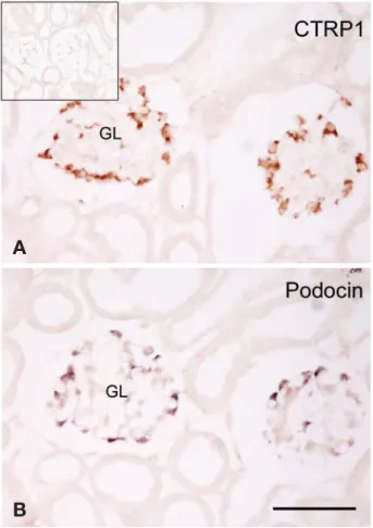
No signal was detected in the absence of primary anti- bodies in kidney sections. Pre-incubation of the antibody against CTRP1 with the corresponding CRTP1 proteins also completely abolished the immunolabeling in kidney sections (data not shown). CTRP1 immunoreactivity was found in the cells located in the peripheral part of the glo- meruli. The location and shape of the CTRP1-immuno- reactive cells strongly suggested that CTRP1 is expressed in podocytes (Fig. 2A). In situ hybridization for podocin in the adjacent tissue section showed that the pattern of podocin-positive cells was very similar to that of CTRP1- immunoreactive cells (Fig. 2B). These two stains suggest that CTRP1 is expressed in podocytes. CTRP1 immuno- reactivity was detected in the cytoplasm of the podocyte cell body, but may not be on the cell membrane of the foot process. This implies that CTRP1 is not related to the fil- tration barrier directly, including the foot processes of podocytes and slit diaphragm.
PAS staining showed that the amount of PAS-positive mesangial matrix was increased in the kidneys of FGS/
Nga mice (Fig. 3A, 3B). Increased amounts of PAS-posi-
tive mesangial matrix indicate a thickened basement membrane in the glomeruli. This is one of the features of glomerular filtration membrane damage and increased protein permeability. Urine analysis with a urine analysis strip showed proteinuria in the FGS/Nga mice (data not shown). Given that the FGS/Nga mice developed protein- uria and glomerulosclerosis beginning at the age of 3 months, this experimental animal provides an available animal model of glomerulosclerosis. Both the number of CTRP1 immunoreactive cells and the intensity of CTRP1 immunoreactivity were decreased in the sclerotic glomeruli regions, compared with the normal region in the FGS/Nga mice (Fig. 3C, 3D). These findings suggest that CTRP1 is Fig. 1. Tissue expression pattern of the CTRP1 gene in the mouse
(Northern blot analysis). CTPR1 mRNA is expressed abundantly in the mouse heart, liver, and kidney.
CTRP1 9.5
7.5
4.4
2.4
1.35
0.24
kb
Heart Brain Spleen Lung Liver Skeletal muscle Kidney Testis
Fig. 2. Immunohistochemistry for CTRP1 (A) and in situ hybri- dization for podocin (B) in the C57/BL6 mouse kidney. Serially sectioned kidney slides were used for the colocalization of CTRP1 and podocin. (A) CTRP1 immunoreactivity is seen in probable podocytes in the kidney. Square in the figure A is immunoreactivity of negative control in the absence of primary antibody. (B) Podocin, a specific marker for podocytes, is expressed in the podo- cytes in the kidney. GL; glomerulus, Scale bar in B==50 μm in A.
A
B
involved in the normal activity of podocytes related to glomerular filtration.
Discussion
Our data show that CTRP1 is expressed in the podocytes of the mouse kidney, and that CTRP1 expression is down- regulated in the glomeruli in the kidney accompanying focal glomerulosclerosis. This study provides the first description of the localization of CTRP1 expression in the normal kidney and in the FGS/Nga mouse, a model of glomerulosclerosis.
Adiponectin has collagen repeat and complement C1q domains [15,16], can bind to collagen, and shows anti- inflammatory and vasoprotective activities [17-20], in addi- tion to an anti-diabetic effect. CTRP1 also has collagen
repeat and C1q domains [7], which are features of adipo- nectin paralogs. Consequently, we expect that CTRP1 has a function similar to that of adiponectin. A recent report showed that CTRP1 prevents collagen-induced platelet aggregation by specifically blocking von Willebrand’s factor (vVW) binding to collagen [8]. In this study, we showed that CTRP1 is expressed in podocytes but not in endothelial cells or smooth muscle cells of the intrarenal arteries/arterioles, or in the endothelium of the glomerular and peritubular capillaries in the normal kidney [2]. In addition, CTRP1 expression is attenuated in the podocytes of an animal model of glomerulosclerosis. These facts sug- gest that CTRP1 is involved in the normal function of the glomerular filtration barrier.
The filtration barrier of the kidney consists of three spe- cialized layers: the capillary endothelium, the glomerular basement membrane, and a single-cell layer of glomerular Fig. 3. Histopathology and CTRP1 immunoreactivity in normal and pathologic (FGS) regions in FGS/Nga mice. An FGS/Nga mouse showing focal glomerulosclerosis (rectangular area in B) in the kidney (A and B, PAS stain). CTRP1 immunoreactivity was decreased in the pathologic glomeruli compared with the normal region in the FGS/Nga mouse (C and D, counterstained with Mayer’s Hematoxylin).
Arrows, CTRP1 immunoreactive cells, GL; glomerulus, Scale bar in D==50 μm in A~C.
A B
C D
epithelial cells or podocytes. The foot processes of neigh- boring podocytes interdigitate regularly, leaving filtration slits between them that are bridged by an extracellular structure known as the slit diaphragm. A growing number of proteins, including nephrin, CD2AP, FAT, ZO-1, P- cadherin, podocin, and Neph 1-3 have been shown to be expressed in the slit diaphragm, and some of these mole- cules play a major role in maintaining the structural and functional integrity of the slit diaphragm [21,22]. However, our data show that CTRP1 is expressed in the cytoplasm of podocyte cell body, not in the cell membrane of foot process or slit diaphragm. Although CTRP1 is not express- ed in the slit diaphragm, we cannot rule out the possibility that CTRP1 acts in the slit diaphragm because CTRP1 is a secretory protein [7] released from the podocytes in the glomeruli and can reach the slit diaphragm. Further in vivo and in vitro study is needed to elucidate the role of CTRP1 in the kidney.
In conclusion, CTRP1 is expressed in the podocytes of the mouse kidney and may be involved in the role of podo- cytes in glomerular filtration in the kidney.
References
1. Shapiro L, Scherer PE. The crystal structure of a comple- ment-1q family protein suggests an evolutionary link to tumor necrosis factor. Curr Biol. 1998; 8:335-8.
2. Rovin BH, Song H, Hebert LA, Nadasdy T, Nadasdy G, Birmingham DJ, et al. Plasma, urine, and renal expression of adiponectin in human systemic lupus erythematosus.
Kidney Int. 2005; 68:1825-33.
3. Heimburger O, Stenvinkel P. Adipokines in chronic kidney disease-fat tissue gives nephrologists a message. Perit Dial Int. 2005; 25:340-2.
4. Isobe T, Saitoh S, Takagi S, Takeuchi H, Chiba Y, Katoh N, et al. Influence of gender, age and renal function on plasma adiponectin level: the Tanno and Sobetsu study. Eur J Endo- crinol. 2005; 153:91-8.
5. Ignacy W, Chudek J, Adamczak M, Funahashi T, Matsu- zawa Y, Kokot F, et al. Reciprocal association of plasma adiponectin and serum C-reactive protein concentration in haemodialysis patients with end-stage kidney disease--a follow-up study. Nephron Clin Pract. 2005; 101:c18-c24.
6. Schalkwijk CG, Chaturvedi N, Schram MT, Fuller JH, Ste- houwer CD. EURODIAB Prospective Complications Study Group. Adiponectin is inversely associated with renal func- tion in type 1 diabetic patients. J Clin Endocrinol Metab.
2006; 91:129-35.
7. Wong GW, Wang J, Hug C, Tsao TS, Lodish HF. A family of Acrp30/adiponectin structural and functional paralogs.
Proc Natl Acad Sci USA. 2004; 101:10302-7.
8. Lasser G, Guchhait P, Ellsworth JL, Sheppard P, Lewis K, Bishop P, et al. C1qTNF-related protein-1 (CTRP-1): a vascular wall protein that inhibits collagen-induced platelet aggregation by blocking VWF binding to collagen. Blood.
2006; 107:423-30.
9. Yoshida F, Matsuo S, Fujishima H, Kim HK, Tomita T.
Renal lesions of the FGS strain of mice: a spontaneous ani- mal model of progressive glomerulosclerosis. Nephron.
1994; 66:317-25.
10. Kim EH, Choi KS, Lee KW, Suh JG, Choi YK, Hyun BH, et al. Changes of renal lesion-related parameters in FGS/
Nga and the parental mouse strains, CBA/N and RFM/Nga.
Exp Anim. 2004; 53:97-102.
11. Jiang T, Liebman SE, Lucia MS, Li J, Levi M. Role of altered renal lipid metabolism and the sterol regulatory ele- ment binding proteins in the pathogenesis of age-related renal disease. Kidney Int. 2005; 68:2608-20.
12. Pedrycz A, Wieczorski M, Czerny K. Histological and histo- chemical assessment of the effects of a single dose adriamy- cin on fetal rat kidney. Acta Histochem. 2005; 107:215-20.
13. Jeon JH, Kim KY, Kim JH, Baek A, Cho H, Lee YH, et al.
A novel adipokine CTRP1 stimulates aldosterone produc- tion. FASEB J. 2008; 22:1502-11.
14. Kim KY, Kim HY, Kim JH, Lee CH, Kim DH, Lee YH, et al. Tumor necrosis factor-alpha and interleukin-1beta incre- ases CTRP1 expression in adipose tissue. FEBS Lett. 2006;
580:3953-60.
15. Ishikawa Y, Akasaka Y, Ishii T, Yoda-Murakami M, Choi- Miura NH, Tomita M, et al. Changes in the distribution pattern of gelatin-binding protein of 28 kDa (adiponectin) in myocardial remodelling after ischaemic injury. Histo- pathology. 2003; 42:43-52.
16. Diez JJ, Iglesias P. The role of the novel adipocyte-derived hormone adiponectin in human disease. Eur J Endocrinol.
2003; 148:293-300.
17. Ouchi N, Kihara S, Arita Y, Maeda K, Kuriyama H, Oka- moto Y, et al. Novel modulator for endothelial adhesion molecules: adipocyte-derived plasma protein adiponectin.
Circulation. 1999; 100:2473-6.
18. Kawanami D, Maemura K, Takeda N, Harada T, Nojiri T, Imai Y, et al. Direct reciprocal effects of resistin and adipo- nectin on vascular endothelial cells: a new insight into adi- pocytokine-endothelial cell interactions. Biochem Biophys Res Commun. 2004; 314:415-9.
19. Kumada M, Kihara S, Ouchi N, Kobayashi H, Okamoto Y, Ohashi K, et al. Adiponectin specifically increased tissue
inhibitor of metalloproteinase-1 through interleukin-10 expression in human macrophages. Circulation. 2004; 109:
2046-9.
20. Chen H, Montagnani M, Funahashi T, Shimomura I, Quon MJ. Adiponectin stimulates production of nitric oxide in vascular endothelial cells. J Biol Chem. 2003; 278:45021-6.
21. Asanuma K, Mundel P. The role of podocytes in glomeru- lar pathobiology. Clin Exp Nephrol. 2003; 7:255-9.
22. Levidiotis V, Power DA. New insights into the molecular biology of the glomerular filtration barrier and associated disease. Nephrology. 2005; 10:157-66.
생쥐에서 C1q/TNF-α Related Protein 1의 발세포 특이적 발현
조근자
1,2, 양 영
3, 이영호
4,5, 김수일
4,5공주대학교 1응급구조학과, 2건강산업연구센터, 3숙명여자대학교 생명과학과, 충남대학교 4의학전문대학원 해부학교실, 5의학연구소
간추림 : C1q/TNF-α Related Protein 1 (CTRP1)은 adiponectin의 파라로그로서 C1q/TNF-α Related Protein 계열 의 새로운 물질이다. CTRP1은 콩팥에서 발현되지만 콩팥에서의 국소발현과 기능에 대해서는 알려져 있지 않 다. 본 연구에서는 CTRP1의 콩팥에서 발현과 기능을 알아보고자 하였다.
C57/BL6쥐와 FGS/Nga 생쥐의 콩팥에서 CTRP1에 대한 면역조직화학염색과 PAS 염색을 하였다. Podocin에 대한 in situ hybridization도 생쥐 콩팥에서 시행하였다.
CTRP1 양성세포가 콩팥의 토리에서 발견되었으며, 이들 세포가 발세포임을 확인하였다. CTRP1 양성세포 수 와 면역강도는 진행형 단백뇨와 국소토리굳음증을 나타내는 FGS/Nga 생쥐 토리에서 감소하였다.
CTRP1은 생쥐 콩팥의 발세포에서 발현되는 단백질이며 콩팥에서 토리 여과와 관련된 역할을 할 것으로 본다.
찾아보기 낱말 : 발세포, CTRP1, 토리굳음증, 단백뇨, 토리 여과
교신저자 : 김수일(충남대학교 의학전문대학원 해부학교실) 전자우편 : [email protected]